. 2022 Oct 1;79(10):975-985.
doi: 10.1001/jamaneurol.2022.2379.

Association of Elevated Amyloid and Tau Positron Emission Tomography Signal With Near-Term Development of Alzheimer Disease Symptoms in Older Adults Without Cognitive Impairment

Collaborators, Affiliations

Association of Elevated Amyloid and Tau Positron Emission Tomography Signal With Near-Term Development of Alzheimer Disease Symptoms in Older Adults Without Cognitive Impairment

Cherie Strikwerda-Brown et al. JAMA Neurol. .

Abstract

Importance: National Institute on Aging-Alzheimer's Association (NIA-AA) workgroups have proposed biological research criteria intended to identify individuals with preclinical Alzheimer disease (AD).

Objective: To assess the clinical value of these biological criteria to identify older individuals without cognitive impairment who are at near-term risk of developing symptomatic AD.

Design, setting, and participants: This longitudinal cohort study used data from 4 independent population-based cohorts (PREVENT-AD, HABS, AIBL, and Knight ADRC) collected between 2003 and 2021. Participants were older adults without cognitive impairment with 1 year or more of clinical observation after amyloid β and tau positron emission tomography (PET). Median clinical follow-up after PET ranged from 1.94 to 3.66 years.

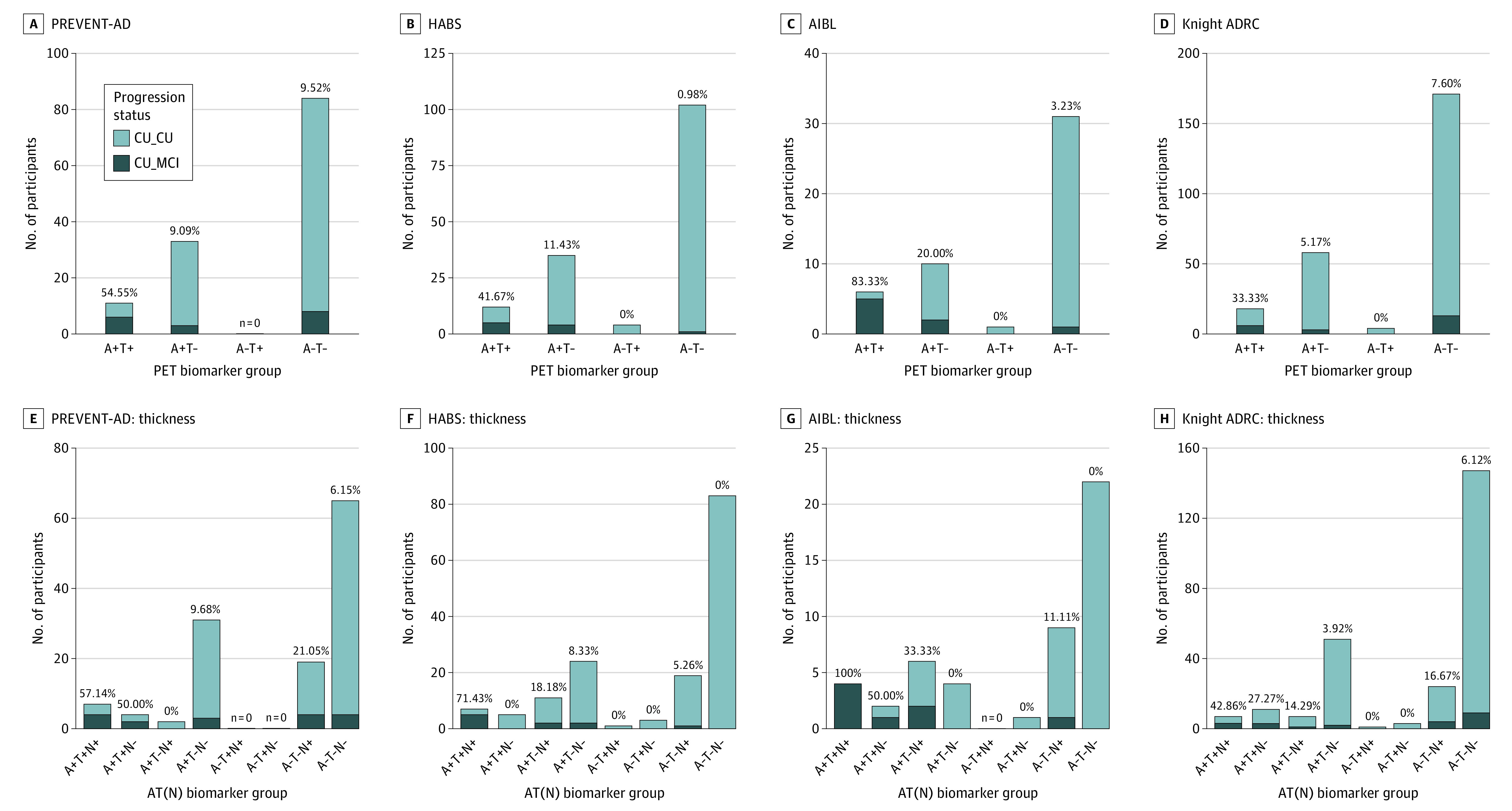
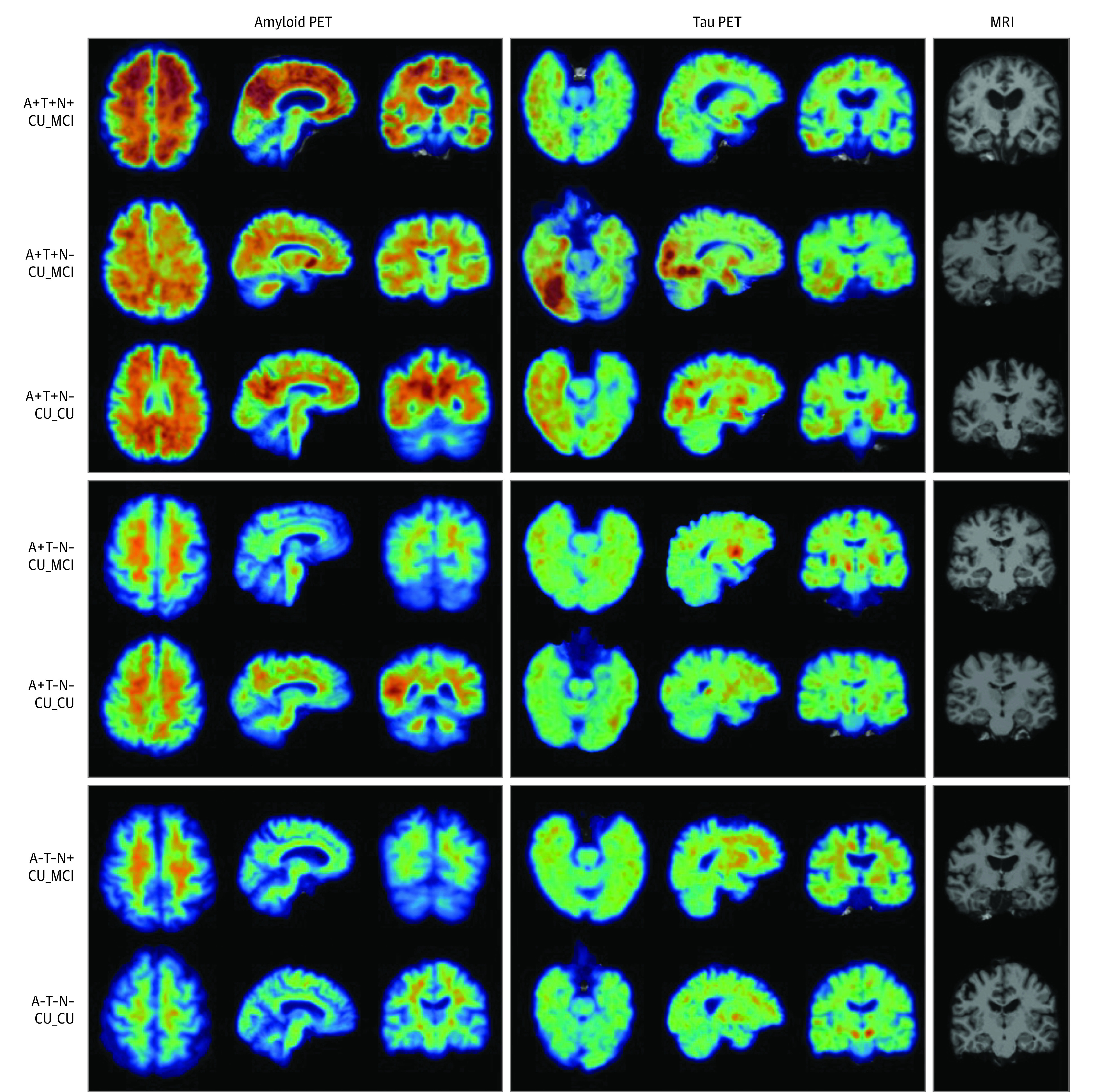
Exposures: Based on binary assessment of global amyloid burden (A) and a composite temporal region of tau PET uptake (T), participants were stratified into 4 groups (A+T+, A+T-, A-T+, A-T-). Presence (+) or absence (-) of neurodegeneration (N) was assessed using temporal cortical thickness.

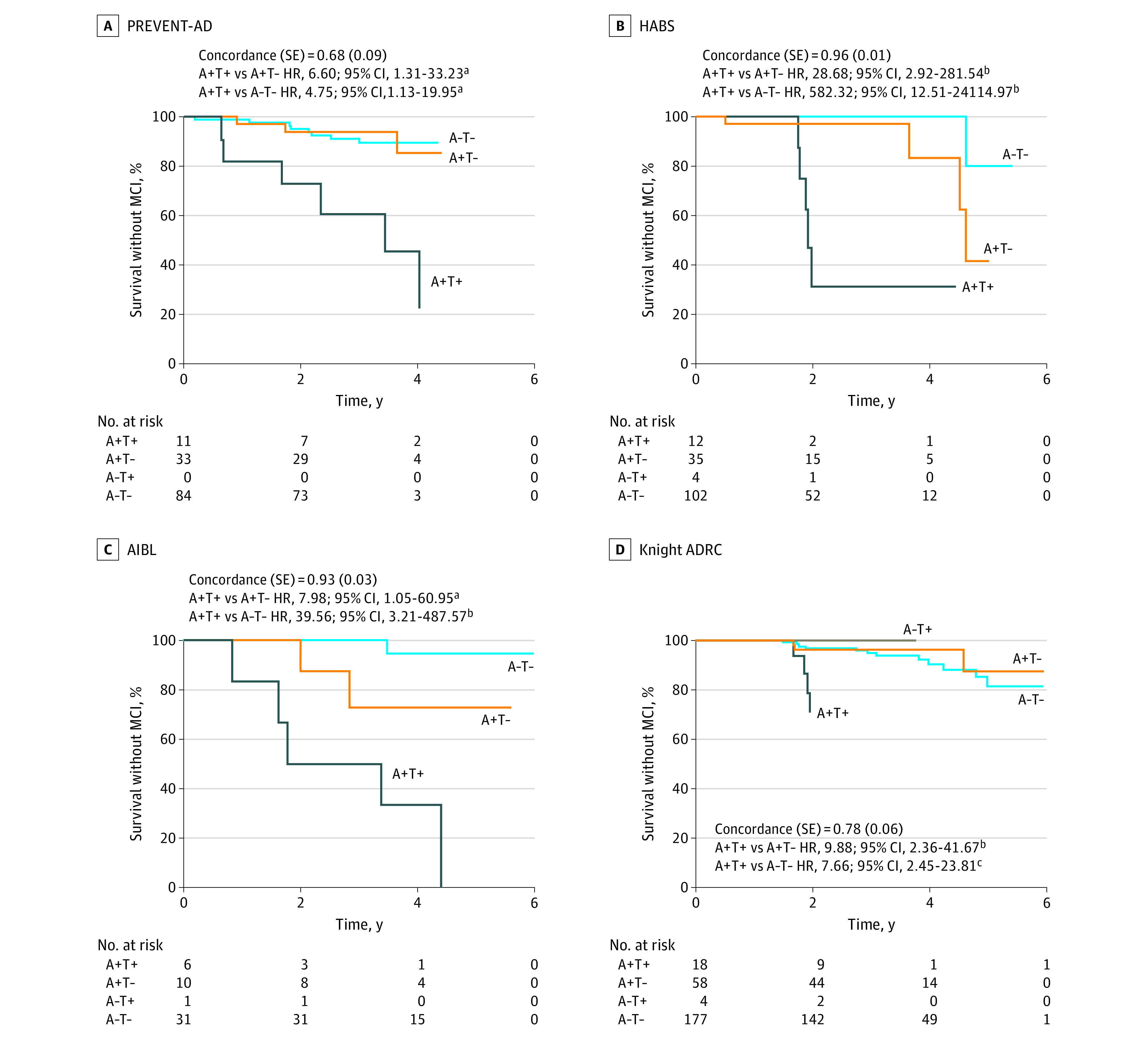
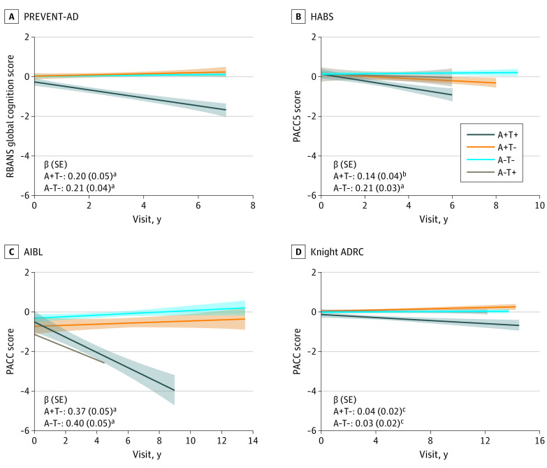
Main outcomes and measures: Each cohort was analyzed separately. Primary outcome was clinical progression to mild cognitive impairment (MCI), identified by a Clinical Dementia Rating score of 0.5 or greater in Knight ADRC and by consensus committee review in the other cohorts. Clinical raters were blind to imaging, genetic, and fluid biomarker data. A secondary outcome was cognitive decline, based on a slope greater than 1.5 SD below the mean of an independent subsample of individuals without cognitive impairment. Outcomes were compared across the biomarker groups.

Results: Among 580 participants (PREVENT-AD, 128; HABS, 153; AIBL, 48; Knight ADRC, 251), mean (SD) age ranged from 67 (5) to 76 (6) years across cohorts, with between 55% (137/251) and 74% (95/128) female participants. Across cohorts, 33% to 83% of A+T+ participants progressed to MCI during follow-up (mean progression time, 2-2.72 years), compared with less than 20% of participants in other biomarker groups. Progression further increased to 43% to 100% when restricted to A+T+(N+) individuals. Cox proportional hazard ratios for progression to MCI in the A+T+ group vs other biomarker groups were all 5 or greater. Many A+T+ nonprogressors also showed longitudinal cognitive decline, while cognitive trajectories in other groups remained predominantly stable.

Conclusions and relevance: The clinical prognostic value of NIA-AA research criteria was confirmed in 4 independent cohorts, with most A+T+(N+) older individuals without cognitive impairment developing AD symptoms within 2 to 3 years.


Conflict of interest statement

Conflict of Interest Disclosures: Dr Strikwerda-Brown reported grants from the Canadian Institutes of Health Research (CIHR), Fonds de Recherche du Québec–Santé (FRQS), and Alzheimer’s Society of Canada during the conduct of the study. Dr Gonneaud reported grants from the StoP-AD Center, Quebec Bio-Imaging Network, and Fondation Alzheimer and Fondation de France during the conduct of the study. Dr Soucy reported fees from Biogen Canada and being involved in pro bono projects with Optina Diagnostics outside the submitted work. Dr Benzinger reported grants from the National Institutes of Health (NIH) during the conduct of the study; nonfinancial support from Avid Radiopharmaceuticals, Eli Lilly, Cerveau, and Life Molecular Imaging; grants from Siemens; and consultant and advisor fees from Biogen, Eisai, and Siemens outside the submitted work. Dr Morris reported grants from the NIH during the conduct of the study. Dr Villemagne reported grants from Cerveau during the conduct of the study and consultant fees from Eli Lilly and Life Molecular Imaging outside the submitted work. Dr Sperling reported research funding from Eisai, Eli Lilly, the National Institute on Aging, and the Alzheimer’s Association; consultant fees from AC Immune, Acumen, Genentech, Janssen, Neuraly, Oligomerix, Prothena, Renew, Alnylam, Cytox, Japanese Organization for Medical Device Development, Nervgen, Neurocentria, Shionogi, Vigil Neuroscience, and Ionis; and being an employee of Brigham and Women’s Hospital. Dr Johnson reported grants from the NIH during the conduct of the study. Dr Rowe reported grants from National Health and Medical Research Council (APP1011689, APP10475151, APP1071430) and Cerveau Technologies during the conduct of the study; grants from Biogen, Eisai, Actinogen, and AbbVie outside the submitted work; speaker fees from Nutricia, Biogen, and Roche; scientific advisory board fees from Cerveau Technology and Prothena; and payment for preparation of educational materials from Biogen. Dr Poirier reported grants from Fonds de la Research en Santé Infrastructure outside the submitted work. Dr Villeneuve reported grants from the CIHR during the conduct of the study. No other disclosures were reported.

Figures

Figure 1.
Figure 1.. Participants Progressing to Mild Cognitive Impairment (MCI) After Positron Emission Tomography (PET) vs Those Remaining Cognitively Unimpaired (CU) in Each Biomarker Group, Across Cohorts
Percentage values represent the proportion of MCI progressors within the group. Note: The amyloid-negative, tau-positive (A−T+) group is displayed for visualization purposes but was not included in statistical analysis because of the small sample size. While the MCI classifications were based on clinical consensus in the PREVENT-AD, HABS, and AIBL cohorts, they were based on a Clinical Dementia Rating of 0.5 or greater for the Knight ADRC cohort. Neurodegeneration (N) was defined by temporal cortical thickness. ADRC indicates Alzheimer Disease Research Center; AIBL, Australian Imaging, Biomarker & Lifestyle study; CU_CU, cognitively unimpaired at time of PET, remaining cognitively unimpaired during follow-up; CU_MCI, cognitively unimpaired at time of PET, progressing to MCI during follow-up; HABS, Harvard Aging Brain Study; PREVENT-AD, Pre-symptomatic Evaluation of Experimental or Novel Treatments for Alzheimer Disease study.
Figure 2.
Figure 2.. Sample Amyloid β (A) and Tau (T) Positron Emission Tomography (PET) and Magnetic Resonance Imaging (MRI) Scans From Different Biomarker Groups in the PREVENT-AD Cohort
Demographic data in descending order are as follows: 73-year-old female participant; 70-year-old female participant; 65-year-old female participant; 64-year-old female participant; 66-year-old female participant; 61-year-old female participant; 69-year-old female participant. CU_CU indicates cognitively unimpaired at time of PET, remaining cognitively unimpaired during follow-up; CU_MCI, cognitively unimpaired at time of PET, progressing to mild cognitive impairment during follow-up; N, evidence of neurodegeneration; PREVENT-AD, Pre-symptomatic Evaluation of Experimental or Novel Treatments for Alzheimer Disease study.
Figure 3.
Figure 3.. Survival Curves Reflecting Time From Positron Emission Tomography (PET) Scan to Mild Cognitive Impairment (MCI) Classification for the 4 Biomarker Groups, Across Cohorts
The temporal meta–region of interest was used to define tau positivity (T+). Concordance values and SE are reported for the Cox regression models, which included age at the time of tau PET, sex, education, and APOE e4 status as covariates. Hazard ratios (HRs) for the PET biomarker groups are in reference to the amyloid-positive, tau-positive (A+T+) group. Inverted HRs are reported for ease of interpretation (ie, reflecting risk of progression to MCI in the A+T+ group relative to the other groups). The A−T+ group is displayed for visualization purposes but was not included in statistical analysis because of the small sample size. ADRC indicates Alzheimer Disease Research Center; AIBL, Australian Imaging, Biomarker & Lifestyle study; HABS, Harvard Aging Brain Study; PREVENT-AD, Pre-symptomatic Evaluation of Experimental or Novel Treatments for Alzheimer Disease study. aP < .05. bP < .01. cP < .001.
Figure 4.
Figure 4.. Longitudinal Cognitive Slopes for Each Biomarker Group Across Cohorts
Models included random slopes and intercepts for each participant and covariates of age, sex, and years of education. For all cohorts, positron emission tomography (PET) was added midstudy and was therefore performed at different cognitive follow-up visits for each participant. Longitudinal cognition analyses included time points both before and after PET scanning. The amyloid-negative, tau-positive (A−T+) group is displayed for visualization purposes but was not included in statistical analyses because of the small sample size. β estimates and SE are for the interaction between biomarker group and visit date, with A+T+ as the reference group. Cognitive scores are reported as cohort-derived z scores. ADRC indicates Alzheimer Disease Research Center; AIBL, Australian Imaging, Biomarker & Lifestyle study; HABS, Harvard Aging Brain Study; PACC, Preclinical Alzheimer Cognitive Composite; PREVENT-AD, Pre-symptomatic Evaluation of Experimental or Novel Treatments for Alzheimer Disease study; RBANS, Repeatable Battery for the Assessment of Neuropsychological Status. aP < .05. bP < .01. cP < .001.

Comment in

  • doi: 10.1001/jamaneurol.2022.2967

References

    1. Jack CR Jr, Bennett DA, Blennow K, et al. ; Contributors . NIA-AA research framework: toward a biological definition of Alzheimer’s disease. Alzheimers Dement. 2018;14(4):535-562. doi:10.1016/j.jalz.2018.02.018 - DOI - PMC - PubMed
    1. Dubois B, Villain N, Frisoni GB, et al. . Clinical diagnosis of Alzheimer’s disease: recommendations of the International Working Group. Lancet Neurol. 2021;20(6):484-496. doi:10.1016/S1474-4422(21)00066-1 - DOI - PMC - PubMed
    1. Lowe VJ, Bruinsma TJ, Min HK, et al. . Elevated medial temporal lobe and pervasive brain tau-PET signal in normal participants. Alzheimers Dement (Amst). 2018;10:210-216. doi:10.1016/j.dadm.2018.01.005 - DOI - PMC - PubMed
    1. Driscoll I, Troncoso J. Asymptomatic Alzheimer’s disease: a prodrome or a state of resilience? Curr Alzheimer Res. 2011;8(4):330-335. doi:10.2174/156720511795745348 - DOI - PMC - PubMed
    1. Folstein MF, Folstein SE, McHugh PR. “Mini-mental state”: a practical method for grading the cognitive state of patients for the clinician. J Psychiatr Res. 1975;12(3):189-198. doi:10.1016/0022-3956(75)90026-6 - DOI - PubMed

Publication types