. 2022 Aug 4;109(8):1436-1457.
doi: 10.1016/j.ajhg.2022.06.011. Epub 2022 Jul 30.

ADGRL1 haploinsufficiency causes a variable spectrum of neurodevelopmental disorders in humans and alters synaptic activity and behavior in a mouse model

Affiliations

ADGRL1 haploinsufficiency causes a variable spectrum of neurodevelopmental disorders in humans and alters synaptic activity and behavior in a mouse model

Antonio Vitobello et al. Am J Hum Genet. .

Abstract

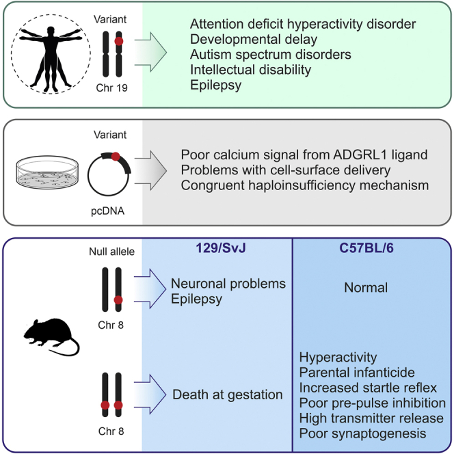
ADGRL1 (latrophilin 1), a well-characterized adhesion G protein-coupled receptor, has been implicated in synaptic development, maturation, and activity. However, the role of ADGRL1 in human disease has been elusive. Here, we describe ten individuals with variable neurodevelopmental features including developmental delay, intellectual disability, attention deficit hyperactivity and autism spectrum disorders, and epilepsy, all heterozygous for variants in ADGRL1. In vitro, human ADGRL1 variants expressed in neuroblastoma cells showed faulty ligand-induced regulation of intracellular Ca2+ influx, consistent with haploinsufficiency. In vivo, Adgrl1 was knocked out in mice and studied on two genetic backgrounds. On a non-permissive background, mice carrying a heterozygous Adgrl1 null allele exhibited neurological and developmental abnormalities, while homozygous mice were non-viable. On a permissive background, knockout animals were also born at sub-Mendelian ratios, but many Adgrl1 null mice survived gestation and reached adulthood. Adgrl1-/- mice demonstrated stereotypic behaviors, sexual dysfunction, bimodal extremes of locomotion, augmented startle reflex, and attenuated pre-pulse inhibition, which responded to risperidone. Ex vivo synaptic preparations displayed increased spontaneous exocytosis of dopamine, acetylcholine, and glutamate, but Adgrl1-/- neurons formed synapses in vitro poorly. Overall, our findings demonstrate that ADGRL1 haploinsufficiency leads to consistent developmental, neurological, and behavioral abnormalities in mice and humans.

Keywords: ADGRL1; ADHD; ASD; Adgrl1 knockout mice; attention deficit hyperactivity disorder; autism spectrum disorder; developmental delay; epilepsy; intellectual disability; malfunctional behavior in mice; neuropsychiatric disorders; variable expressivity.


Conflict of interest statement

Declaration of interests The Department of Molecular and Human Genetics at Baylor College of Medicine receives revenue from clinical genetic testing completed at Baylor Genetics Laboratories. A.C. is an employee of GeneDx, Inc.

Figures

None
Graphical abstract
Figure 1
Figure 1
Individuals and variants identified in our cohort (A) Schematic representation of ADGRL1 and distribution of the pathogenic variants reported in the study. Galactose-binding lectin domain (GL), olfactomedin-like domain (OLF), hormone receptor domain (HRM), GPCR-autoproteolysis-inducing domain (GAIN), GPCR proteolysis site domain (GPS), 7 transmembrane domain (7TM) and cytosolic latrophilin domain are depicted. Nonsense and frameshift variants are indicated in red. Missense variants are indicated in black. (B) Variant segregation analysis in the families described in this cohort. Individuals with documented evaluation are indicated as i1 through i10. Arrows indicate the first family member coming to medical attention. E1 indicates exome results regarding ADGRL1. E2 refers to the pathogenic TAOK1 variant (encoding p.Trp188) identified in family F9 (Table 1). DD, developmental delay; ID, intellectual disability; ASD, autism spectrum disorder; ADHD, attention deficit hyperactivity disorder. (C) Individuals with pathogenic ADGRL1 variants.
Figure 2
Figure 2
In vitro analysis of mutations in ADGRL1 (A) Expression and surface delivery of the mutated constructs in NB2a cells. Cells transfected with the vector expressing WT ADGRL1, its variants (as indicated), or no ADGRL1 (Vector) were incubated with αLTX, lysed, and immunoblotted with antibodies against the NTF of ADGRL1, αLTX, and β-actin. In a separate experiment, surface exposure of p.Met1152Thr and p.Ser1164Phe was detected by biotinylation of live cells and staining of the lysate with streptavidin. The blots represent n = 3 experiments, with similar results. M, molecular mass markers. (B) Quantification of surface expression of the ADGRL1 mutants relative to native ADGRL1 (n = 3). (C) Cytosolic calcium signaling induced in individual NB2a cells transfected with ADGRL1, its variants, or an empty vector and detected using confocal microscopy and an intracellular fluorescent Ca2+ sensor, Fluo-4. As indicated by arrowheads, the cells were first stimulated by buffer or 2 nM LTXN4C in the absence of extracellular calcium, then 2 mM Ca2+e was added, and at the end of the procedure, cells were treated with 1 nM αLTX to form membrane pores and detect maximal fluorescence. Two exemplary traces are shown for each variant; the number of cells analyzed was 72–93 in n = 3–7 independent experiments. (D) Quantification of integrated calcium signals in the cells expressing ADGRL1 or its mutants. All data are the means ± SEM; ∗, p < 0.05; ∗∗, p < 0.01; ∗∗∗, p < 0.001; NS, non-significant.
Figure 3
Figure 3
Cognitive and behavioral abnormalities in Adgrl1−/− mice (A and B) Neurological deficits in the offspring on the mixed 129/SvJ-C57BL/6 background. (A) Left: An example of a loss of righting reflex in a P4 HET pup, compared to its WT littermate. Center: P3 HET pup underdeveloped due to suckling problems, with its WT littermate. Right: An example of a P21 HET pup experiencing arrest and seizures after transfer into a new environment (open space). (B) The frequency of neurodevelopmental deficits in WT, HET, and KO pups (circles, mean values; bars, ± 95% confidence intervals, CI; whiskers, ± 99% CIs. WT, n = 23; HET, n = 28 normal, 6 compromised; KO, n = 1 normal, estimated 23 dead in utero). (C–J) Behavioral abnormalities in the Adgrl1−/− colony on the compensatory C57BL/6 background. (C) Percent of breeding pairs of specified genotype committing parental infanticide (X = WT or HET; circles, mean values; bars, ± 95% CIs; whiskers, ± 99% CIs). (D) Consecutive litters killed by parents of indicated genotype. (E) Parity in breeding pairs of specified genotypes (X = WT or HET; circles, mean values; bars, ± 95% CI; all KO pairs n = 156, nulliparous n = 72; all WT pairs n = 20, nulliparous n = 4). (F) Examples of running wheel activity of WT and KO littermates (ticks correspond to revolutions per min). (G) Average wheel-running activity in mice of indicated genotypes (KO animals are plotted as two groups of high- or low-locomotor activity; WT or HET, n = 8; KO, n = 4 and 4). (H) Typical auditory startle reflex responses in WT and KO mice under indicated protocols (respective individual traces overlaid). Trial types: PN, pre-pulse, no startle stimulus; NS, no pre-pulse, startle stimulus; PS, pre-pulse, startle stimulus; RIS, 1 mg/kg risperidone, 30 min prior to test. (WT animals: vehicle group, n = 6; risperidone group, n = 4; KO animals: vehicle group, n = 4; risperidone group, n = 4). (I) Quantification of startle responses as in (H). (J) PPI in PS trials in control and risperidone-treated animals. (K) Overall time spent self-grooming by WT and KO mice over a 10-min period in a new environment. (WT animals, n = 9; KO animals, n = 10). All data are the means ± SEM; ∗, p < 0.05; ∗∗, p < 0.01; ∗∗∗, p < 0.001; NS, non-significant.
Figure 4
Figure 4
Protein expression, synaptic activity, and synapse formation in Adgrl1−/− mice (A) Expression of the ADGRL family proteins and TEN2 in WT, HET, and KO mouse brains. A representative western blot of brain membranes, stained for the NTFs of ADGRL proteins and the C-terminal fragment of TEN2. β-actin was used to control gel loading. (B) Quantification of receptor expression, as in (E) (ADGRL1, n = 5; ADGRL2, n = 5–9; ADGRL3, n = 5–6; TEN2, n = 3–5; NRXN1, n = 3–6). (C) Increased spontaneous release of acetylcholine (electrophysiologically recorded as mEPPs) at KO mouse NMJs. Left, representative raw traces; right, quantification of mEPP frequency at WT and KO mouse NMJs (p < 0.012; WT, n = 8; KO, n = 11). (D) Increased release of glutamate (Glu) and dopamine (DA) from synaptosomes isolated from KO mouse brain. Synaptosomes were preloaded with [14C]Glu and [3H]DA and incubated for 15 min without stimulation (Glu release, p < 0.0008, n = 6; DA release, p < 0.0041, n = 6). NT, neurotransmitter. (E) Specific Ca2+-independent binding of 125I-LTX to cerebrocortical synaptosomes from the WT and KO mice (n = 6). (F) LTXN4C (1 nM) increases glutamate release from the WT but not KO synaptosomes in the presence of 2 mM Ca2+ (n = 6). (G) LTXN4C increases the mEPP frequency at the NMJs of WT and HET mice, but not KO mice. Left, representative original recordings; right, frequencies of LTXN4C-evoked mEPPs (WT, n = 6; HET, n = 3; KO, n = 5 independent animals). (H) Spontaneous synaptic activity is greatly decreased in KO hippocampal cultures. Patch-clamp recordings demonstrate a regular occurrence of both mIPSCs (upward spikes) and mEPSCs (downward spikes) in WT hippocampal cultures, and a much rarer detection of mIPSCs and especially mEPSCs in KO cultures. (I) The amplitudes and shapes of average miniature postsynaptic currents are similar in hippocampal cultures from WT and KO mice. (J) The frequencies of mIPSCs and mEPSCs in WT and KO neuronal cultures (n = 28 cells from 6 experiments for each condition). (K) KO neurons in culture form a lower number of inhibitory and especially excitatory synapses compared to WT neurons. Hippocampal cultures from P1 WT and KO brains were grown for 21 days and stained with antibodies against VGAT (inhibitory synapses) or VGLUT (excitatory synapses). The images are representative of n = 5 experiments. (L) Quantification of inhibitory and excitatory synapse formation in WT and KO hippocampal cultures (n = 5 for both conditions). All data are the means ± SEM; ∗, p < 0.05; ∗∗, p < 0.01; ∗∗∗, p < 0.001; NS, non-significant.

Similar articles

Cited by

References

    1. Hauser A.S., Attwood M.M., Rask-Andersen M., Schiöth H.B., Gloriam D.E. Trends in GPCR drug discovery: new agents, targets and indications. Nat. Rev. Drug Discov. 2017;16:829–842. doi: 10.1038/nrd.2017.178. - DOI - PMC - PubMed
    1. Grammatopoulos D.K. Regulation of G-protein coupled receptor signalling underpinning neurobiology of mood disorders and depression. Mol. Cell. Endocrinol. 2017;449:82–89. doi: 10.1016/j.mce.2017.02.013. - DOI - PubMed
    1. Leung C., Wong Y. Role of G Protein-Coupled receptors in the regulation of structural Plasticity and cognitive function. Molecules. 2017;22:1239. doi: 10.3390/molecules22071239. - DOI - PMC - PubMed
    1. Kooistra A.J., Mordalski S., Pándy-Szekeres G., Esguerra M., Mamyrbekov A., Munk C., Keserű G.M., Gloriam D.E. GPCRdb in 2021: integrating GPCR sequence, structure and function. Nucleic Acids Res. 2021;49:D335–D343. doi: 10.1093/nar/gkaa1080. - DOI - PMC - PubMed
    1. Byrne K.F., Pal A., Curtin J.F., Stephens J.C., Kinsella G.K. G-protein-coupled receptors as therapeutic targets for glioblastoma. Drug Discovery Today. 2021:2858–2870. doi: 10.1016/j.drudis.2021.07.008. - DOI - PubMed