. 2022 Jul 30;20(1):351.
doi: 10.1186/s12951-022-01565-9.

Ultra-thin layered double hydroxide-mediated photothermal therapy combine with asynchronous blockade of PD-L1 and NR2F6 inhibit hepatocellular carcinoma

Affiliations

Ultra-thin layered double hydroxide-mediated photothermal therapy combine with asynchronous blockade of PD-L1 and NR2F6 inhibit hepatocellular carcinoma

Yuan-Fei Lu et al. J Nanobiotechnology. .

Abstract

Background: The efficacy of immune checkpoint blockade (ICB), in the treatment of hepatocellular carcinoma (HCC), is limited due to low levels of tumor-infiltrating T lymphocytes and deficient checkpoint blockade in this immunologically "cool" tumor. Thus, combination approaches are needed to increase the response rates of ICB and induce synergistic antitumor immunity.

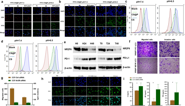
Methods: Herein, we designed a pH-sensitive multifunctional nanoplatform based on layered double hydroxides (LDHs) loaded with siRNA to block the intracellular immune checkpoint NR2F6, together with the asynchronous blockade surface receptor PD-L1 to induce strong synergistic antitumor immunity. Moreover, photothermal therapy (PTT) generated by LDHs after laser irradiation modified an immunologically "cold" microenvironment to potentiate Nr2f6-siRNA and anti-PD-L1 immunotherapy. Flow cytometry was performed to assess the immune responses initiated by the multifunctional nanoplatform.

Results: Under the slightly acidic tumor extracellular environment, PEG detached and the re-exposed positively charged LDHs enhanced tumor accumulation and cell uptake. The accumulated siRNA suppressed the signal of dual protumor activity in both immune and H22 tumor cells by silencing the NR2F6 gene, which further reduced the tumor burden and enhanced systemic antitumor immunity. The responses include enhanced tumor infiltration by CD4+ helper T cells, CD8+ cytotoxic T cells, and mature dendritic cells; the significantly decreased level of immune suppressed regulator T cells. The therapeutic responses were also attributed to the production of IL-2, IFN-γ, and TNF-α. The prepared nanoparticles also exhibited potential magnetic resonance imaging (MRI) ability, which could serve to guide synergistic immunotherapy treatment.

Conclusions: In summary, the three combinations of PTT, NR2F6 gene ablation and anti-PD-L1 can promote a synergistic immune response to inhibit the progression of primary HCC tumors and prevent metastasis. This study can be considered a proof-of-concept for the targeting of surface and intracellular immune checkpoints to supplement the existing HCC immunotherapy treatments.

Keywords: Hepatocellular carcinoma; NR2F6; PD-L1 blockade; Photothermal therapy.


Conflict of interest statement

The authors declare that they have no competing interests.

Figures

Fig. 1
Fig. 1
Schematic diagram of synthesis (a) and application in cancer treatment (b) of CS@P nanoparticles. a Preparation of pH-sensitive Nanoparticles CS@P used to deliver Nr2f6 siRNA to both T cells and tumor cells. b Under NIR laser irradiation, fever temperature (~ 42–45 °C) reveres immunosuppressive TME and activates T cells. Via Nr2f6 siRNA-mediated gene silencing, the pH- triggered nanoparticles CS@P not only suppress HCC cell proliferation and metastasis but also strongly enhance cytokines secretion of activated T cells, such as IL-2, IFN-γ, and TNF-α. The loss of NR2F6 and mild PTT further increase the response rates of established PD-1/PD-L1 checkpoint blockade to suppress primary and distant tumors, and prevent tumor metastasis
Fig. 2
Fig. 2
Characterizations of CS@P. a HRTEM image of the CCF-LDHs. Scale bar = 50 nm. b AFM image and c represented the thickness of CCF-LDHs monolayer nanosheets. d EDS mapping of the CCF-LDHs nanosheets. XPS spectra of e Co 2p, f Cu 2p and g Fe 2p. h T2 relaxation versus Fe concentration of CCF-LDHs and inset shows the corresponding T2-weighted images. i Zeta potential of CCF-LDHs and C@P in pH 7.4 and 6.5 media. Error bars stand for ± SD (n = 3). Photothermal curves of C@P dispersion at different concentrations irradiated by 880 nm laser at 1.0 W/cm2 for 5 min in pH = 7.4 (j) and 6.5 (k), respectively. l siRNA loading analysis at different w/w ratios of CCF-LDHs to siRNA
Fig. 3
Fig. 3
H22 and T cell dual cellular uptake and gene silencing. CLSM images of a T cells and b H22 tumor cells treated with CS@P in media at pH 7.4 or 6.5, respectively. Scale bar: 50 µm. Flow cytometry images of cellular uptake of FITC-labeled nanoparticles by c T cells and d H22 tumor cells at pH 7.4 and pH 6.5 conditions for 4 h. e Western blot analysis of the indicated proteins in T cells (left) and H22 tumor cells (right) 48 h after the incubated with CS. f, g Transwell results of the effect of NR2F6 on migration (left) and invasion (right). h CLSM images of PD-L1 expression in H22 cell after the indicated treatments. i Cytokine levels in the supernatant on 48 h after the indicated treatments (n = 3)
Fig. 4
Fig. 4
In vitro immune responses mediated by CS@P with laser irradiation. In vitro cell viability of H22 tumor cells incubated with CS@P for 24 h without (a) and with (b) laser irradiation (300 s, 1 W cm−2). Error bars stand for ± SD (n = 3). c Calcein AM/PI staining result of H22 tumor cells after the indicated treatments (C@P concentration: 100 μg mL−1, siRNA 100 nM). d Flow cytometry analysis of apoptosis levels in H22 cells after treatment with PBS, laser, C@P, C@P + laser, CS@P, or CS@P + laser (100 μg/mL C@P, 100 nM siRNA) for 24 h with 808 nm laser irradiation (300 s, 1 W cm−2) or not. e The percentages of mature DCs (CD11c+CD86+CD80+) through flow cytometry after indicated treatments in vitro DCs/H22 co-culture system. Error bars stand for ± SD (n = 3). f The flow cytometry plots and proportions of CD4+ and CD8+ T cells in vitro T lymphocytes/DCs/H22 cells (50:10:1) triple co-culture system (gated on the CD3+). Error bars stand for ± SD (n = 3). g The Treg flow cytometry analysis and frequencies in vitro T lymphocytes/DCs/H22 cells (50:10:1) triple co-culture system. Error bars stand for ± SD (n = 3)
Fig. 5
Fig. 5
Immune response of CS@P together with laser and PD-L1 in vivo. a Schematic diagram of the model and therapeutic schedule of primary and distant H22 tumor model. b Primary tumor growth tendency of H22 tumor-bearing mice with various treatments. Tumor sizes were normalized to initial sizes. Error bars stand for ± SD (n = 6). c Primary tumor growth curves in the H22 tumor-bearing BALB/c mice (n = 6). d Representative flow cytometry plots showing different groups of T cells in primary tumors (gated on CD3+ T cells) after indicated treatments. e The Treg flow cytometry analysis in primary tumors after various treatments. f Representative flow cytometry plots showing matured DC cells in primary tumors after various treatments. g Immunohistochemical staining of NR2F6 and PD-L1 in primary tumor sections (top). H&E and TUNEL examination of tumor sections (bottom). Scale bar: 100 µm. *P < 0.05, **P < 0.01, and ***P < 0.001
Fig. 6
Fig. 6
Abscopal effect of CS@P combined with NIR irradiation and aPD-L1. a Distant tumor growth tendency of H22 tumor-bearing mice with various treatments. Tumor sizes were normalized to initial sizes. Error bars stand for ± SD (n = 6). b Distant tumor growth curves in the H22 tumor-bearing BALB/c mice (n = 6). c Images of the distant tumor harvested on day 21. d The amount of CD4+ and CD8+ T cells in distant tumors detected upon various treatments detected by flow cytometry (gated on the CD3+). Data represented mean ± SD (n = 3). e The amount of Treg cells in distant tumors upon various treatments detected by flow cytometry Data represent mean ± SD (n = 3). f Frequency of the CD4+ and CD8+ T cells in spleens after indicated treatments. g Frequency of the Treg cells in spleens after indicated treatments. The levels of IFN-γ (h), TNF-α (i), and IL-2 (j) in the primary tumor after the various treatments (n = 3), expressed as the concentration per gram of tumor (pg/per g tumor). *P < 0.05, **P < 0.01, and ***P < 0.001
Fig. 7
Fig. 7
Anti-metastasis effect of CS@P combined with NIR irradiation and aPD-L1. a Schematic diagram of the model and therapeutic schedule of metastatic H22 tumor model. b Number of pulmonary metastatic lesions after different treatments (n = 3). c Survival curves of H22 tumor-bearing mice after different treatments (n = 6). d In vivo bioluminescence images of the lungs on day 5, 10, 15 and 20 (n = 3). e Representative photographs of metastatic nodules after different treatments. f H&E staining of harvested lung tissues on day 21. Scale bars: 100 μm. g Immunohistochemical staining of Ki67 and CD8+ of isolated lung sections. Scale bars: 100 μm

Similar articles

Cited by

References

    1. Sung H, Ferlay J, Siegel RL, Laversanne M, Soerjomataram I, Jemal A, Bray F. Global Cancer Statistics 2020: GLOBOCAN estimates of incidence and mortality worldwide for 36 cancers in 185 countries. A Cancer J Clin. 2021;71(3):209–249. doi: 10.3322/caac.21660. - DOI - PubMed
    1. Llovet JM, Zucman-Rossi J, Pikarsky E, Sangro B, Schwartz M, Sherman M, Gores G. Hepatocellular carcinoma. Nat Rev Dis Primers. 2016;2:16018. doi: 10.1038/nrdp.2016.18. - DOI - PubMed
    1. Xu J, Zheng Q, Cheng X, Hu S, Zhang C, Zhou X, Sun P, Wang W, Su Z, Zou T, Song Z, Xia Y, Yi X, Gao Y. Chemo-photodynamic therapy with light-triggered disassembly of theranostic nanoplatform in combination with checkpoint blockade for immunotherapy of hepatocellular carcinoma. J Nanobiotechnol. 2021;19(1):355–355. doi: 10.1186/s12951-021-01101-1. - DOI - PMC - PubMed
    1. Riley RS, June CH, Langer R, Mitchell MJ. Delivery technologies for cancer immunotherapy. Nat Rev Drug Discov. 2019;18(3):175–196. doi: 10.1038/s41573-018-0006-z. - DOI - PMC - PubMed
    1. Lu J, Liu X, Liao YP, Salazar F, Sun B, Jiang W, Chang CH, Jiang J, Wang X, Wu AM, Meng H, Nel AE. Nano-enabled pancreas cancer immunotherapy using immunogenic cell death and reversing immunosuppression. Nat Commun. 2017;8(1):1811. doi: 10.1038/s41467-017-01651-9. - DOI - PMC - PubMed

MeSH terms