. 2022 Jul 30;13(1):385.
doi: 10.1186/s13287-022-03047-z.

Bone morphogenetic protein 4 rescues the bone regenerative potential of old muscle-derived stem cells via regulation of cell cycle inhibitors

Affiliations

Bone morphogenetic protein 4 rescues the bone regenerative potential of old muscle-derived stem cells via regulation of cell cycle inhibitors

Haizi Cheng et al. Stem Cell Res Ther. .

Abstract

Background: Bone morphogenetic protein 4 (BMP4) promotes the osteogenic differentiation and the bone regenerative potential of muscle-derived stem cells (MDSCs). BMP4 also promotes the self-renewal of both embryonic and somatic stem cells; however, BMP4 signaling activity significantly decreases with age. Cyclin-dependent kinase inhibitors P16INK4A (P16) and P18INK4C (P18) induce early G1-phase cell cycle blockade by targeting cyclin-dependent kinase 4/6. It is still unclear if BMP4 affects the bone regenerative potential of old MDSCs through regulation of P16 and P18 expression.

Methods: Young and old MDSCs were isolated from 3 week (young) and 2-year-old (old) mice. In vitro cell proliferation and multipotent differentiation were performed for young and old MDSCs both before and after BMP4/GFP transduction. Cell cycle genes were analyzed using Q-PCR. The bone regenerative potential of young and old MDSCs transduced with BMP4/GFP were compared using Micro-CT and histological analysis. The bone regenerative potential of young and old MDSCs was also compared between single and double transduction (higher BMP4 levels expression). The cell proliferation, mitochondrial function and osteogenic differentiation was also compared in vitro between cells that have been transduced with BMP4GFP (single and double transduction). The correlation of bone regeneration capacity of young and old MDSCs with P16 and P18 expression was further evaluated at 10 days after cell transplantation using histology and western blot analysis.

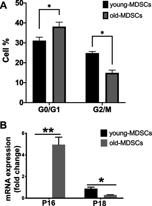
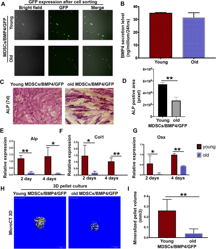
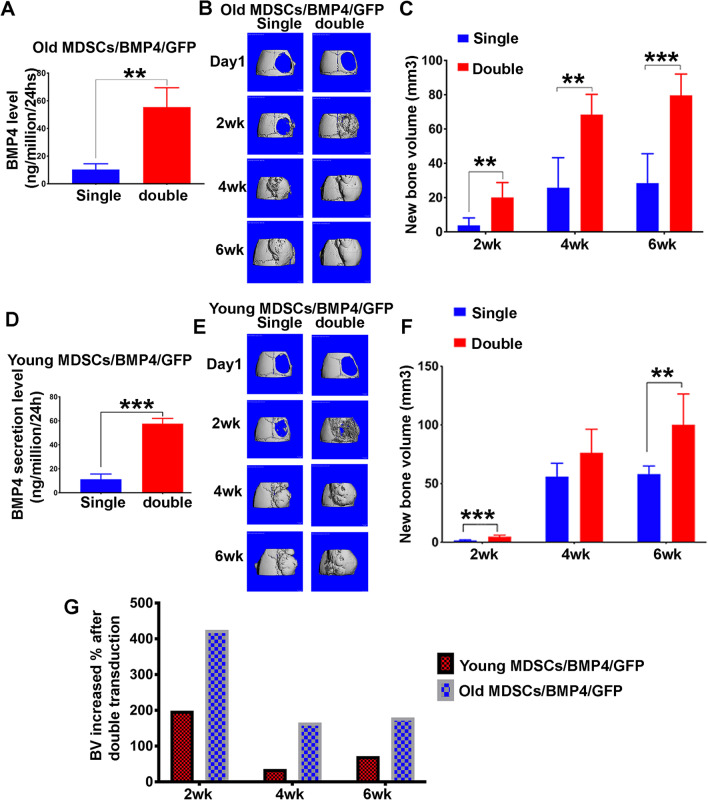
Results: Old murine MDSCs (MDSCs) exhibit reduced proliferation and multi-lineage differentiation potential with or without BMP4 stimulation, when compared to young murine MDSCs. Old MDSCs express significantly higher P16 and lower P18, with more cells in the G0/1 phase and fewer cells in the G2/M phase, compared to young MDSCs. Old MDSCs retrovirally transduced to express BMP4 regenerated less bone in a critical size skull defect in CD-1 nude mice when compared to young retrovirally transduced MDSCs expressing similar BMP4 levels and contribute less to the new regenerated new bone. Importantly, both young and old MDSCs can regenerate more bone when BMP4 expression levels are increased by double-transduction with the retroviral-BMP4/GFP. However, the bone regeneration enhancement with elevated BMP4 was more profound in old MDSCs (400% at 2 weeks) compared to young MDSCs (200%). Accordingly, P18 is upregulated while P16 is downregulated after BMP4 transduction. Double transduction did not further increase cell proliferation nor mitochondrial function but did significantly increase Osx expression in both young and old MDSCs. Old MDSCs had even significant higher Osx levels as compared to young MDSCs following double transduction, while a similar Alp expression was observed between young and old MDSCs after double transduction. In addition, at 10 days after cell transplantation, old MDSCs having undergone double transduction regenerated bone more rapidly as showed by Alcian blue and Von Kossa staining. Western blot assays demonstrated that old MDSCs after retro-BMP4/GFP double transduction have significantly lower P18 expression levels when compared to young BMP4-transduced MDSCs. In addition, P18 expression was slightly increased in old MDSCs after double transduction when compared to single transduction. P16 expression was not detectable for both young and two old BMP4/GFP transduced MDSCs groups.

Conclusions: In summary, BMP4 can offset the adverse effect of aging on the osteogenic differentiation and the bone regenerative potential of old MDSCs via up-regulation of P18 and down-regulation P16 expression.

Keywords: Aging; Bone morphogenetic protein 4; Bone regeneration; Muscle-derived stem cells; P16; P18.


Conflict of interest statement

J.H received Royalties from Cooke Myocytes Inc. All authors declare no competing interest.

Figures

Fig. 1
Fig. 1
Old MDSCs demonstrated reduced cell proliferation and multipotent differentiation when compared to young MDSCs. A Proliferation of young and old MDSCs measured by live-cell imaging system. Old MDSCs showed decreased cell numbers at 36 and 48 h after seeding (P = 0.031 and 0.004, respectively). B–C Myogenic differentiation. fMHC positive cells were significantly decreased in old MDSCs compared to young MDSCs after 7 days differentiation (P < 0.0001). fMHC stained red and nuclei stained blue. D–E Osteogenic differentiation. ALP positive staining percentage was significantly lower in old MDSC compared to young MDSCs (P = 0.0097). F–G Chondrogenic differentiation using pellet culture. Alcian Blue positive area was significantly lower in pellets formed by old MDSCs compared to that of young MDSCs (P = 0.0005). H–I Adipogenesis. Oil red positive (dark red) area percentage was significantly lower in old MDSCs group compared to that of young MDSCs group (P = 0.0502)
Fig. 2
Fig. 2
Old MDSCs exhibit a greater percentage of cells in the G0/1 phase and higher P16 and lower P18 expression, when compared to young MDSCs. A Propidium iodide (PI) staining of DNA detected by flow cytometry of young and old MDSCs. Percentage of cells in G0/1 phase is significantly higher while percentage of cells in G2/M is significantly lower in old MDSCs compared to young MDSCs (*P < 0.05). B qPCR analysis of P16 and P18. P16 mRNA expression is undetectable low in young MDSCs, but high in old MDSCs (**P < 0.01). P18 mRNA expression is significantly lower in old MDSCs than in young MDSCs (*P < 0.05)
Fig. 3
Fig. 3
The osteogenic potential of old MDSCs remained significantly lower compared to young MDSCs, after BMP4 transduction. A Transduction efficiency of BMP4/GFP transduction for both young and old MDSCs. The efficiency of BMP4/GFP transduction on both young and old MDSCs is less than 100%. After cell sorting based on GFP positivity, GFP positive cells is at 100% for both young and old MDSCs. B BMP4 secretion level of young and old MDSCs after GFP cells sorting. No significant difference on the secretion levels of BMP4 between young and old MDSCs/BMP4/GFP. C–D Osteogenic differentiation of monolayer culture for young and old BMP4/GFP-transduced MDSCs. ALP positive staining area (purple pixel) is significantly lower in the old MDSC group compared to young MDSCs group. **P < 0.05. E–G qPCR analysis of Alp, Col1, and Osx.*P<0.05, **P<0.01. H–I MicroCT scan for osteogenic pellets of BMP4/GFP transduced MDSCs. Old MDSCs/BMP4/GFP could undergo osteogenic differentiation as demonstrated by mineralization using microCT scanning. However, the mineralized pellet volume of old MDSCs/BMP4/GFP was significantly smaller than young MDSCs/BMP4/GFP-transduced despite expressing similar levels of BMP4 at around 30 ng/million cells/24 h.**P < 0.01
Fig. 4
Fig. 4
The bone regenerative capacity of old MDSCs is inferior to young MDSCs despite expressing similar levels of BMP4. A Micro-CT 3D images at different time points after cell transplantation. B Quantification of new bone volume in the skull defect at different time points showed significantly less new bone formed by old MDSCs/BMP4/GFP compared to young MDSCs/BMP4/GFP in the skull defect area when both young and old MDSCs/ BMP4/GFP have BMP4 secretion level of around 30 ng/million cells/24 h. *P < 0.05, **P < 0.01. C H&E staining for the new bone on the skull defect. The new bone is mainly trabecular bone (TB) constituted of new bone matrix and bone marrow including myeloid cells (green arrows, red blood cells (black arrows) and megakaryocytes (Blue arrows). No significant differences between young and old MDSCs/BMP4/GFP groups were found. Scale bars = 200 µm. D–E Herovici’s staining for the new bone on the skull defect and quantification. COL1 stained red/pink color. COL3 3 stained blue color. The newly formed bone tissues showed cancellous bone structure in both groups. Quantification of COL1 area percentage showed no significant difference between young and old MDSCs/BMP4/GFP groups. Scale bars = 200 µm. F–G Immunohistochemistry of GFP to trace donor cells for the new bone on the skull defect with associated quantification. The new bone area showed that most of the osteoblasts and osteocytes are GFP positive in both groups. Quantification for GFP positive area of the new bone on the skull defect showed lower GFP+ area percentage in the newly regenerated bone in old MDSCs/BMP4/GFP group compared to young MDSCs/BMP4/GFP group. P = 0.0009. Scale bars = 100 µm
Fig. 5
Fig. 5
Increased BMP4 expression levels in old MDSCs improve their bone regenerative potential to levels comparable to young MDSCs. A BMP4 secretion levels after double transduction. **P < 0.01. B Micro-CT 3D images at different time points of single and double transduction of old MDSCs/BMP4/GFP. Bone formation enhancement is more obvious at the 2 weeks timepoint. Both groups showed complete defect healing at 4 weeks with similar bone density for both groups. C Quantification of new bone volume. **P < 0.01. ***P < 0.001. D BMP4 secretion level of single and double transduction of young MDSCs/BMP4/GFP. ***P < 0.001. E Micro-CT 3D images at different time points. Double transduction showed obvious accelerated bone regeneration at two weeks and nearly completely healed defects at 2 weeks after cell transplantation and complete healing of defect at 4 and 6 weeks after cell transplantation. F Quantification of new bone volume at different time points. At the 2 and 6 weeks timepoints, double transduction significantly increased new bone formation, but not at 4 weeks. **P < 0.01, ***P < 0.001. G Bone regeneration increase percentage after double transduction of young and old MDSCs/BMP4/GFP at different time points. Old MDSCs/BMP4/GFP showed an increase of 400% versus 200% at 2 weeks in young MDSCs/BMP4/GFP group and more than 150% at 4 and 6 weeks compared to less than 100% in young MDSCs/BMP4/GFP group after double transduction
Fig. 6
Fig. 6
BMP4 reverse aged-related decline in cell proliferation via regulating P18 and P16 after single BMP4/GFP transduction. A Doubling time for old MDSCs before and after BMP4 transduction. BMP4/GFP transduction decreased the population doubling time of old MDSCs. *P < 0.05. B qPCR for P18 in transduced and un-transduced MDSCs. BMP4 transduction significantly increased P18 expression for both young and old MDSCs. *P < 0.05, **P < 0.01. C Q-PCR for P16 in BMP4 transduced and un-transduced MDSCs. Young MDSCs did not express detectable P16 before and after BMP4 transduction. BMP4 transduction significantly decreased P16 expression in old MDSCs. *P < 0.05. D QPCR for P18 in transduced and un-transduced MDSCs cultured in proliferation medium or osteogenesis medium. *P < 0.05, **P < 0.01. E Q-PCR for P16 in transduced and un-transduced MDSCs cultured in proliferation medium or osteogenesis medium
Fig. 7
Fig. 7
Dose-dependent effect of BMP4 after double transduction on cell proliferation, mitochondria function and osteogenic differentiation. A, B Ki67 staining and quantification. Ki67+ stained red in nuclei. Scale bars = 200 µm. Exact values are shown between group comparisons. C Population doubling time quantification. Exact values are showed between group comparisons. D Live mitochondrial staining with Mitotracker Red FM to show oxidation activity of mitochondria. Mitochondria are stained red (Mitotracker Red) and nuclei are stained blue with Hoechst 33342. Insets are the enlarged boxed area of cells to better display mitochondria in the cytoplasm. Scale bars = 200 µm. E Quantification of Mitotracker red fluorescence intensity relative to cell number using Hoechst 33342. F Quantification of LDS-751 fluorescence intensity relative to cell number using Hoechst 33342. G Q-PCR analysis of osteogenic differentiation of single and double transduced young and old MDSCs. *P < 0.05, **P < 0.01, ***P < 0.001. ****P < 0.0001
Fig. 8
Fig. 8
Bone regeneration at day 10 after in vivo transplantation of young and old BMP4/GFP-transduced MDSCs. A Alcian blue staining. Chondrocytes and cartilage stained blue. 20X magnification shows the entire defect area. Transplanted cells proliferated and formed thick cell layers on top of the critical sized defect. Old MDSCs with double transduction showed early endochondral bone formation at day 10 after transplantation with obvious chondrogenic nodule in blue as did young MDSCs. Scale bars = 1 mm for 20× and 200 µm for 100×, respectively. B Von Kossa Staining. Bone stained brown-black. Both young and old BMP4/GFP transduced MDSCs showed mineralization, old MDSCs with double BMP4/GFP transduction showed more mineralization than single transduction. These findings indicate that higher levels of BMP4 reversed the impairment of bone regeneration of old MDSCs. Scale bars = 1 mm for 20× and 200 µm for 100×, respectively. C Fluorescent western blot for P18 (green) and GFP (blue) with β-actin (red) as loading control. There is a green non-specific band in the P18 channel merging with the β-actin (red) channel. D Quantification of P18 expression relative to GFP showed old MDSCs/BMP4/GFP expressed less P18 compared to young MDSCs/BMP4/GFP, while increased BMP4 expression by double transduction slightly increased P18 expression. E Western blot of P16 (Red) and GFP (blue) and loading control β-actin (red) at day 10 after cell transplantation. P16 is not detectable by western blot at this time point of bone regeneration in both young and old MDSCs/BMP4/GFP group with single or double transduction
Fig. 9
Fig. 9
Schematic representation of the role of BMP4 on the regulation of stem cell function and bone regeneration

Similar articles

Cited by

References

    1. Deasy BM, Schugar RC, Huard J. Sex differences in muscle-derived stem cells and skeletal muscle. Crit Rev Eukaryot Gene Expr. 2008;18(2):173–188. doi: 10.1615/CritRevEukarGeneExpr.v18.i2.60. - DOI - PubMed
    1. Payne TR, Oshima H, Okada M, Momoi N, Tobita K, Keller BB, Peng H, Huard J. A relationship between vascular endothelial growth factor, angiogenesis, and cardiac repair after muscle stem cell transplantation into ischemic hearts. J Am Coll Cardiol. 2007;50(17):1677–1684. doi: 10.1016/j.jacc.2007.04.100. - DOI - PubMed
    1. Pan L, Chen S, Weng C, Call G, Zhu D, Tang H, Zhang N, Xie T. Stem cell aging is controlled both intrinsically and extrinsically in the Drosophila ovary. Cell Stem Cell. 2007;1(4):458–469. doi: 10.1016/j.stem.2007.09.010. - DOI - PubMed
    1. Lee PY, Cobain E, Huard J, Huang L. Thermosensitive hydrogel PEG-PLGA-PEG enhances engraftment of muscle-derived stem cells and promotes healing in diabetic wound. Mol Ther. 2007;15(6):1189–1194. doi: 10.1038/sj.mt.6300156. - DOI - PubMed
    1. Corsi KA, Pollett JB, Phillippi JA, Usas A, Li G, Huard J. Osteogenic potential of postnatal skeletal muscle-derived stem cells is influenced by donor sex. J Bone Miner Res. 2007;22(10):1592–1602. doi: 10.1359/jbmr.070702. - DOI - PubMed

Publication types

Substances

LinkOut - more resources