. 2022 Jul 30;20(1):342.
doi: 10.1186/s12967-022-03543-z.

Modulation of hepatic stellate cells by Mutaflor® probiotic in non-alcoholic fatty liver disease management

Affiliations

Modulation of hepatic stellate cells by Mutaflor® probiotic in non-alcoholic fatty liver disease management

Noha M Hany et al. J Transl Med. .

Abstract

Background: NAFLD and NASH are emerging as primary causes of chronic liver disease, indicating a need for an effective treatment. Mutaflor® probiotic, a microbial treatment of interest, was effective in sustaining remission in ulcerative colitis patients.

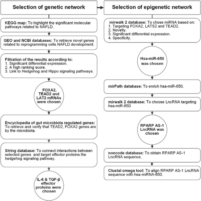
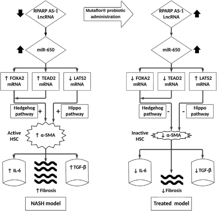
Objective: To construct a genetic-epigenetic network linked to HSC signaling as a modulator of NAFLD/NASH pathogenesis, then assess the effects of Mutaflor® on this network.

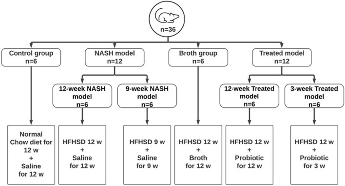
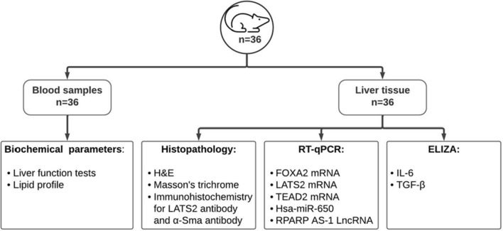
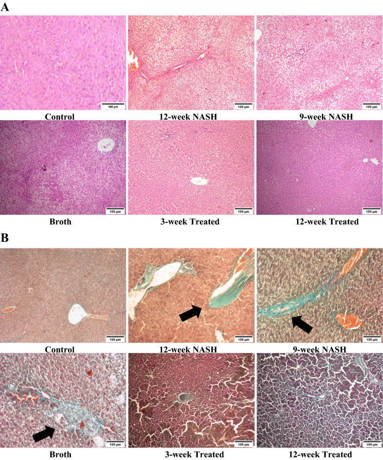
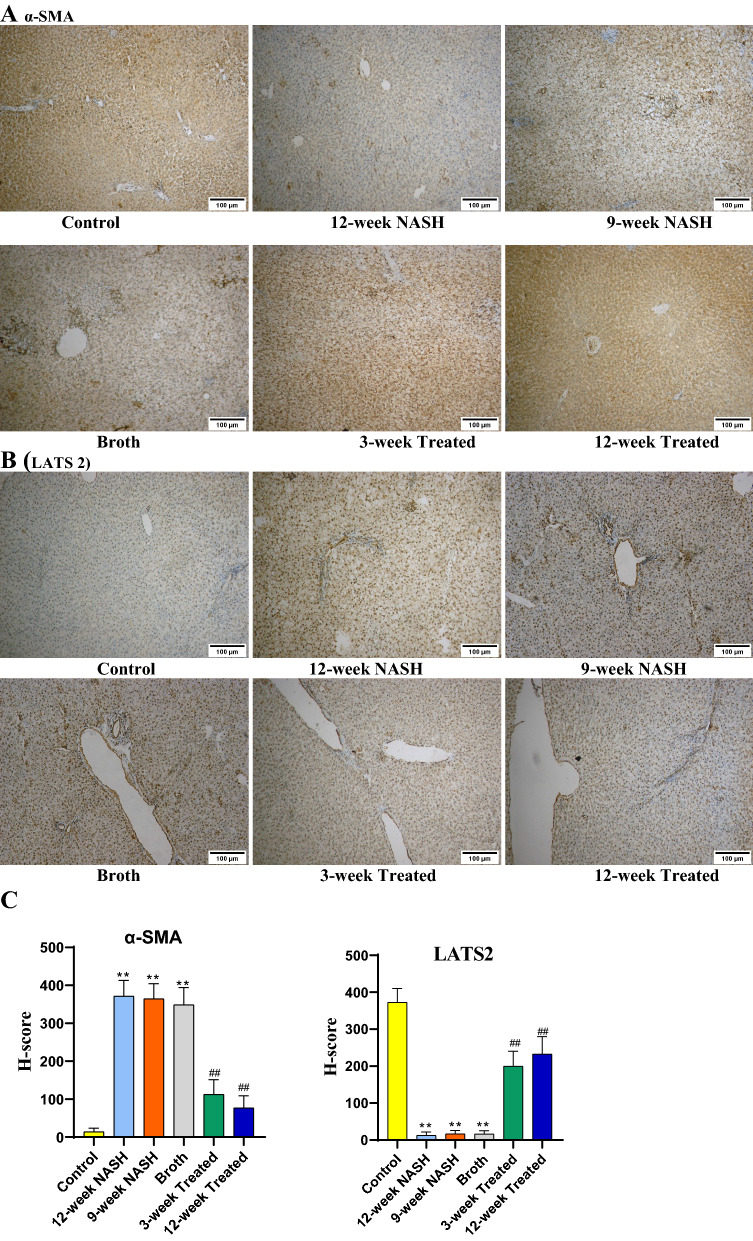
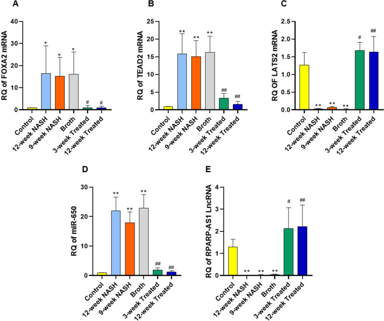
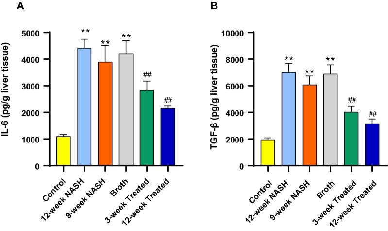
Methods: First, in silico analysis was used to construct a genetic-epigenetic network linked to HSC signaling. Second, an investigation using rats, including HFHSD induced NASH and Mutaflor® treated animals, was designed. Experimental procedures included biochemical and histopathologic analysis of rat blood and liver samples. At the molecular level, the expression of genetic (FOXA2, TEAD2, and LATS2 mRNAs) and epigenetic (miR-650, RPARP AS-1 LncRNA) network was measured by real-time PCR. PCR results were validated with immunohistochemistry (α-SMA and LATS2). Target effector proteins, IL-6 and TGF-β, were estimated by ELISA.

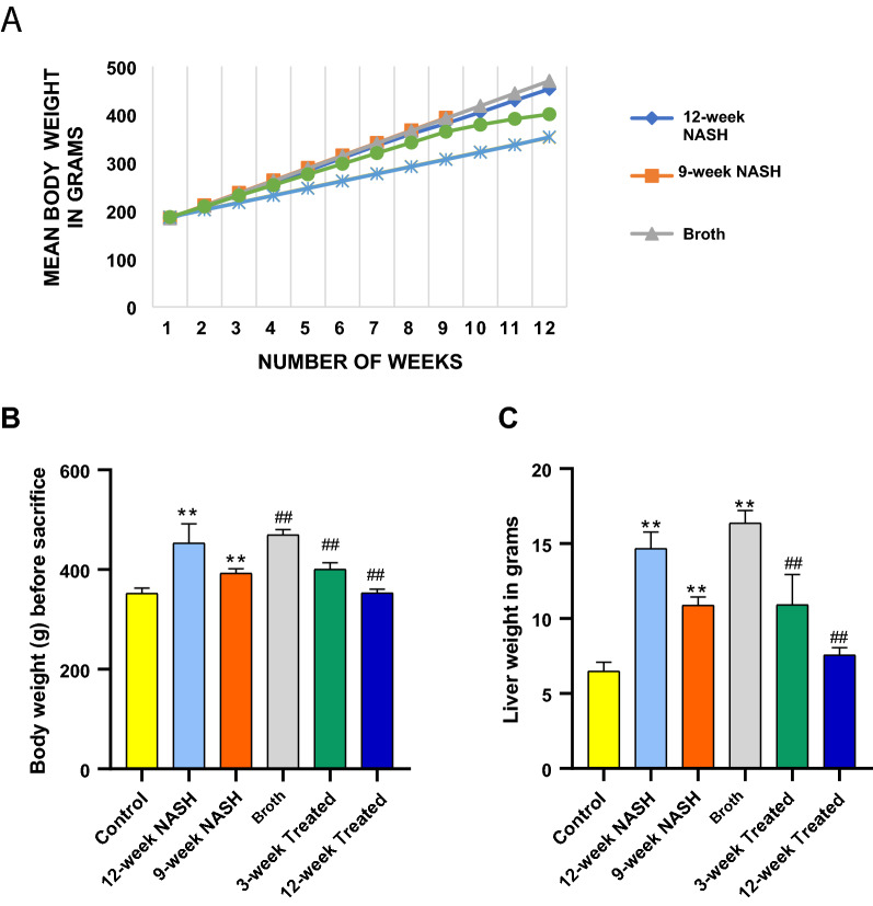
Results: Mutaflor® administration minimized biochemical and histopathologic alterations caused by NAFLD/NASH. HSC activation and expression of profibrogenic IL-6 and TGF-β effector proteins were reduced via inhibition of hedgehog and hippo pathways. Pathways may have been inhibited through upregulation of RPARP AS-1 LncRNA which in turn downregulated the expression of miR-650, FOXA2 mRNA and TEAD2 mRNA and upregulated LATS2 mRNA expression.

Conclusion: Mutaflor® may slow the progression of NAFLD/NASH by modulating a genetic-epigenetic network linked to HSC signaling. The probiotic may be a useful modality for the prevention and treatment of NAFLD/NASH.

Keywords: E. coli; Hedgehog; Hepatic Stellate cells; Hippo; Liver fibrosis; NAFLD; NASH; Probiotic.


Conflict of interest statement

The authors report no declarations of interest.

Figures

Fig. 1
Fig. 1
Workflow of bioinformatics steps
Fig. 2
Fig. 2
Workflow chart representing the experimental design of the animal groups. NASH nonalcoholic steatohepatitis, HFHSD high-fat high sucrose diet
Fig. 3
Fig. 3
Experimental procedures carried out on rat blood and tissue samples to validate Insilco hypothesis
Fig. 4
Fig. 4
Effect of NASH induction and Probiotic administration on body weight and liver weight expressed in grams. A Mean body weight of each group for 12 weeks except 9-week NASH group. B Mean body weight of each groupat the end of the study, calculated in grams before sacrifaction at 12 weeks for all groups except for the 9-week NASH group, values are expressed as means ± SD. C Liver weight, values are expressed as means ± SD (n = 6). *p < 0.05 **p < 0.01 symbols are used when groups are compared with Control group while #p < 0.05 and ##p < 0.01 symbols are used when groups are compared to 12-week NASH group
Fig. 5
Fig. 5
The effect of NASH induction and Probiotic use on steatosis, inflammation and fibrosis of the liver using HE (A) and Masson’s trichrome staining (B) (Magnifications: ×100). Liver histological examinations showed both macro- and microvesicular steatosis, lobular inflammation, focal necrosis, hepatocellular ballooning, and extensive fibrosis (Arrow) in NASH models. These effects were minimized with Probiotic were remarkable especially in 12-week Treated model
Fig. 6
Fig. 6
A Immunohistochemical staining (IHC) of α-SMA and B LATS 2 in liver sections from separate groups as indicated in the figures (Magnifications: ×100). C Statistical difference in α-SMA and LATS2 H-score among study groups. IHC assays demonstrated that liver tissue of NASH rats had more α-SMA-positive cells were localized in areas with inflammatory cells and areas with remarkable perisinusoidal fibrosis. The expression of α-SMA showed a significant increase in 12-week NASH group, 9-week NASH group and Broth group when compared with the Control group, However, in 12-week Treated and 9-week Treated groups, there was an evident decrease in the α-SMA expression. IHC assays of LATS2 were markedly decreased in 12-week NASH, 9-week NASH and Broth groups when compared to the Control group. However, 12-week Treated and 9-week Treated groups showed a significant increase in LATS2 expression when compared to the 12-week NASH group
Fig. 7
Fig. 7
Effect of NASH induction and Probiotic administration on the expression level of hepatic FOXA2 mRNA, TEAD2 mRNA and LATS2 mRNA (AC), hepatic miR-650 (D) and hepatic RPARP-AS1 LncRNA (E). Values are mean ± SD; number of animals = 6 rats/each group. *p < 0.05 **p < 0.01 symbols are used when groups are compared with Control group while #p < 0.05 and ##p < 0.01 symbols are used when groups are compared to 12-week NASH group. RQ, relative quantification
Fig. 8
Fig. 8
Effect of NASH induction and Probiotic administration on A Relative concentration of IL-6 and B TGF-β/g liver tissue. Values are mean ± SD; number of animals = 6 rats/each group. *p < 0.05 **p < 0.01 symbols are used when groups are compared with Control group while #p < 0.05 and ##p < 0.01 symbols are used when groups are compared to 12-week NASH group. (pg/g) picogram per gram liver tissue
Fig. 9
Fig. 9
Proof of Concept map of the study hypothesis

References

    1. Pydyn N, Miękus K, Jura J, Kotlinowski J. New therapeutic strategies in nonalcoholic fatty liver disease: a focus on promising drugs for nonalcoholic steatohepatitis. Pharmacol Rep. 2020;72(1):1–12. doi: 10.1007/s43440-019-00020-1. - DOI - PubMed
    1. Perumpail BJ, Khan MA, Yoo ER, Cholankeril G, Kim D, Ahmed A. Clinical epidemiology and disease burden of nonalcoholic fatty liver disease. World J Gastroenterol. 2017;23(47):8263–8276. doi: 10.3748/wjg.v23.i47.8263. - DOI - PMC - PubMed
    1. Negro F. Natural history of NASH and HCC. Liver Int. 2020;40(Suppl 1):72–76. doi: 10.1111/liv.14362. - DOI - PubMed
    1. Burra P, Becchetti C, Germani G. NAFLD and liver transplantation: disease burden, current management and future challenges. JHEP Rep. 2020;2(6):100192. doi: 10.1016/j.jhepr.2020.100192. - DOI - PMC - PubMed
    1. Fang YL, Chen H, Wang CL, Liang L. Pathogenesis of non-alcoholic fatty liver disease in children and adolescence: from "two hit theory" to "multiple hit model". World J Gastroenterol. 2018;24(27):2974–2983. doi: 10.3748/wjg.v24.i27.2974. - DOI - PMC - PubMed

Publication types