

Advances in cell therapies using stem cells/progenitors as a novel approach for neurovascular repair of the diabetic retina
- PMID: 35907890
- PMCID: PMC9338609
- DOI: 10.1186/s13287-022-03073-x
Advances in cell therapies using stem cells/progenitors as a novel approach for neurovascular repair of the diabetic retina
Abstract
Background: Diabetic retinopathy, a major complication of diabetes mellitus, is a leading cause of sigh-loss in working age adults. Progressive loss of integrity of the retinal neurovascular unit is a central element in the disease pathogenesis. Retinal ischemia and inflammatory processes drive interrelated pathologies such as blood retinal barrier disruption, fluid accumulation, gliosis, neuronal loss and/or aberrant neovascularisation. Current treatment options are somewhat limited to late-stages of the disease where there is already significant damage to the retinal architecture arising from degenerative, edematous and proliferative pathology. New preventive and interventional treatments to target early vasodegenerative and neurodegenerative stages of the disease are needed to ensure avoidance of sight-loss.
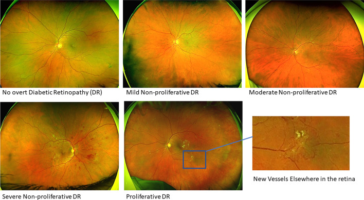
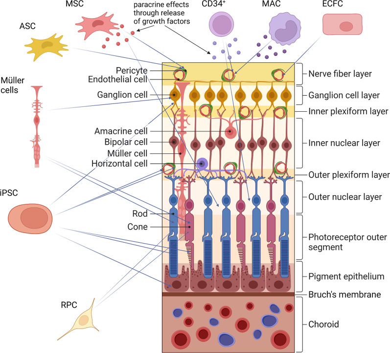
Main body: Historically, diabetic retinopathy has been considered a primarily microvascular disease of the retina and clinically it is classified based on the presence and severity of vascular lesions. It is now known that neurodegeneration plays a significant role during the pathogenesis. Loss of neurons has been documented at early stages in pre-clinical models as well as in individuals with diabetes and, in some, even prior to the onset of clinically overt diabetic retinopathy. Recent studies suggest that some patients have a primarily neurodegenerative phenotype. Retinal pigment epithelial cells and the choroid are also affected during the disease pathogenesis and these tissues may also need to be addressed by new regenerative treatments. Most stem cell research for diabetic retinopathy to date has focused on addressing vasculopathy. Pre-clinical and clinical studies aiming to restore damaged vasculature using vasoactive progenitors including mesenchymal stromal/stem cells, adipose stem cells, CD34+ cells, endothelial colony forming cells and induced pluripotent stem cell derived endothelial cells are discussed in this review. Stem cells that could replace dying neurons such as retinal progenitor cells, pluripotent stem cell derived photoreceptors and ganglion cells as well as Müller stem cells are also discussed. Finally, challenges of stem cell therapies relevant to diabetic retinopathy are considered.
Conclusion: Stem cell therapies hold great potential to replace dying cells during early and even late stages of diabetic retinopathy. However, due to the presence of different phenotypes, selecting the most suitable stem cell product for individual patients will be crucial for successful treatment.
Keywords: Adult stem cells; DME; Diabetic retinopathy; Endothelial progenitor cells; Induced pluripotent stem cells; Macular ischemia; Microvascular disease; Neurodegeneration; PDR; Retinal progenitor cells.
© 2022. The Author(s).
Conflict of interest statement
JL and NL have no competing interests. RJM and AWS are founders and scientific advisors for VascVersa Ltd.
Figures
Similar articles
-
Mesenchymal stromal/stem cells as potential therapy in diabetic retinopathy.Immunobiology. 2018 Dec;223(12):729-743. doi: 10.1016/j.imbio.2018.01.001. Epub 2018 Feb 15. Immunobiology. 2018. PMID: 29402461 Review.
-
Cell Therapy Applications for Retinal Vascular Diseases: Diabetic Retinopathy and Retinal Vein Occlusion.Invest Ophthalmol Vis Sci. 2016 Apr 1;57(5):ORSFj1-ORSFj10. doi: 10.1167/iovs.15-17594. Invest Ophthalmol Vis Sci. 2016. PMID: 27116667 Review.
-
Retinal Cell Damage in Diabetic Retinopathy.Cells. 2023 May 8;12(9):1342. doi: 10.3390/cells12091342. Cells. 2023. PMID: 37174742 Free PMC article. Review.
-
Prognostic factors for the development and progression of proliferative diabetic retinopathy in people with diabetic retinopathy.Cochrane Database Syst Rev. 2023 Feb 22;2(2):CD013775. doi: 10.1002/14651858.CD013775.pub2. Cochrane Database Syst Rev. 2023. PMID: 36815723 Free PMC article. Review.
-
Inflammation in diabetic retinopathy: possible roles in pathogenesis and potential implications for therapy.Neural Regen Res. 2023 May;18(5):976-982. doi: 10.4103/1673-5374.355743. Neural Regen Res. 2023. PMID: 36254977 Free PMC article. Review.
Cited by
-
Pericytes as Key Players in Retinal Diseases: A Comprehensive Narrative Review.Biology (Basel). 2025 Jun 20;14(7):736. doi: 10.3390/biology14070736. Biology (Basel). 2025. PMID: 40723297 Free PMC article. Review.
-
Classical and Innovative Evidence for Therapeutic Strategies in Retinal Dysfunctions.Int J Mol Sci. 2024 Feb 9;25(4):2124. doi: 10.3390/ijms25042124. Int J Mol Sci. 2024. PMID: 38396799 Free PMC article. Review.
-
Exosomes derived from human umbilical cord blood mesenchymal stem cells protect against blue light-induced damage to retinal pigment epithelial cells by inhibiting FGF2 expression.Cytotechnology. 2025 Jun;77(3):88. doi: 10.1007/s10616-025-00752-4. Epub 2025 Apr 9. Cytotechnology. 2025. PMID: 40225792
-
Effects of mesenchymal stromal cells and human recombinant Nerve Growth Factor delivered by bioengineered human corneal lenticule on an innovative model of diabetic retinopathy.Front Endocrinol (Lausanne). 2024 Oct 15;15:1462043. doi: 10.3389/fendo.2024.1462043. eCollection 2024. Front Endocrinol (Lausanne). 2024. PMID: 39473506 Free PMC article.
-
Retinal Ganglion Cell Replacement in Glaucoma Therapy: A Narrative Review.J Clin Med. 2024 Nov 27;13(23):7204. doi: 10.3390/jcm13237204. J Clin Med. 2024. PMID: 39685661 Free PMC article. Review.
References
-
- Stitt AW, Curtis TM, Chen M, Medina RJ, McKay GJ, Jenkins A, et al. The progress in understanding and treatment of diabetic retinopathy. Vol. 51, Progress in Retinal and Eye Research. Elsevier Ltd; 2016. p. 156–86. - PubMed
-
- International Diabetes Federation. IDF Diabetes Atlas, 10th edn. . Brussels, Belgium: 2021. Available at: https://www.diabetesatlas.org. 2021.
-
- Barber AJ. A new view of diabetic retinopathy: a neurodegenerative disease of the eye. Progr Neuro-Psychopharmacol Biol Psychiatry. 2003;27(2). - PubMed
Publication types
MeSH terms
Grants and funding
LinkOut - more resources
Full Text Sources
Medical
