. 2022 Jul 30;12(1):13131.
doi: 10.1038/s41598-022-16274-4.

Curcumin piperidone derivatives induce anti-proliferative and anti-migratory effects in LN-18 human glioblastoma cells

Affiliations

Curcumin piperidone derivatives induce anti-proliferative and anti-migratory effects in LN-18 human glioblastoma cells

Nur Syahirah Che Razali et al. Sci Rep. .

Abstract

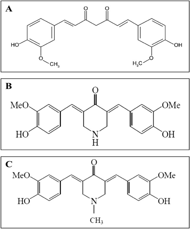
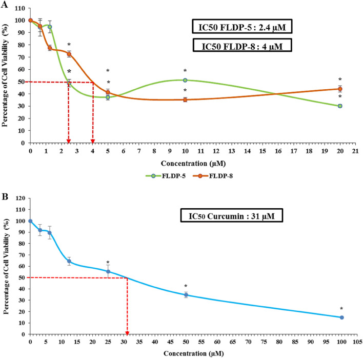
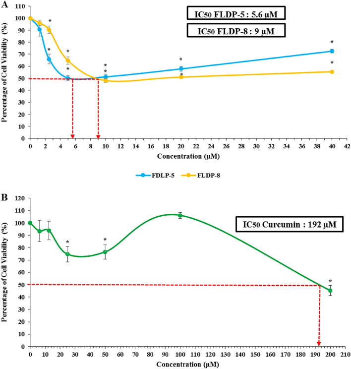
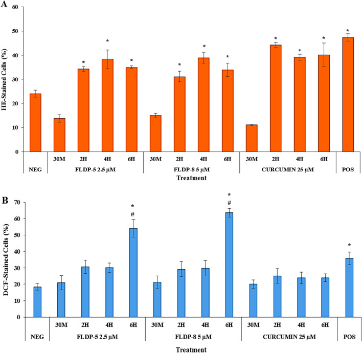
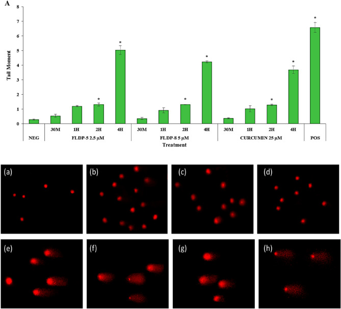
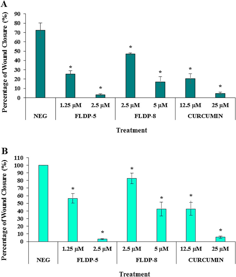
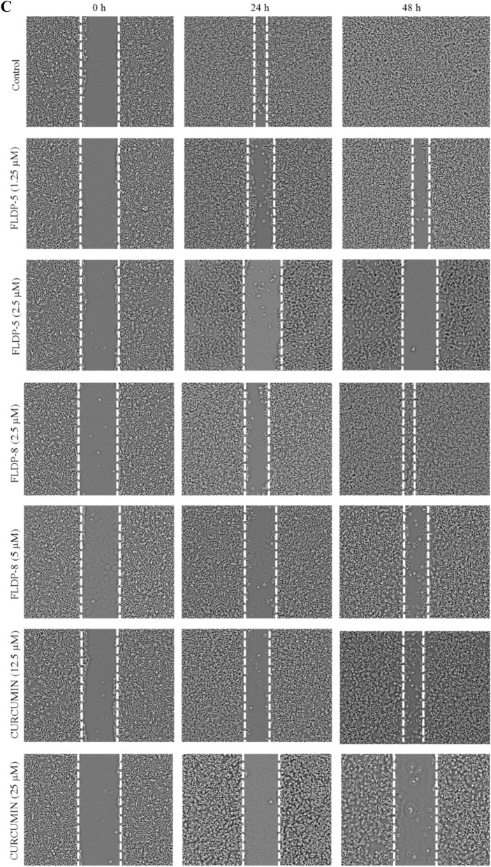
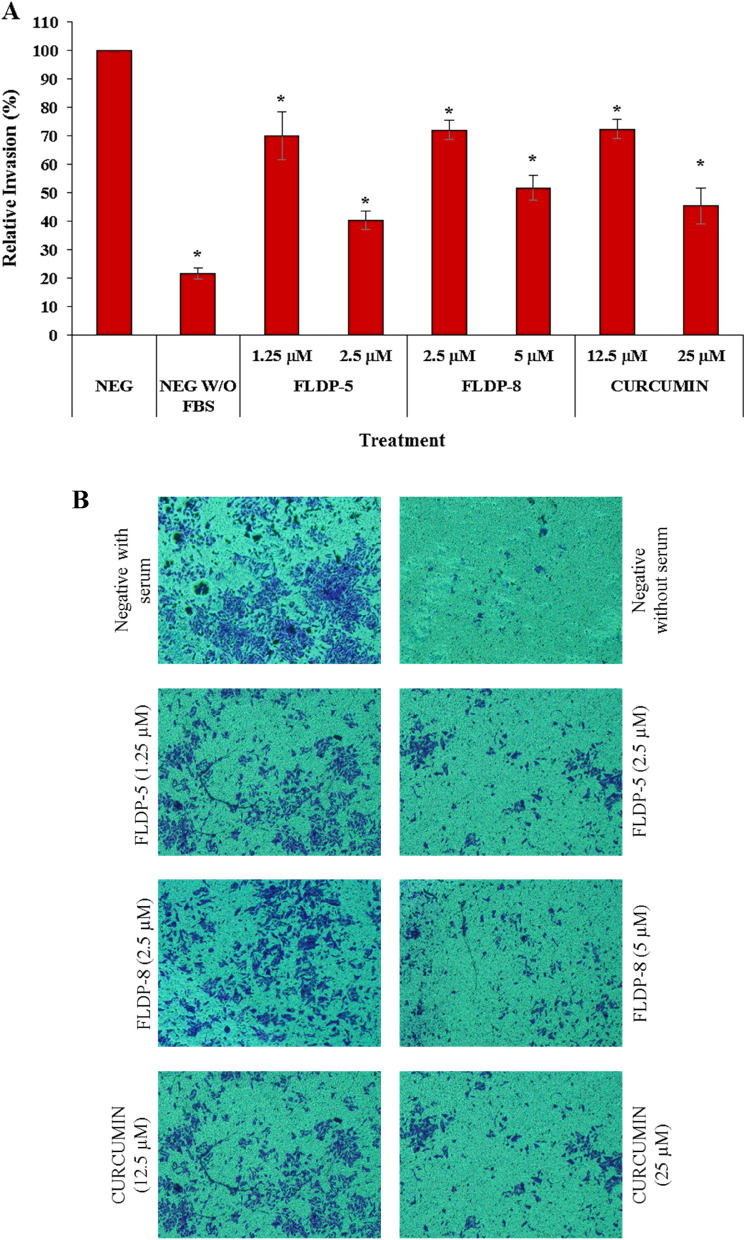
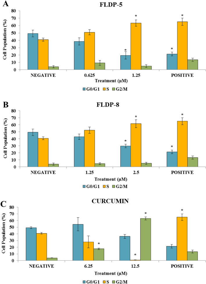
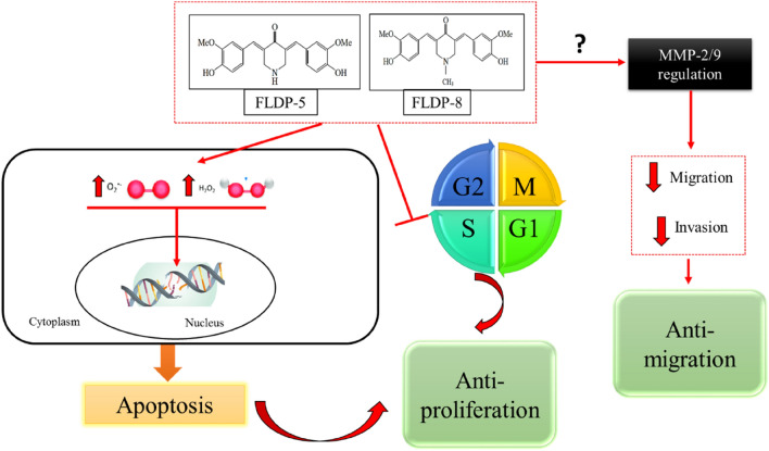
Curcumin has demonstrated potential cytotoxicity across various cell lines despite its poor bioavailability and rapid metabolism. Therefore, our group have synthesized curcuminoid analogues with piperidone derivatives, FLDP-5 and FLDP-8 to overcome these limitations. In this study, the analogues were assessed on LN-18 human glioblastoma cells in comparison to curcumin. Results from cytotoxicity assessment showed that FLDP-5 and FLDP-8 curcuminoid analogues caused death in LN-18 cells in a concentration-dependent manner after 24-h treatment with much lower IC50 values of 2.5 µM and 4 µM respectively, which were more potent compared to curcumin with IC50 of 31 µM. Moreover, a significant increase (p < 0.05) in the level of superoxide anion and hydrogen peroxide upon 2-h and 6-h treatment confirmed the oxidative stress involvement in the cell death process induced by these analogues. These analogues also showed potent anti-migratory effects through inhibition of LN-18 cells' migration and invasion. In addition, cell cycle analysis showed that these analogues are capable of inducing significant (p < 0.05) S-phase cell cycle arrest during the 24-h treatment as compared to untreated, which explained the reduced proliferation indicated by MTT assay. In conclusion, these curcuminoid analogues exhibit potent anti-cancer effects with anti-proliferative and anti-migratory properties towards LN-18 cells as compared to curcumin.


Conflict of interest statement

The authors declare no competing interests.

Figures

Figure 1
Figure 1
Chemical structures of test compounds in this study (A) Chemical structure of curcumin (B) Chemical structure of curcuminoid analogue FLDP-5 with molecular name 4-Peperidinone,3,5-bis[(4-hydroxy-3-methoxyphenyl) methylene]-,(3E,5E) (Molecular weight: 367.40 g/mol) (C) Chemical structure of curcuminoid analogue FLDP-8 with molecular name 4-Peperidinone,3,5-bis[(4-hydroxy-3-methoxyphenyl)methylene]-1-Methyl(3E,5E) (Molecular weight: 381.42 g/mol).
Figure 2
Figure 2
The cytotoxicity assessment of curcuminoid analogues (FLDP-5 and FLDP-8) and curcumin on LN-18 cells. (A) Cytotoxicity of FLDP-5 and FLDP-8 curcuminoid analogues treated LN-18 cells with concentrations from 0.625 μM till 20 μM was observed after 24-h treatment. IC50 values of 2.4 μM and 4 μM were observed respectively in FLDP-5 curcuminoid analogues and FLDP-8. (B) Cytotoxicity of curcumin-treated LN-18 cells with concentrations from 3.125 μM till 100 μM was observed after 24-h treatment. An IC50 value of 31 μM was observed. Each data point was obtained from three independent experimental replicates and expressed as mean ± SEM of percentage of cell viability. *p < 0.05 against negative control (untreated cell).
Figure 3
Figure 3
The cytotoxicity assessment of curcuminoid analogues (FLDP-5 and FLDP-8) and curcumin on HBEC-5i cells. (A) Cytotoxicity of FLDP-5 and FLDP-8 curcuminoid analogues treated HBEC-5i cells with concentrations from 1.25 μM till 40 μM was observed after 24-h treatment. IC50 values of 5.6 μM and 9 μM were observed respectively in FLDP-5 and FLDP-8 curcuminoid analogues. (B) Cytotoxicity of curcumin-treated HBEC-5i cells with concentrations from 6.25 μM till 200 μM was observed after 24-h treatment. An IC50 value of 192 μM was observed. Each data point was obtained from three independent experimental replicates and expressed as mean ± SEM of percentage of cell viability. *p < 0.05 against negative control (untreated cell).
Figure 4
Figure 4
ROS production assessment in LN-18 cells. (A) Flow cytometric analysis of superoxide level using HE staining. (B) Flow cytometric analysis of hydrogen peroxide level using DCFH-DA staining. Cells were treated respectively with IC50 values of FLDP-5, FLDP-8 and curcumin at different time-points ranging from 30 min until 6-h. Both assays used HQ treatment at 12.5 μM for 6-h as positive control (POS). Each data point was obtained from three independent experimental replicates and expressed as mean ± SEM of HE- or DCF-stained cells (%). *p < 0.05 against negative control, NEG and # p < 0.05 against curcumin.
Figure 5
Figure 5
Assessment of DNA damage in LN-18 using alkaline comet assay. (A) DNA damage expressed as tail moment in cells treated respectively with IC50 values of FLDP-5, FLDP-8 and curcumin at different time-points ranging from 30 min until 6-h. Fluorescence microscopic images stained with EtBr stain of untreated cells (a), cells treated with FLDP-5 at 2.5 μM for 4-h (b), FLDP-8 at 5 μM for 4-h (c), curcumin at 25 μM for 4-h (d), FLDP-5 at 2.5 μM for 6-h (e), FLDP-8 at 5 μM for 6-h (f), curcumin at 25 μM for 6-h (g) and positive control (h). Each data was obtained from three independent experimental replicates and each data point in (A) was expressed as mean ± SEM of tail moment. *p < 0.05 against negative control, NEG and # p < 0.05 against curcumin.
Figure 6
Figure 6
Assessment of cell migration in LN-18 cells using scratch/wound-healing assay. (A) LN-18 cells were treated respectively with IC50 and IC25 of FLDP-5, FLDP-8 curcuminoid analogues and curcumin at 24-h and the percentage of wound closure was measured. (B) LN-18 cells were treated respectively with IC50 and IC25 of FLDP-5, FLDP-8 curcuminoid analogues and curcumin at 48-h and the percentage of wound closure was measured. (C) Microscopic images of untreated cells, cells treated with FLDP-5 curcuminoid analogue (1.25 μM and 2.5 μM), FLDP-8 curcuminoid analogue (2.5 μM and 5 μM) and curcumin (12.5 μM and 25 μM) for 0 h, 24-h and 48-h. Each data was obtained from three independent experimental replicates and each data point in (A) and (B) was expressed as mean ± SEM of wound closure percentage. *p < 0.05 against negative control.
Figure 6
Figure 6
Assessment of cell migration in LN-18 cells using scratch/wound-healing assay. (A) LN-18 cells were treated respectively with IC50 and IC25 of FLDP-5, FLDP-8 curcuminoid analogues and curcumin at 24-h and the percentage of wound closure was measured. (B) LN-18 cells were treated respectively with IC50 and IC25 of FLDP-5, FLDP-8 curcuminoid analogues and curcumin at 48-h and the percentage of wound closure was measured. (C) Microscopic images of untreated cells, cells treated with FLDP-5 curcuminoid analogue (1.25 μM and 2.5 μM), FLDP-8 curcuminoid analogue (2.5 μM and 5 μM) and curcumin (12.5 μM and 25 μM) for 0 h, 24-h and 48-h. Each data was obtained from three independent experimental replicates and each data point in (A) and (B) was expressed as mean ± SEM of wound closure percentage. *p < 0.05 against negative control.
Figure 7
Figure 7
Assessment of cell invasion in LN-18 cells using Boyden chamber transwell assay. (A) LN-18 cells were treated respectively with IC50 and IC25 of FLDP-5, FLDP-8 curcuminoid analogues and curcumin at 24-h and the number of invaded of cells was measured at 560 nm. (B) Microscopic images of untreated cells with serum, untreated cells without serum, cells treated with FLDP-5 curcuminoid analogue (1.25 μM and 2.5 μM), FLDP-8 curcuminoid analogue (2.5 μM and 5 μM) and curcumin (12.5 μM and 25 μM) for 24-h. Each data was obtained from three independent experimental replicates and each data point was expressed as mean ± SEM of percentage of relative invasion. *p < 0.05 against negative control, NEG.
Figure 8
Figure 8
Assessment of cell cycle arrest in LN-18 using PI/RNase staining. (A) LN-18 cells were treated respectively with IC25 and IC12.5 of FLDP-5 at 24-h and the percentage cell population in each phase is measured. (B) LN-18 cells were treated respectively with IC25 and IC12.5 of FLDP-8 at 24-h and the percentage cell population in each phase is measured. (C) LN-18 cells were treated respectively with IC25 and IC12.5 of curcumin at 24-h and the percentage cell population in each phase is measured. Each data was obtained from three independent experimental replicates and each data point in (A), (B) and (C) was expressed as mean ± SEM of cell population percentage. *p < 0.05 against negative control.
Figure 9
Figure 9
Schematic representation of FLDP-5 and FLDP-8 curcuminoid analogues induced anti-proliferative and anti-migratory effects in LN-18 human GBM cells. Curcuminoid analogues (FLDP-5 and FLDP-8) induced DNA damage through oxidative stress with the increase in the production of intracellular ROS. The analogues also induced S-phase cell cycle arrest leading to anti-proliferative effect on LN-18 cells. Moreover, FLDP-5 and FLDP-8 curcuminoid analogues potentiate anti-migratory effect on LN-18 cells through inhibition in the migration and invasion of this cell line.

References

    1. Banu Z. Glioblastoma multiforme: a review of its pathogenesis and treatment. Int. Res. J. Pharm. 2019;9:7–12. doi: 10.7897/2230-8407.0912283. - DOI
    1. Nørøxe DS, Poulsen HS, Lassen U. Hallmarks of glioblastoma: a systematic review. ESMO Open. 2016;1:1–9. doi: 10.1136/esmoopen-2016-000144. - DOI - PMC - PubMed
    1. Shahar T, et al. The impact of enrollment in clinical trials on survival of patients with glioblastoma. J. Clin. Neurosci. 2012;19:1530–1534. doi: 10.1016/j.jocn.2012.04.005. - DOI - PubMed
    1. Atkins RJ, Ng W, Stylli SS, Hovens CM, Kaye AH. Repair mechanisms help glioblastoma resist treatment. J. Clin. Neurosci. 2015;22:14–20. doi: 10.1016/j.jocn.2014.09.003. - DOI - PubMed
    1. Van Tellingen O, et al. Overcoming the blood-brain tumor barrier for effective glioblastoma treatment. Drug Resist. Updat. 2015;19:1–12. doi: 10.1016/j.drup.2015.02.002. - DOI - PubMed

Publication types