. 2022 Jul 30;10(1):115.
doi: 10.1186/s40168-022-01303-1.

Host-microbiota interaction-mediated resistance to inflammatory bowel disease in pigs

Affiliations

Host-microbiota interaction-mediated resistance to inflammatory bowel disease in pigs

Xuan Zhao et al. Microbiome. .

Abstract

Background: Disease resistance phenotypes are associated with immune regulatory functions and immune tolerance and have implications for both the livestock industry and human health. Microbiota plays an essential role in regulating immunity and autoimmunity in the host organism, but the influence of host-microbiota interactions on disease resistance phenotypes remains unclear. Here, multiomics analysis was performed to identify potential regulatory mechanisms of disease resistance at both the microbiome and host levels in two pig breeds.

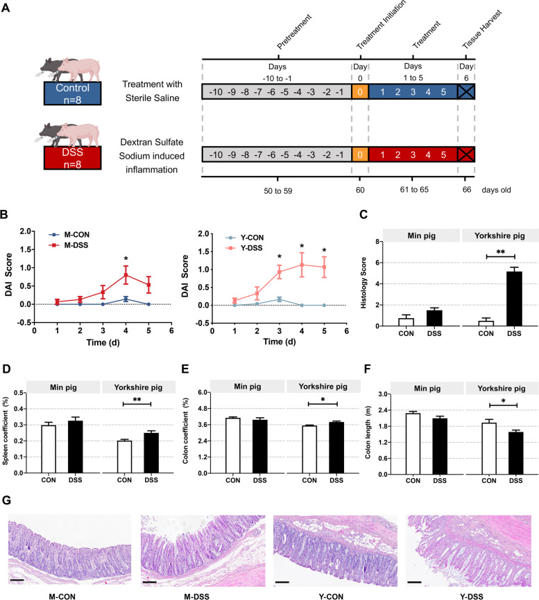
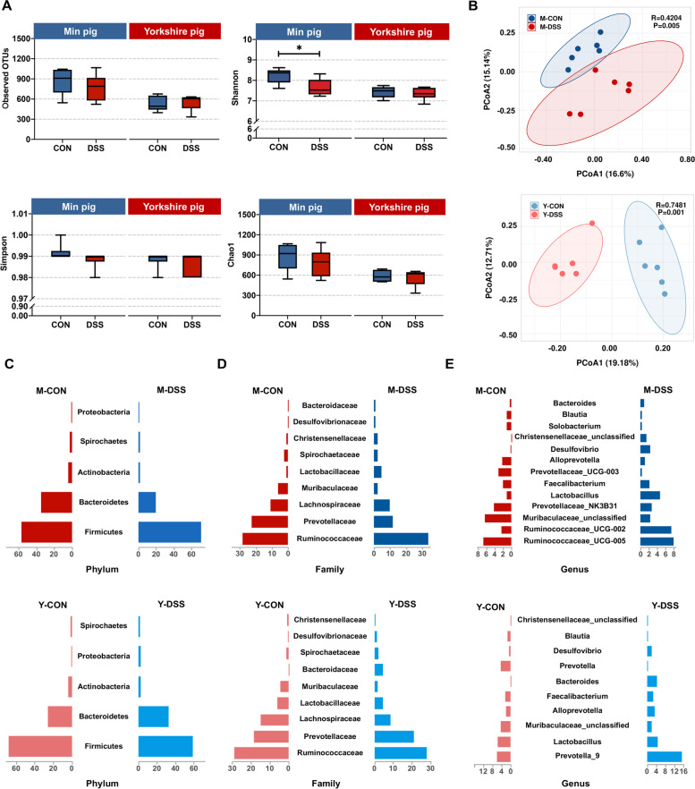
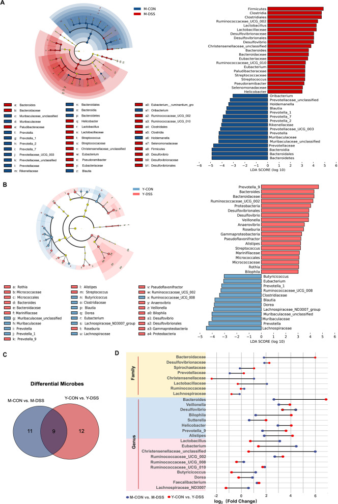
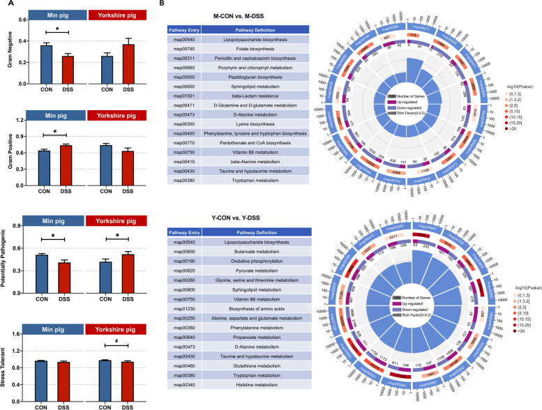
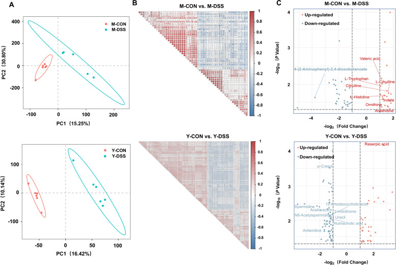
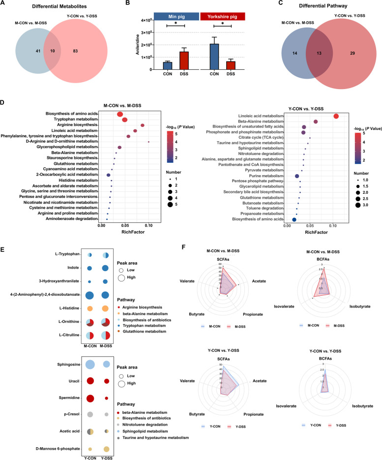
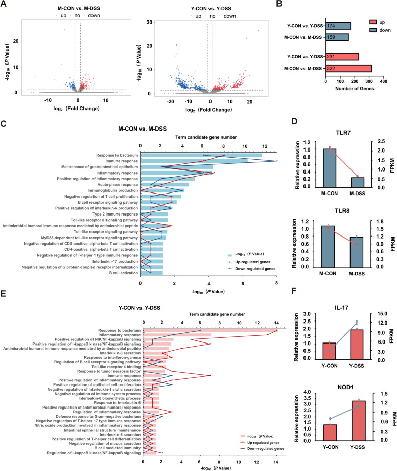
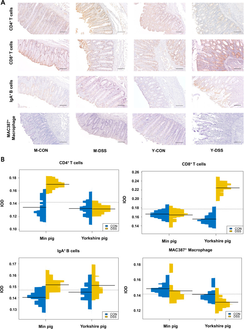
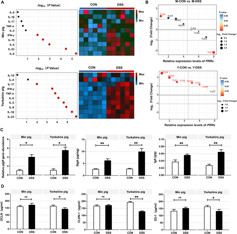
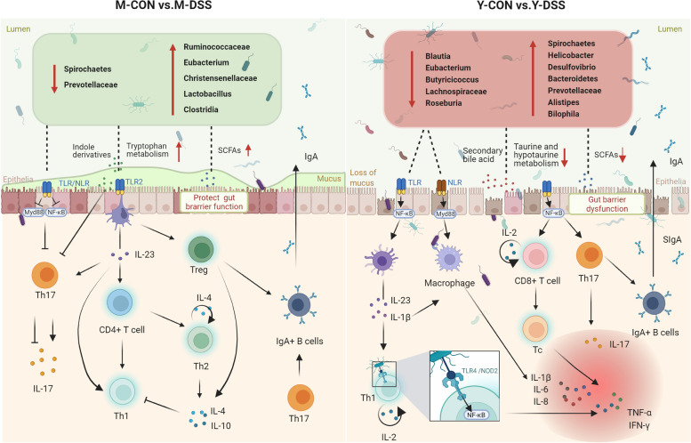
Results: Acute colitis models were established in Min pigs and Yorkshire pigs, and control and diseased individuals were compared. Compared with Yorkshire pigs under the same nutritional and management conditions, Min pigs exhibited strong disease resistance, as indicated by a low disease activity index (DAI) and a low histological activity index (HAI). Microbiota sequencing analysis showed that potentially harmful microbes Desulfovibrio, Bacteroides and Streptococcus were enriched in diseased individuals of the two breeds. Notably, potentially beneficial microbes, such as Lactobacillus, Clostridia and Eubacterium, and several genera belonging to Ruminococcaceae and Christensenellaceae were enriched in diseased Min pigs and were found to be positively associated with the microbial metabolites related to intestinal barrier function. Specifically, the concentrations of indole derivatives and short-chain fatty acids were increased in diseased Min pigs, suggesting beneficial action in protecting intestinal barrier. In addition, lower concentrations of bile acid metabolites and short-chain fatty acids were observed in diseased Yorkshire pigs, which were associated with increased potentially harmful microbes, such as Bilophila and Alistipes. Concerning enrichment of the immune response, the increase in CD4+ T cells in the lamina propria improved supervision of the host immunity response in diseased Min pigs, contributing to the maintenance of Th2-type immune superiority and immune tolerance patterns and control of excessive inflammation with the help of potentially beneficial microbes. In diseased Yorkshire pigs, more terms belonging to biological processes of immunity were enriched, including Toll-like receptors signalling, NF-κB signalling and Th1 and Th17-type immune responses, along with the increases of potentially harmful microbes and damaged intestinal barrier.

Conclusions: Cumulatively, the results for the two pig breeds highlight that host-microbiota crosstalk promotes a disease resistance phenotype in three ways: by maintaining partial PRR nonactivation, maintaining Th2-type immune superiority and immunological tolerance patterns and recovering gut barrier function to protect against colonic diseases. Video abstract.

Keywords: Disease resistance; Immunity response; Intestinal barrier function; Metabolome; Microbiota.


Conflict of interest statement

The authors declare that they have no competing interests.

Figures

Fig. 1
Fig. 1
Response of Min pigs and Yorkshire pigs to DSS-induced colitis. a Min pigs and Yorkshire pigs were administered CON (control, sterile saline) or DSS (4% DSS in 100 mL of water) via gavage for 5 days. The first dose had a volume of 200 mL. b Plot of the disease activity index (DAI) values of Min pigs and Yorkshire pigs against the number of days (n = 8 samples/group). DAI values were calculated as (weight loss rate score + stool consistency score + bloody stool score)/3. c Histological activity index (HAI) values of Min pigs and Yorkshire pigs after DSS treatment. Comparison of d spleen coefficient, e colon coefficient and f colon length in Min pigs and Yorkshire pigs after DSS treatment (n = 8 samples/group). g Distal colon inflammation and histopathology score of Min pigs and Yorkshire pigs after DSS treatment (n = 5 samples/group). Scale bar, 200 μm. Data are presented as the mean ± SEM and were analysed by the t-test; significance is reported as *P < 0.05, **P < 0.01
Fig. 2
Fig. 2
Shifts in the colonic microbiota composition of Min pigs and Yorkshire pigs after DSS treatment. a Comparison of the observed OTUs and Shannon, Simpson and Chao1 indices of the gut microbiota between control and diseased Min pigs and Yorkshire pigs (n = 6 samples/group). b Principal coordinate analysis (PCoA) plot of the microbial compositional profiles between control and diseased Min pigs and Yorkshire pigs (n = 6 samples/group). Relative abundance of the colonic microbiota at the c phylum, d family and e genus levels in Min pigs and Yorkshire pigs after DSS treatment (n = 6 samples/group)
Fig. 3
Fig. 3
Differences in microbial abundance between control and diseased Min pigs and Yorkshire pigs. Cladogram. LDA distribution. Linear discriminate analysis effect size (LEfSe) was used to analyse the differences in microbial abundance in a Min pigs and b Yorkshire pigs after DSS treatment (n = 6 samples/group). c Venn diagram for differential microbes in the comparisons M-CON vs. M-DSS and Y-CON vs. Y-DSS (n = 6). d Fold changes of differential microbes in the comparisons M-CON vs. M-DSS and Y-CON vs. Y-DSS (n = 6)
Fig. 4
Fig. 4
Differential functions of the microbiome between control and diseased Min pigs and Yorkshire pigs. a Bacterial community phenotypes of control and diseased Min pigs and Yorkshire pigs were predicted using BugBase (n = 6 samples/group). b Comparison of microbial KEGG modules between M-CON and M-DSS pigs and Y-CON and Y-DSS pigs (n = 6)
Fig. 5
Fig. 5
Colon metabolome changes. a Partial least squares discriminant analysis (PLS-DA) of metabolite composition in Min pigs and Yorkshire pigs after DSS treatment (n = 6 samples/group). b Correlations of the metabolites between control and diseased Min pigs and Yorkshire pigs (n = 6 samples/group). c Changes in colonic metabolites in Min pigs and Yorkshire pigs after DSS treatment (n = 6 samples/group)
Fig. 6
Fig. 6
Comparison of colonic metabolites in control and diseased Min pigs and Yorkshire pigs. a Venn diagram for differential metabolites in the comparisons M-CON vs. M-DSS and Y-CON vs. Y-DSS. b Common differential metabolites between control and diseased Min pigs and Yorkshire pigs. c Venn diagram for differential pathways of the comparisons M-CON vs. M-DSS and Y-CON vs. Y-DSS. d Pathway enrichment analysis was performed using the significantly different metabolites between control and diseased Min pigs and Yorkshire pigs (n = 6 samples/group). e The major pathways of significantly different metabolites in Min pigs and Yorkshire pigs after DSS treatment. f Concentrations of short-chain fatty acids (SCFAs) in Min pigs and Yorkshire pigs after DSS treatment (n = 8 samples/group)
Fig. 7
Fig. 7
RNA-seq analysis of the colon of Min pigs and Yorkshire pigs. a Volcano plot of differentially expressed genes between control and diseased Min pigs and Yorkshire pigs. b Numbers of upregulated and downregulated differentially expressed genes in Min pigs and Yorkshire pigs after DSS treatment. GO functional enrichment analysis of differentially expressed genes and qRT–PCR validation of the biological process of immunity and intestinal structure in cd Min pigs and ef Yorkshire pigs after DSS treatment
Fig. 8
Fig. 8
Changes in immune cells in the pig colon after DSS treatment. a Immunohistochemical staining (IHC) shows the numbers and distribution of CD4+ T cells, CD8+ T cells, IgA+ B cells and MAC387+ macrophages in control and diseased Min pigs and Yorkshire pigs (n = 4 samples/group). b Qualitative comparisons of the numbers of immune cells. The results are expressed as integrated optical density (IOD)
Fig. 9
Fig. 9
Analysis of the response of the gut barrier to inflammation based on transcriptome data. a Heat map showing the changes in inflammatory cytokines associated with different types of immunity in colon homogenate isolated from Min pigs and Yorkshire pigs after DSS treatment (n = 6 samples/group). b Relative expression levels of pattern recognition receptors (PRRs) in Min pigs and Yorkshire pigs after DSS treatment as detected by qRT-PCR (n = 6 samples/group). c Confirmation of autoantibody secretion in Min pigs and Yorkshire pigs after DSS treatment. d To evaluate the mechanical barrier, tight junction (TJ) proteins in Min pigs and Yorkshire pigs treated with DSS were quantified by ELISA
Fig. 10
Fig. 10
Host-microbiota interaction-mediated resistance to inflammatory bowel disease in Min pigs and Yorkshire pigs. The comparison of the responses of the two breeds to disease and inflammatory immune stimuli showed that colon microbial taxonomies, functions and metabolites and their interactions with host physiological suitability were associated with resistance. Many potentially beneficial microbes, such as Lactobacillus, Clostridia, Eubacterium and several Ruminococcaceae and Christensenellaceae genera, were increased in diseased Min pigs, which were positively correlated with the improvement of microbial metabolites, including indole derivatives and short-chain fatty acids, and thus may recover the intestinal barrier. In addition, core bacteria altered in abundance, and partial PRR nonactivation was maintained, thereby effectively inhibiting the inflammatory response. The increase in CD4+ T cells in the lamina propria improved the supervision of the host immunity response, contributing to maintenance of Th2-type immune superiority and immune tolerance patterns and controlling excessive inflammatory reactions. In Yorkshire pigs, concentrations of bile acid metabolites and short-chain fatty acids were lower in diseased individuals, associated with the increases in potentially harmful microbes, such as Bilophila, Alistipes and Bacteroidetes, and the depletion of potentially beneficial microbes, such as Blautia, Eubacterium, Dorea, Butyricicoccus, Lachnospiraceae and Roseburia. The increases in potentially harmful microbes and damaged intestinal barrier in Yorkshire pigs might be a factor in activating PRRs, thereby enriching Toll-like receptor 4 binding, NF-κB signalling and T-helper 1- and 17-type immune responses

Similar articles

Cited by

References

    1. Zhernakova A, Kurilshikov A, Bonder MJ, Tigchelaar EF, Schirmer M, Vatanen T, Mujagic Z, Vila AV, Falony G, Vieira-Silva S, et al. Population-based metagenomics analysis reveals markers for gut microbiome composition and diversity. Science. 2016;352:565–569. doi: 10.1126/science.aad3369. - DOI - PMC - PubMed
    1. Chen LM, Collij V, Jaeger M, van den Munckhof ICL, Vila AV, Kurilshikov A, Gacesa R, Sinha T, Oosting M, Joosten LAB, et al. Gut microbial co-abundance networks show specificity in inflammatory bowel disease and obesity. Nat Commun. 2020;11:1–12. doi: 10.1038/s41467-019-13993-7. - DOI - PMC - PubMed
    1. Chen L, Wang D, Garmaeva S, Kurilshikov A, Vich Vila A, Gacesa R, Sinha T, Lifelines Cohort S, Segal E, Weersma RK, et al. The long-term genetic stability and individual specificity of the human gut microbiome. Cell. 2021;184:2302–2315. doi: 10.1016/j.cell.2021.03.024. - DOI - PubMed
    1. Ijssennagger N, Belzer C, Hooiveld GJ, Dekker J, van Mil SW, Muller M, Kleerebezem M, van der Meer R. Gut microbiota facilitates dietary heme-induced epithelial hyperproliferation by opening the mucus barrier in colon. Proc Natl Acad Sci U S A. 2015;112:10038–10043. doi: 10.1073/pnas.1507645112. - DOI - PMC - PubMed
    1. Yano JM, Yu K, Donaldson GP, Shastri GG, Ann P, Ma L, Nagler CR, Ismagilov RF, Mazmanian SK, Hsiao EY. Indigenous bacteria from the gut microbiota regulate host serotonin biosynthesis. Cell. 2015;161:264–276. doi: 10.1016/j.cell.2015.02.047. - DOI - PMC - PubMed

Publication types