. 2022 Jul 30;12(1):13136.
doi: 10.1038/s41598-022-17501-8.

A maternal high-fat diet induces fetal origins of NASH-HCC in mice

Affiliations

A maternal high-fat diet induces fetal origins of NASH-HCC in mice

Takao Takiyama et al. Sci Rep. .

Abstract

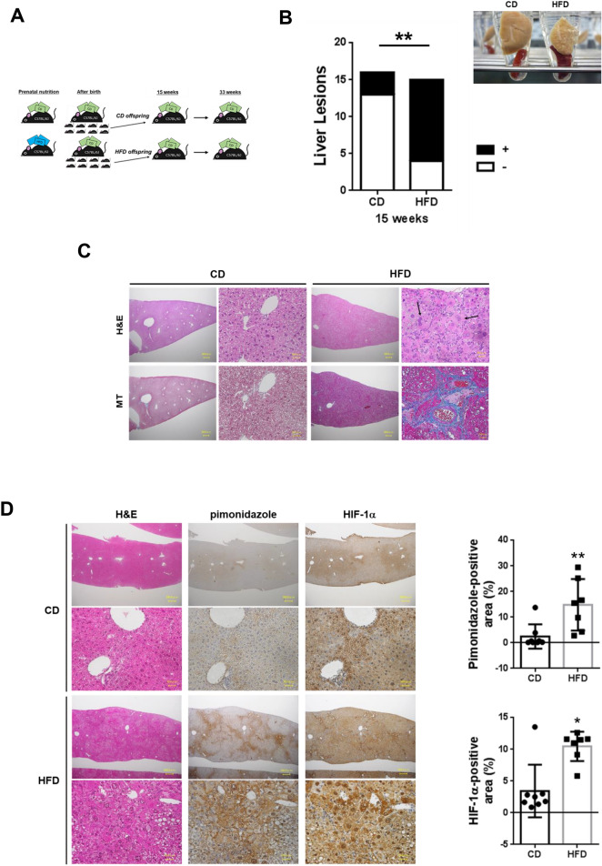
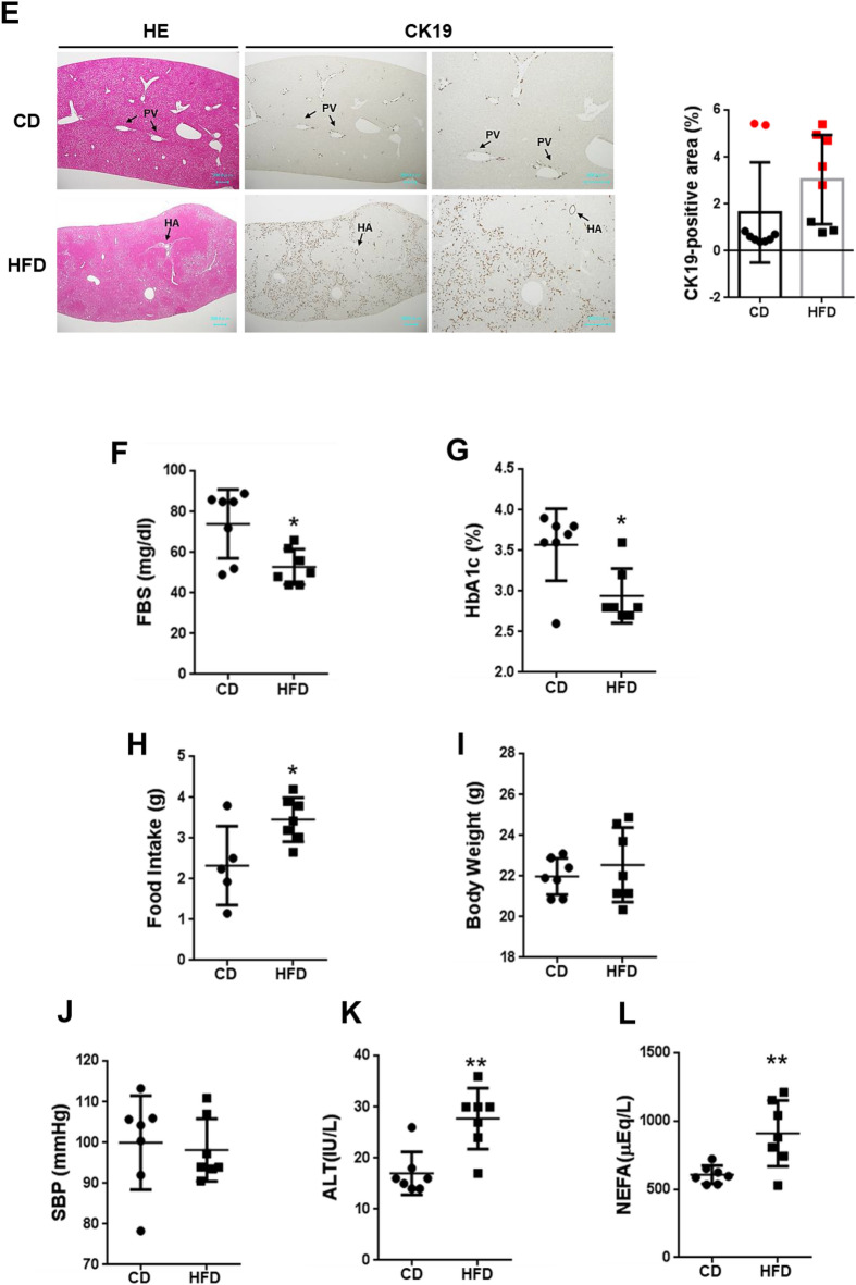
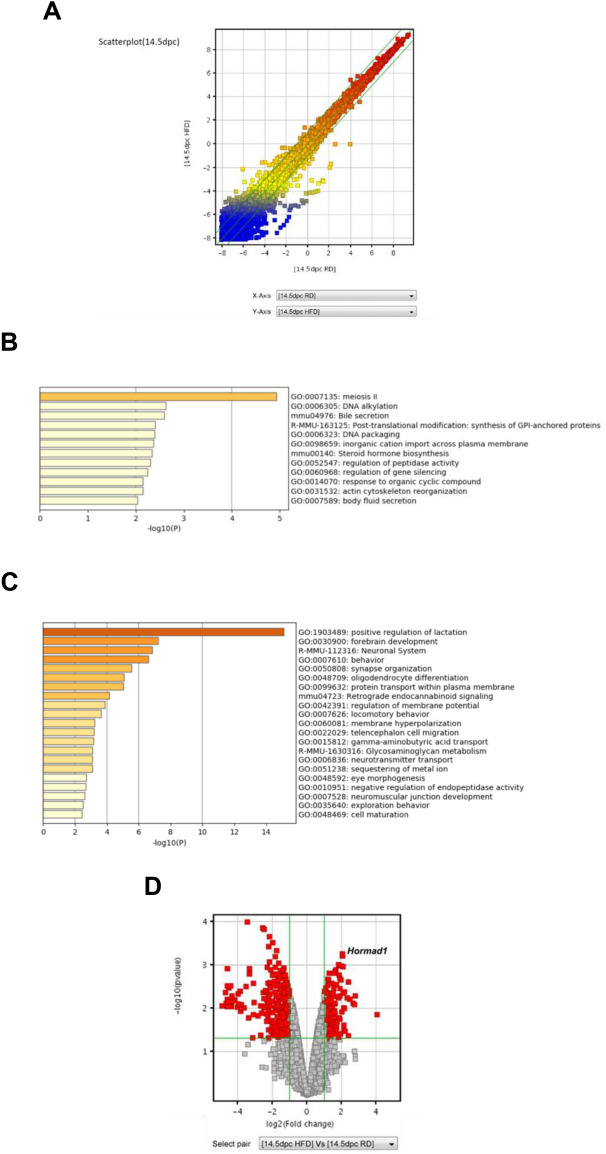
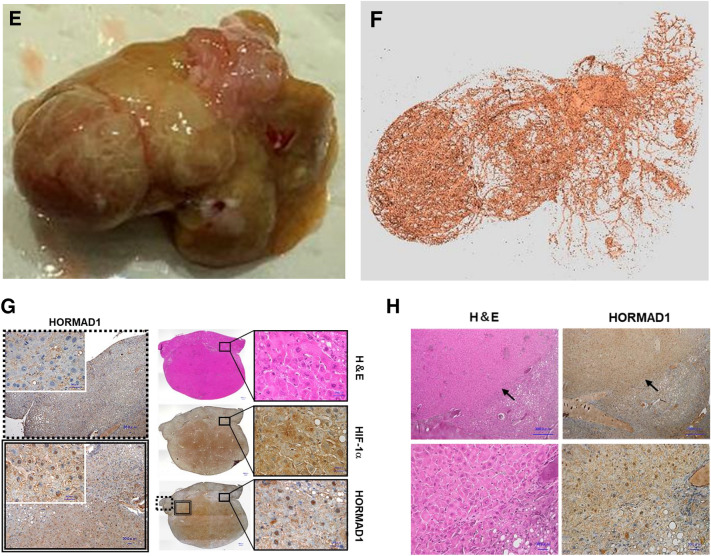
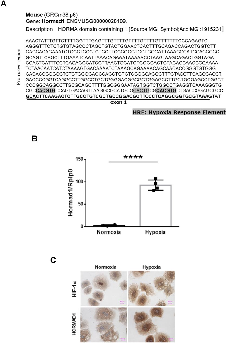
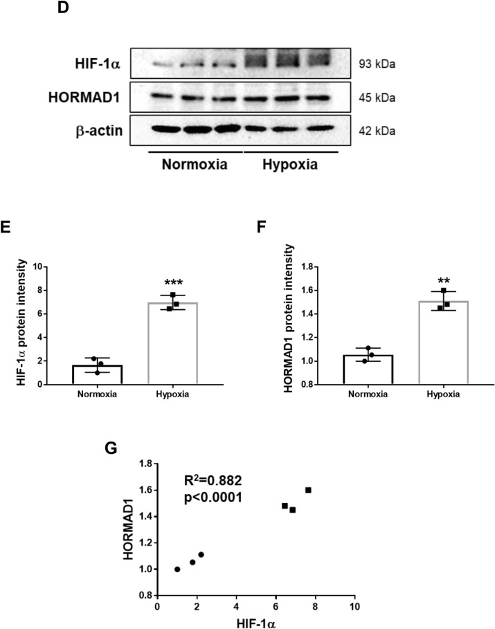
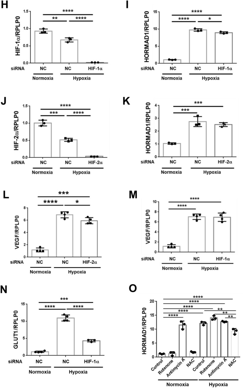
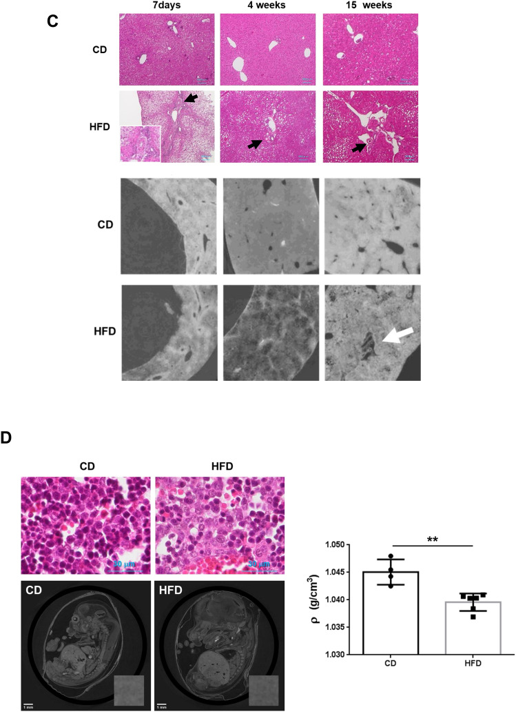
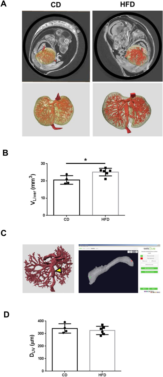
Maternal overnutrition affects offspring susceptibility to nonalcoholic steatohepatitis (NASH). Male offspring from high-fat diet (HFD)-fed dams developed a severe form of NASH, leading to highly vascular tumor formation. The cancer/testis antigen HORMA domain containing protein 1 (HORMAD1), one of 146 upregulated differentially expressed genes in fetal livers from HFD-fed dams, was overexpressed with hypoxia-inducible factor 1 alpha (HIF-1alpha) in hepatoblasts and in NASH-based hepatocellular carcinoma (HCC) in offspring from HFD-fed dams at 15 weeks old. Hypoxia substantially increased Hormad1 expression in primary mouse hepatocytes. Despite the presence of three putative hypoxia response elements within the mouse Hormad1 gene, the Hif-1alpha siRNA only slightly decreased hypoxia-induced Hormad1 mRNA expression. In contrast, N-acetylcysteine, but not rotenone, inhibited hypoxia-induced Hormad1 expression, indicating its dependency on nonmitochondrial reactive oxygen species production. Synchrotron-based phase-contrast micro-CT of the fetuses from HFD-fed dams showed significant enlargement of the liver accompanied by a consistent size of the umbilical vein, which may cause hypoxia in the fetal liver. Based on these findings, a maternal HFD induces fetal origins of NASH/HCC via hypoxia, and HORMAD1 is a potential therapeutic target for NASH/HCC.


Conflict of interest statement

The authors declare no competing interests.

Figures

Figure 1
Figure 1
Maternal HFD consumption induces hepatic steatosis in offspring. (A) Experimental setup and time points that were analyzed. Female C57BL/6J mice were fed either a HFD or a CD during gestation. During lactation and after weaning, all mice were fed a CD. (B) The number of 15-week-old offspring with liver lesions indicating steatohepatitis (steatosis, lobular inflammation, hepatocellular ballooning, and fibrosis) in the CD (n = 16) and HFD groups (n = 15). Photographs of liver sections and spleens from 15-week-old offspring of the CD- and HFD-fed dams. Statistical analysis: Fisher’s exact test. (C) Hematoxylin and eosin staining (upper panels) and Masson’s trichrome (MT) staining (lower panels) of the livers from offspring of HFD-fed dams. The atypical cells exhibited severe dysplasia, with an increased nuclear-cytoplasmic ratio and enlarged nuclei with hyperploidization (black arrows) accompanied by fat deposition. Scale bars: 300 μm (low magnification) and 30 μm (high magnification). (D) The liver sections from offspring of CD- and HFD-fed dams were stained for pimonidazole and HIF-1α. Representative images of pimonidazole and HIF-1α staining (cells stained brown) are shown. Scale bars: 300 μm (low magnification) and 30 μm (high magnification) in each group. Quantification of the pimonidazole-positive area and HIF-1α-positive area (CD = 8 offspring from 3 dams, HFD = 7 offspring from 3 dams). *p < 0.05 and **p < 0.01 using the Mann–Whitney test. (E) CK19 immunohistochemistry in the liver sections from offspring of CD- and HFD-fed dams. Scale bars: 300 μm. PV portal vein, HA hepatic artery. Quantification of the CK19-positive area. p > 0.05 using the Mann–Whitney test (CD = 9 offspring from 3 dams, HFD = 8 offspring from 3 dams). Two red circles and five red rectangles show the results for offspring with liver lesions. The stained sections were observed and visualized using a light microscope system (BZ-8100; Keyence, Osaka, Japan. https://www.keyence.com). (F) FBS, (G) HbA1c, (H) food intake/day, (I) body weight, (J) SBP, (K) ALT, and (L) NEFA levels. The values represent individual measurements and are presented as the means ± SD. The significance of the differences between groups was determined using unpaired Student’s t tests. Welch’s corrections were used when the variances between groups were unequal. *p < 0.05 and **p < 0.01.
Figure 1
Figure 1
Maternal HFD consumption induces hepatic steatosis in offspring. (A) Experimental setup and time points that were analyzed. Female C57BL/6J mice were fed either a HFD or a CD during gestation. During lactation and after weaning, all mice were fed a CD. (B) The number of 15-week-old offspring with liver lesions indicating steatohepatitis (steatosis, lobular inflammation, hepatocellular ballooning, and fibrosis) in the CD (n = 16) and HFD groups (n = 15). Photographs of liver sections and spleens from 15-week-old offspring of the CD- and HFD-fed dams. Statistical analysis: Fisher’s exact test. (C) Hematoxylin and eosin staining (upper panels) and Masson’s trichrome (MT) staining (lower panels) of the livers from offspring of HFD-fed dams. The atypical cells exhibited severe dysplasia, with an increased nuclear-cytoplasmic ratio and enlarged nuclei with hyperploidization (black arrows) accompanied by fat deposition. Scale bars: 300 μm (low magnification) and 30 μm (high magnification). (D) The liver sections from offspring of CD- and HFD-fed dams were stained for pimonidazole and HIF-1α. Representative images of pimonidazole and HIF-1α staining (cells stained brown) are shown. Scale bars: 300 μm (low magnification) and 30 μm (high magnification) in each group. Quantification of the pimonidazole-positive area and HIF-1α-positive area (CD = 8 offspring from 3 dams, HFD = 7 offspring from 3 dams). *p < 0.05 and **p < 0.01 using the Mann–Whitney test. (E) CK19 immunohistochemistry in the liver sections from offspring of CD- and HFD-fed dams. Scale bars: 300 μm. PV portal vein, HA hepatic artery. Quantification of the CK19-positive area. p > 0.05 using the Mann–Whitney test (CD = 9 offspring from 3 dams, HFD = 8 offspring from 3 dams). Two red circles and five red rectangles show the results for offspring with liver lesions. The stained sections were observed and visualized using a light microscope system (BZ-8100; Keyence, Osaka, Japan. https://www.keyence.com). (F) FBS, (G) HbA1c, (H) food intake/day, (I) body weight, (J) SBP, (K) ALT, and (L) NEFA levels. The values represent individual measurements and are presented as the means ± SD. The significance of the differences between groups was determined using unpaired Student’s t tests. Welch’s corrections were used when the variances between groups were unequal. *p < 0.05 and **p < 0.01.
Figure 2
Figure 2
Maternal HFD consumption upregulates Hormad1 gene expression in the fetal liver. (A) Scatterplot of the gene expression profiles for hepatocytes from fetuses (14.5 dpc). Green lines indicate the cutoffs for twofold up- and downregulation. Enrichment scores obtained from the Gene Ontology enrichment analysis of the selected mRNAs. (B) GO analysis of the upregulated genes in livers from fetuses of HFD-fed dams compared with CD-fed dams. (C) GO analysis of the downregulated genes in livers from fetuses of HFD-fed dams compared with CD-fed dams. (D) Volcano plots of the gene expression data. The horizontal axis represents the log2 (fold change), and the vertical axis represents the  − log 10 (p value). The red plots represent the selected DEGs. Volcano plot showing Hormad1, which had the lowest p value (p = 0.0006), and a greater than four-fold change (log2 = 2.037) in expression in livers from fetuses of HFD-fed dams compared with CD-fed dams. Data are presented using GeneSpring software 12.1 (Agilent Technologies. https://www.agilent.com).
Figure 3
Figure 3
HORMAD1 colocalizes with HIF-1α in fetal livers. (A) Representative photomicrographs of immunohistochemical staining for HIF-1α and HORMAD1 in fetal livers at 14.5 dpc. Scale bars: 300 μm (low magnification) and 30 μm (high magnification). Insets show the magnified images from the left panels. (B) Western blot analysis showed positive correlations between HORMAD1 and HIF-1α levels in the fetal livers of mice (R2 = 0.8469, p = 0.0012). (C) Immunohistochemical staining for HIF-1α and HORMAD1 in the livers of offspring at 15 weeks of age. Scale bars: 300 μm (low magnification) and 30 μm (high magnification). Quantification of HORMAD1-positive area (CD = 8 offspring from 3 dams, HFD = 7 offspring from 3 dams). **p < 0.01 using the Mann–Whitney test. (D) Western blot analysis of HIF-1α and HORMAD1 levels in the livers of 15-week-old offspring. MWM: 117 kDa band in the molecular weight marker (HiMark Prestained protein standard, Thermo Fisher Scientific). (E) Macroscopic image of the liver after barium perfusion of offspring from HFD-fed dams at 33 weeks old. (F) Visualization of 3D vessels in the liver of offspring from HFD-fed dams at 33 weeks old using synchrotron micro-CT and Amira software (version 1.4.0, https://www.thermofisher.com). (G) Immunohistochemical staining for HIF-1α and HORMAD1 in hepatic tumors from 33-week-old offspring of HFD-fed dams. Insets in the middle panels show the magnified images (right panels). HORMAD1 expression in peritumoral lesions (dotted line) and intratumoral lesions (double lines) in hepatic tumors from 33-week-old offspring of HFD-fed dams. Insets in the left panels show the magnified image of each lesion. (H) Capsular invasion of a hepatic tumor. Arrows show the area of invasion. Bars: 300 μm (low magnification) and 30 μm (high magnification).
Figure 3
Figure 3
HORMAD1 colocalizes with HIF-1α in fetal livers. (A) Representative photomicrographs of immunohistochemical staining for HIF-1α and HORMAD1 in fetal livers at 14.5 dpc. Scale bars: 300 μm (low magnification) and 30 μm (high magnification). Insets show the magnified images from the left panels. (B) Western blot analysis showed positive correlations between HORMAD1 and HIF-1α levels in the fetal livers of mice (R2 = 0.8469, p = 0.0012). (C) Immunohistochemical staining for HIF-1α and HORMAD1 in the livers of offspring at 15 weeks of age. Scale bars: 300 μm (low magnification) and 30 μm (high magnification). Quantification of HORMAD1-positive area (CD = 8 offspring from 3 dams, HFD = 7 offspring from 3 dams). **p < 0.01 using the Mann–Whitney test. (D) Western blot analysis of HIF-1α and HORMAD1 levels in the livers of 15-week-old offspring. MWM: 117 kDa band in the molecular weight marker (HiMark Prestained protein standard, Thermo Fisher Scientific). (E) Macroscopic image of the liver after barium perfusion of offspring from HFD-fed dams at 33 weeks old. (F) Visualization of 3D vessels in the liver of offspring from HFD-fed dams at 33 weeks old using synchrotron micro-CT and Amira software (version 1.4.0, https://www.thermofisher.com). (G) Immunohistochemical staining for HIF-1α and HORMAD1 in hepatic tumors from 33-week-old offspring of HFD-fed dams. Insets in the middle panels show the magnified images (right panels). HORMAD1 expression in peritumoral lesions (dotted line) and intratumoral lesions (double lines) in hepatic tumors from 33-week-old offspring of HFD-fed dams. Insets in the left panels show the magnified image of each lesion. (H) Capsular invasion of a hepatic tumor. Arrows show the area of invasion. Bars: 300 μm (low magnification) and 30 μm (high magnification).
Figure 4
Figure 4
Hypoxia induces HORMAD1 expression in MPHs. (A) Three putative HREs containing the consensus sequence (A/G)CGTG within the mouse Hormad1 gene. (B) Hormad1 mRNA expression in MPHs was examined using qRT‒PCR. Isolated MPHs were exposed to hypoxia (1% O2) overnight. (C) Immunocytochemical staining for HIF-1α and HORMAD1 in MPHs cultured under normoxic or hypoxic conditions. Bars: 30 μm. (D) Western blot analysis of the total cell lysates of MPHs. (E) Effects of hypoxia on HIF-1α protein expression in MPHs. (F) Effects of hypoxia on HORMAD1 protein expression in MPHs. (G) Positive correlation between HIF-1α and HORMAD1 protein expression in MPHs (R2 = 0.882, p < 0.0001). (H) The efficacy of siRNAs targeting HIF-1α in reducing the Hif-1α mRNA expression level in MPHs. (I) The effect of the HIF-1α siRNA on the Hormad1 mRNA expression level in MPHs. (J) The efficacy of the HIF-2α siRNA in reducing the Hif-2α mRNA expression level in MPHs. (K) The effect of the HIF-2α siRNA on the Hormad1 mRNA expression level in MPHs. (L) The effect of the HIF-2α siRNA on the Vegf mRNA expression level in MPHs. (M) The effects of the HIF-1α siRNA on the Vegf mRNA expression level in MPHs. (N) The effect of the HIF-1α siRNA on Glut1 mRNA expression levels in MPHs. (O) MPHs were treated with mitochondrial inhibitors of complex I (rotenone, 1 μM) and complex III Qi sites (antimycin A, 1 μM) and the glutathione precursor N-acetylcysteine (NAC, 1 mM) to determine their effects on Hormad1 mRNA expression. All data are presented as the means ± SD, and they are representative of at least three independent experiments. The significance of differences between groups was determined using unpaired Student’s t tests. Statistical comparisons were analyzed using one-way ANOVA with post hoc Bonferroni multiple comparison tests. *p < 0.05, **p < 0.01, ***p < 0.001, and ****p < 0.0001.
Figure 4
Figure 4
Hypoxia induces HORMAD1 expression in MPHs. (A) Three putative HREs containing the consensus sequence (A/G)CGTG within the mouse Hormad1 gene. (B) Hormad1 mRNA expression in MPHs was examined using qRT‒PCR. Isolated MPHs were exposed to hypoxia (1% O2) overnight. (C) Immunocytochemical staining for HIF-1α and HORMAD1 in MPHs cultured under normoxic or hypoxic conditions. Bars: 30 μm. (D) Western blot analysis of the total cell lysates of MPHs. (E) Effects of hypoxia on HIF-1α protein expression in MPHs. (F) Effects of hypoxia on HORMAD1 protein expression in MPHs. (G) Positive correlation between HIF-1α and HORMAD1 protein expression in MPHs (R2 = 0.882, p < 0.0001). (H) The efficacy of siRNAs targeting HIF-1α in reducing the Hif-1α mRNA expression level in MPHs. (I) The effect of the HIF-1α siRNA on the Hormad1 mRNA expression level in MPHs. (J) The efficacy of the HIF-2α siRNA in reducing the Hif-2α mRNA expression level in MPHs. (K) The effect of the HIF-2α siRNA on the Hormad1 mRNA expression level in MPHs. (L) The effect of the HIF-2α siRNA on the Vegf mRNA expression level in MPHs. (M) The effects of the HIF-1α siRNA on the Vegf mRNA expression level in MPHs. (N) The effect of the HIF-1α siRNA on Glut1 mRNA expression levels in MPHs. (O) MPHs were treated with mitochondrial inhibitors of complex I (rotenone, 1 μM) and complex III Qi sites (antimycin A, 1 μM) and the glutathione precursor N-acetylcysteine (NAC, 1 mM) to determine their effects on Hormad1 mRNA expression. All data are presented as the means ± SD, and they are representative of at least three independent experiments. The significance of differences between groups was determined using unpaired Student’s t tests. Statistical comparisons were analyzed using one-way ANOVA with post hoc Bonferroni multiple comparison tests. *p < 0.05, **p < 0.01, ***p < 0.001, and ****p < 0.0001.
Figure 5
Figure 5
Synchrotron radiation micro-CT images show liver circulation in hepatic tumors and fetal livers. (A) Photographs of livers of 33-week-old offspring from CD- or HFD-fed dams (upper panels). Macroscopic images of livers after barium perfusion (lower panels). (B) CT fluoroscopy (left panels) and visualization of 3D vessels via synchrotron micro-CT using Amira software (version 1.4.0, https://www.thermofisher.com) (right panels) of livers from offspring. The square surrounded by the dotted line shows the typical basket pattern of tumor blood vessels. The computed tomography data from the offspring livers were uploaded to advanced 3D image processing and quantification software (Amira). (C) Hematoxylin and eosin staining (upper panels) and phase-contrast micro-CT imaging (lower panels) of livers of offspring at 7 days (CD; n = 7, HFD; n = 9), 4 weeks (CD; n = 6, HFD; n = 9) and 15 weeks (CD; n = 17, HFD; n = 11) of age. Black and white arrows show abnormal vascular structures, including large and thick hepatic arteries, in the livers from offspring of HFD-fed dams. Scale bars: 100 μm. (D) Hematoxylin and eosin staining of fetal livers (upper panels. Scale bars: 30 μm). Phase-contrast micro-CT imaging of fetuses (CD; n = 4, HFD; n = 6) at 14.5 dpc from CD- and HFD-fed dams. Scale bars: 1 mm. Hepatic tissue density (ρ) of offspring from CD- and HFD-fed dams. All data are presented as the means ± SD. Measured variables were log(e) transformed for statistical analyses. The significance of the differences between groups was determined using unpaired Student’s t tests. **p < 0.01.
Figure 5
Figure 5
Synchrotron radiation micro-CT images show liver circulation in hepatic tumors and fetal livers. (A) Photographs of livers of 33-week-old offspring from CD- or HFD-fed dams (upper panels). Macroscopic images of livers after barium perfusion (lower panels). (B) CT fluoroscopy (left panels) and visualization of 3D vessels via synchrotron micro-CT using Amira software (version 1.4.0, https://www.thermofisher.com) (right panels) of livers from offspring. The square surrounded by the dotted line shows the typical basket pattern of tumor blood vessels. The computed tomography data from the offspring livers were uploaded to advanced 3D image processing and quantification software (Amira). (C) Hematoxylin and eosin staining (upper panels) and phase-contrast micro-CT imaging (lower panels) of livers of offspring at 7 days (CD; n = 7, HFD; n = 9), 4 weeks (CD; n = 6, HFD; n = 9) and 15 weeks (CD; n = 17, HFD; n = 11) of age. Black and white arrows show abnormal vascular structures, including large and thick hepatic arteries, in the livers from offspring of HFD-fed dams. Scale bars: 100 μm. (D) Hematoxylin and eosin staining of fetal livers (upper panels. Scale bars: 30 μm). Phase-contrast micro-CT imaging of fetuses (CD; n = 4, HFD; n = 6) at 14.5 dpc from CD- and HFD-fed dams. Scale bars: 1 mm. Hepatic tissue density (ρ) of offspring from CD- and HFD-fed dams. All data are presented as the means ± SD. Measured variables were log(e) transformed for statistical analyses. The significance of the differences between groups was determined using unpaired Student’s t tests. **p < 0.01.
Figure 6
Figure 6
Maternal HFD consumption induced hepatomegaly without a compensatory increase in blood flow in fetuses. (A) Phase-contrast micro-CT images of fetuses (upper panels) were uploaded to advanced 3D image processing and quantification software (Amira, version 1.4.0, https://www.thermofisher.com) and 3D images of the fetal liver and intrahepatic vascular structure (lower panels) are presented. (B) Liver volume of fetuses (VLiver). The volume of the extracted liver was derived by forming multiple tetrahedrons with the center point as the apex in Amira. CD; n = 4, HFD; n = 6. (C) Hepatic vessels were extracted from fetuses using 3D image processing and quantification software (Amira). Intrahepatic blood vessels were extracted from the umbilical vein (yellow arrowhead) just before they entered the liver and branched. The blood flow rate was determined by deriving the vessel diameter, which is proportional to the multiplier of the vessel diameter according to Murray’s law (left panel). An open-source toolkit (Vascular Modelling Toolkit, version 1.4.0, VMTK, www.vmtk.org) was used to derive the vessel diameter semiautomatically (right panel). (D) Diameter of the umbilical vein (DUV) of fetuses. CD; n = 4, HFD; n = 6. (E) The blood flow rate is proportional to DUV3 per volume of fetal liver (VLiver). CD; n = 4, HFD; n = 6. All measured variables were log(e) transformed for statistical analysis. The significance of the differences between groups was determined using unpaired Student’s t tests. *p < 0.05 and **p < 0.01. (F) Maternal HFD consumption induces hepatomegaly without increasing hepatic blood flow in the fetus, resulting in hypoxia in the fetal liver. Hypoxia induces nonmitochondrial ROS production, leading to the upregulation of HORMAD1 in the fetal liver. After birth, breathing induced normoxia in the livers of offspring, attenuating the increased HORMAD1 expression. Obesity, diabetes and aging, which are known to induce hypoxia and ROS production, might increase HORMAD1 expression. The first hit in NAFLD/NASH/HCC development might be that fetal nutrition causes hepatic hypoxia. Thus, HORMAD1 expression is an indicator of tissue hypoxia resulting from ROS production.
Figure 6
Figure 6
Maternal HFD consumption induced hepatomegaly without a compensatory increase in blood flow in fetuses. (A) Phase-contrast micro-CT images of fetuses (upper panels) were uploaded to advanced 3D image processing and quantification software (Amira, version 1.4.0, https://www.thermofisher.com) and 3D images of the fetal liver and intrahepatic vascular structure (lower panels) are presented. (B) Liver volume of fetuses (VLiver). The volume of the extracted liver was derived by forming multiple tetrahedrons with the center point as the apex in Amira. CD; n = 4, HFD; n = 6. (C) Hepatic vessels were extracted from fetuses using 3D image processing and quantification software (Amira). Intrahepatic blood vessels were extracted from the umbilical vein (yellow arrowhead) just before they entered the liver and branched. The blood flow rate was determined by deriving the vessel diameter, which is proportional to the multiplier of the vessel diameter according to Murray’s law (left panel). An open-source toolkit (Vascular Modelling Toolkit, version 1.4.0, VMTK, www.vmtk.org) was used to derive the vessel diameter semiautomatically (right panel). (D) Diameter of the umbilical vein (DUV) of fetuses. CD; n = 4, HFD; n = 6. (E) The blood flow rate is proportional to DUV3 per volume of fetal liver (VLiver). CD; n = 4, HFD; n = 6. All measured variables were log(e) transformed for statistical analysis. The significance of the differences between groups was determined using unpaired Student’s t tests. *p < 0.05 and **p < 0.01. (F) Maternal HFD consumption induces hepatomegaly without increasing hepatic blood flow in the fetus, resulting in hypoxia in the fetal liver. Hypoxia induces nonmitochondrial ROS production, leading to the upregulation of HORMAD1 in the fetal liver. After birth, breathing induced normoxia in the livers of offspring, attenuating the increased HORMAD1 expression. Obesity, diabetes and aging, which are known to induce hypoxia and ROS production, might increase HORMAD1 expression. The first hit in NAFLD/NASH/HCC development might be that fetal nutrition causes hepatic hypoxia. Thus, HORMAD1 expression is an indicator of tissue hypoxia resulting from ROS production.

Similar articles

Cited by

References

    1. Neri C, Edlow AG. Effects of maternal obesity on fetal programming: Molecular approaches. Cold Spring Harb. Perspect. Med. 2015;6:a026591. doi: 10.1101/cshperspect.a026591. - DOI - PMC - PubMed
    1. Lumey LH, Van Poppel FW. The Dutch famine of 1944–1945: Mortality and morbidity in past and present generations. Soc. Hist. Med. 1994;7:229–246. doi: 10.1093/shm/7.2.229. - DOI - PubMed
    1. Stephenson J, et al. Before the beginning: Nutrition and lifestyle in the preconception period and its importance for future health. Lancet (London, England). 2018;391:1830–1841. doi: 10.1016/S0140-6736(18)30311-8. - DOI - PMC - PubMed
    1. Cho NH, et al. IDF diabetes atlas: Global estimates of diabetes prevalence for 2017 and projections for 2045. Diabetes Res. Clin. Pract. 2018;138:271–281. doi: 10.1016/j.diabres.2018.02.023. - DOI - PubMed
    1. Åmark H, Sirotkina M, Westgren M, Papadogiannakis N, Persson M. Is obesity in pregnancy associated with signs of chronic fetal hypoxia? Acta Obstet. Gynecol. Scand. 2020;99:1649–1656. doi: 10.1111/aogs.13941. - DOI - PubMed

Publication types