. 2023 Jan;39(1):69-82.
doi: 10.1007/s12264-022-00901-1. Epub 2022 Jul 30.

Neural Mechanism Underlying Task-Specific Enhancement of Motor Learning by Concurrent Transcranial Direct Current Stimulation

Affiliations

Neural Mechanism Underlying Task-Specific Enhancement of Motor Learning by Concurrent Transcranial Direct Current Stimulation

Ying Wang et al. Neurosci Bull. 2023 Jan.

Abstract

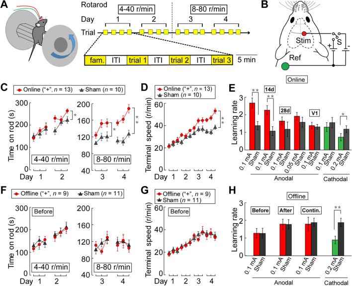
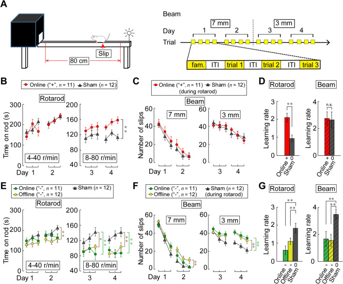
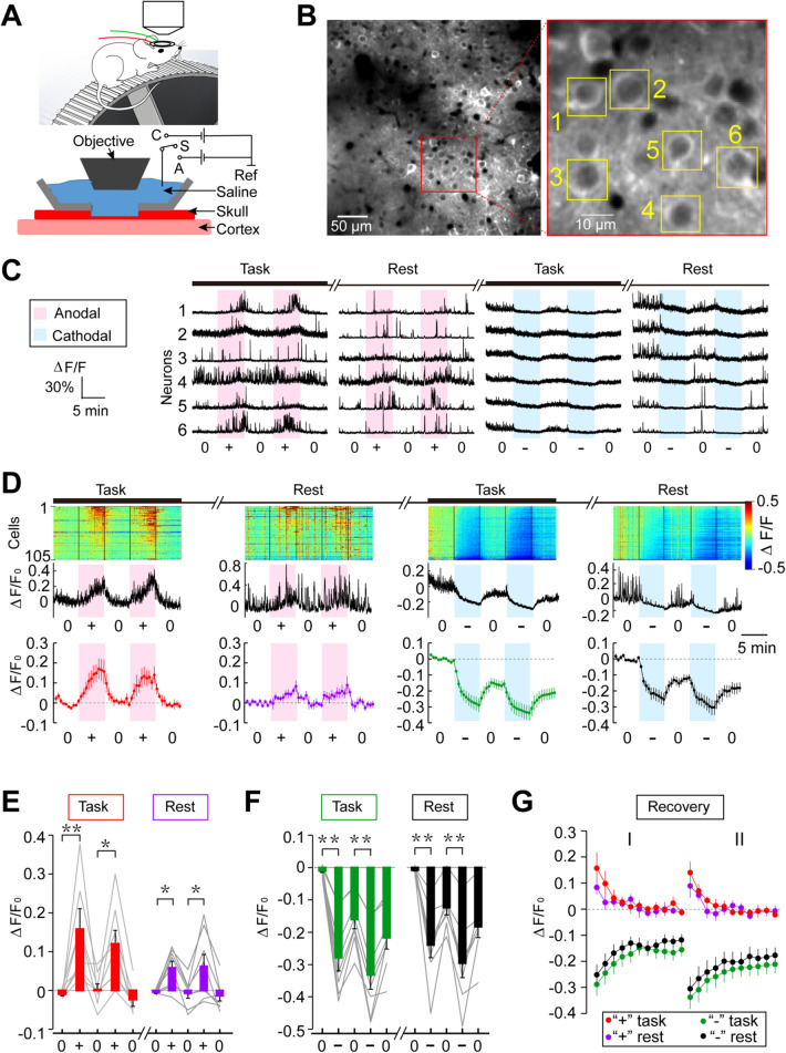
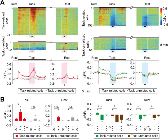
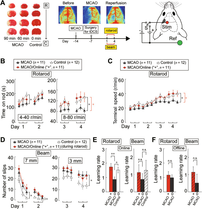
The optimal protocol for neuromodulation by transcranial direct current stimulation (tDCS) remains unclear. Using the rotarod paradigm, we found that mouse motor learning was enhanced by anodal tDCS (3.2 mA/cm2) during but not before or after the performance of a task. Dual-task experiments showed that motor learning enhancement was specific to the task accompanied by anodal tDCS. Studies using a mouse model of stroke induced by middle cerebral artery occlusion showed that concurrent anodal tDCS restored motor learning capability in a task-specific manner. Transcranial in vivo Ca2+ imaging further showed that anodal tDCS elevated and cathodal tDCS suppressed neuronal activity in the primary motor cortex (M1). Anodal tDCS specifically promoted the activity of task-related M1 neurons during task performance, suggesting that elevated Hebbian synaptic potentiation in task-activated circuits accounts for the motor learning enhancement. Thus, application of tDCS concurrent with the targeted behavioral dysfunction could be an effective approach to treating brain disorders.

Keywords: Motor learning; Neural mechanism of tDCS; Neuronal excitability; Stroke model mouse; tDCS effect.


Conflict of interest statement

The authors declare no conflicts of interest.

Figures

Fig. 1
Fig. 1
Effects of tDCS on mouse learning of the rotarod running task. A Training protocol. Each day, the mouse performs a 5-min familiarization trial (fam) at a constant low speed, followed by three 5-min trials [separated by 5 min inter-trial intervals (ITI)] at a linearly-increasing rotation speed (days 1 and 2, 4–40 r/min; days 3 and 4, 8–80 r/min). B Schematic of the electrode configuration [Stim, tDCS electrode; Ref, reference electrode; S, sham (no current); +, anodal; –, cathodal]. C Average time on the rotarod during each trial. D Terminal rotation speed at which mice fall off the rotarod during each trial. Online, anodal tDCS (0.1 mA) is applied during each trial; n, total number of mice. E Summary of results showing the learning rate, as defined by the normalized difference of terminal speed between the last and the first trials of the entire training period. Data depict standard 4-day training with (colored bars) and without (sham, black bars) online anodal or cathodal tDCS applied to M1 at different current amplitudes (14d and 28d, results obtained with 3 additional training trials at 14 and 28 days after training. V1, tDCS applied to primary visual cortex instead of M1). FH As for CE, but tDCS is applied during ITIs. Before and After, average values with tDCS applied during ITIs before and after each trial; Contin., 20-min continuous tDCS applied before the familiarization trial. Error bars, SEM; *P < 0.05, **P < 0.01, two-way ANOVA in C, D, F, G; unpaired t test in E, H.
Fig. 2
Fig. 2
Effects of tDCS-induced modulation of rotarod learning on the learning of beam walking. A Experimental protocol of beam walking. The mouse is subjected to anodal online tDCS as in Fig. 1C, except that the rotarod task is followed by a beam walking learning task in the absence of tDCS. Each mouse is familiarized to a wide beam (25 mm), followed by three trials on a thinner beam (days 1 and 2, 7 mm; days 3 and 4, 3 mm). B, C Data from dual-task experiments. B Average time on the rotarod is presented as in Fig.1C. C The average frequency of hindlimb slips is reduced during the 4-day training for beam walking. Note that online anodal tDCS during rotarod running improves rotarod learning (B), but has no effect on learning beam walking (C). n, total number of mice. D Summary of results showing learning rates for the rotarod and beam walking, as defined by normalized difference of the slip frequencies between the last and the first trials of walking on the 3-mm beam. EG Learning the rotarod and beam walking with cathodal online (or offline) tDCS during rotarod learning. +, anodal tDCS; –, cathodal tDCS; 0, no current. Error bars, SEM; *P < 0.05, **P < 0.01, two-way ANOVA in B, C, E, F; unpaired t test in D, G.
Fig. 3
Fig. 3
Transcranial two-photon imaging of tDCS-induced modulation of cortical neuronal activity. A Schematic depicting the optical window over the thinned skull for two-photon imaging of M1 neurons in a head-fixed mouse on a treadmill that moves at a constant speed during the task. B Example images of Thy1-GCaMP6s-expressing neurons in M1, viewed through the imaging window. Red-boxed region is shown at higher resolution on the right, revealing GCaMP6s fluorescence of individual layer II/III neurons. C Changes of GCaMP6s fluorescence (ΔF/F0) with time monitored in six M1 neurons (marked by boxes in B). Pink, duration of anodal tDCS at 25 µA; blue, duration of cathodal tDCS at 50 μA. D Fluorescence changes of all labelled cells within the image field, recorded from one mouse. Upper panel, amplitude of ΔF/F0 for each cell with time is color-coded (scale on right). The cells are ordered according to the peak values of ΔF/F0. Middle panel, average ΔF/F0 for all cells shown above, Lower panel, average ΔF/F0 for all cells from 8 mice. E, F Summary of tDCS-induced GCaMP6s fluorescence changes for data from all mice (n = 8). Average fluorescence changes (ΔF/F0) during the last 2-min of tDCS are normalized by the average values during the 2-min baseline period prior to tDCS, for two consecutive trials under task and rest conditions. Data for the same set of neurons in each mouse are connected by lines (*P < 0.05, **P < 0.01, paired t test). G Post-treatment persistence of tDCS effects shown by the average fluorescence changes with time, normalized by the values at the time of termination of anodal or cathodal tDCS, for task and rest conditions. Error bars, SEM.
Fig. 4
Fig. 4
Modulation of activity of task-related and task-unrelated cortical cells by tDCS. A Fluorescence changes (ΔF/F0) of task-related cells and task-unrelated cells within the imaged field (definitions in Methods) shown by activity heat maps of M1 cell populations. Upper panels, the amplitude of ΔF/F0 is normalized for each cell by the baseline during the 5-min period before the task onset and color-coded with the scale shown on the right. All cells (anodal: n = 269; cathodal: n = 212) recorded from 4 mice are grouped and ordered according to the peak values of ΔF/F0 within the tDCS time window. Lower panels, changes in the average ΔF/F0 with time during the experiment shown above for task-related and task-unrelated cells. Error bars, SEM. B Summary of tDCS-induced ΔF/F0 for data from all 4 mice. Average ΔF/F0 during the tDCS period (“+” or “-”) were compared with those during the periods before and after tDCS (“0”). Histograms showing the average ΔF/F0 during the last 2 min of each period. Error bars, SEM; *P < 0.05, **P < 0.01, n.s. no significant difference, paired t test.
Fig. 5
Fig. 5
Task-specific restoration of motor learning by online anodal tDCS in MCAO mice. A TTC staining (left) and laser speckle contrast imaging (upper middle) showing the lesion induced by MCAO that was maintained for 90, 60 or 0 min prior to reperfusion. Lower middle, time schedule of MCAO, surgery for tDCS, and training for rotarod running and beam walking. Right, schematic of placement of tDCS electrodes in MCAO mice. The infarct area is marked in gray, and the stimulating electrode (“Stim”) covers parts of M1 and somatosensory cortex. B, C The average time on (B) and terminal speed (C) of the rotarod for MCAO mice with online anodal tDCS, sham stimulation, and sham MCAO surgery (control) in dual-task experiments, in which tDCS is applied only during rotarod running. Data are presented as in Fig. 1C and D. MCAO, mice subjected to 90-min occlusion of the MCA; Control (Sham-MCAO), mice subjected to the same surgery with no occlusion of the MCA; Online, MCAO mice with online tDCS during rotarod running; “n”, total number of mice. D The average frequency of hindlimb slips (contralateral to the lesion) during beam walking. E Learning rates for rotarod running and beam walking in MCAO mice with online anodal tDCS. F Learning rates of rotarod and beam walking in MCAO mice with offline anodal tDCS. Offline, MCAO mice with tDCS before rotarod running; +, anodal tDCS; 0, no current. Error bars, SEM; *P < 0.05, **P < 0.01, n.s. no significant difference, two-way ANOVA in BD, unpaired t test in E, F.

Similar articles

Cited by

References

    1. Bestmann S, Walsh V. Transcranial electrical stimulation. Curr Biol. 2017;27:R1258–R1262. doi: 10.1016/j.cub.2017.11.001. - DOI - PubMed
    1. Polanía R, Nitsche MA, Ruff CC. Studying and modifying brain function with non-invasive brain stimulation. Nat Neurosci. 2018;21:174–187. doi: 10.1038/s41593-017-0054-4. - DOI - PubMed
    1. Kuo MF, Paulus W, Nitsche MA. Therapeutic effects of non-invasive brain stimulation with direct currents (tDCS) in neuropsychiatric diseases. Neuro Image. 2014;85(Pt 3):948–960. - PubMed
    1. Gomez Palacio Schjetnan A, Faraji J, Metz GA, Tatsuno M, Luczak A. Transcranial direct current stimulation in stroke rehabilitation: A review of recent advancements. Stroke Res Treat 2013, 2013: 170256. - PMC - PubMed
    1. Fregni F, Boggio PS, Mansur CG, Wagner T, Ferreira MJL, Lima MC, et al. Transcranial direct current stimulation of the unaffected hemisphere in stroke patients. Neuro Report. 2005;16:1551–1555. - PubMed

MeSH terms

LinkOut - more resources