. 2022 Jul 30;13(1):387.
doi: 10.1186/s13287-022-03076-8.

The therapeutic potential of Camel Wharton jelly mesenchymal stem cells (CWJ-MSCs) in canine chronic kidney disease model

Affiliations

The therapeutic potential of Camel Wharton jelly mesenchymal stem cells (CWJ-MSCs) in canine chronic kidney disease model

Hala M F El Miniawy et al. Stem Cell Res Ther. .

Abstract

Background: Chronic kidney disease (CKD) is a worldwide health problem that its incidence increases nowadays with the increase in the risk of environmental pollution. CKD can progress to end-stage renal disease (ESRD) which usually ends fatally. This study aimed to examine the therapeutic potential of Camel Wharton jelly-mesenchymal stem cells (CWJ-MSCs) in chronic kidney disease model induced in dogs.

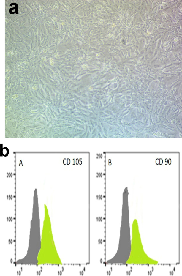
Methods: CWJ-MSCs were injected directed to the kidney with ultrasonographic guidance in dogs with 5/6 nephrectomy to evaluate its therapeutic potency in such cases. Analysis of variance was applied in normally distributed quantitative variables while a non-parametric Mann-Whitney test was used for non-normally distributed quantitative variables.

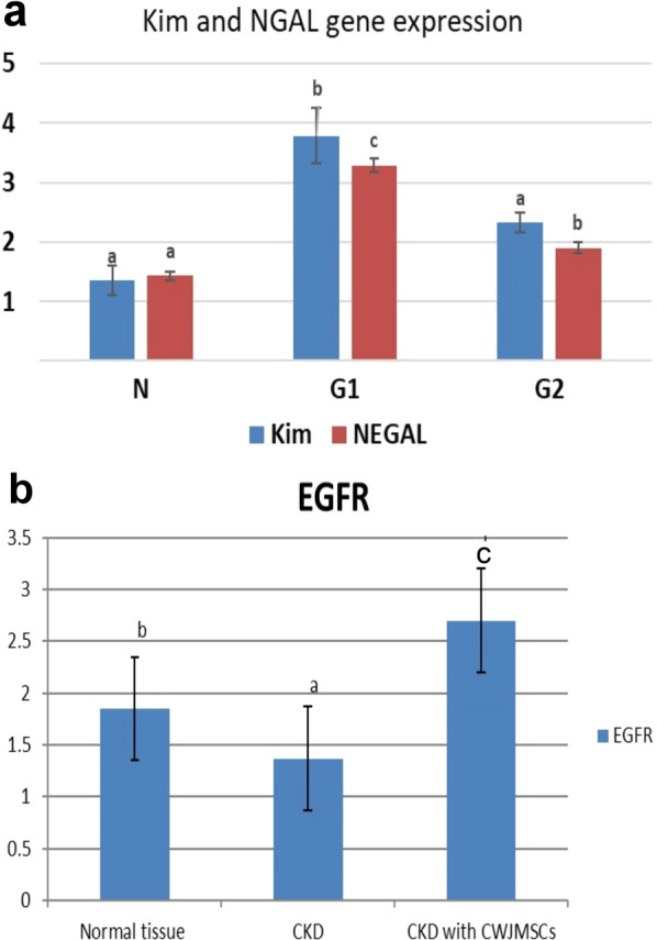
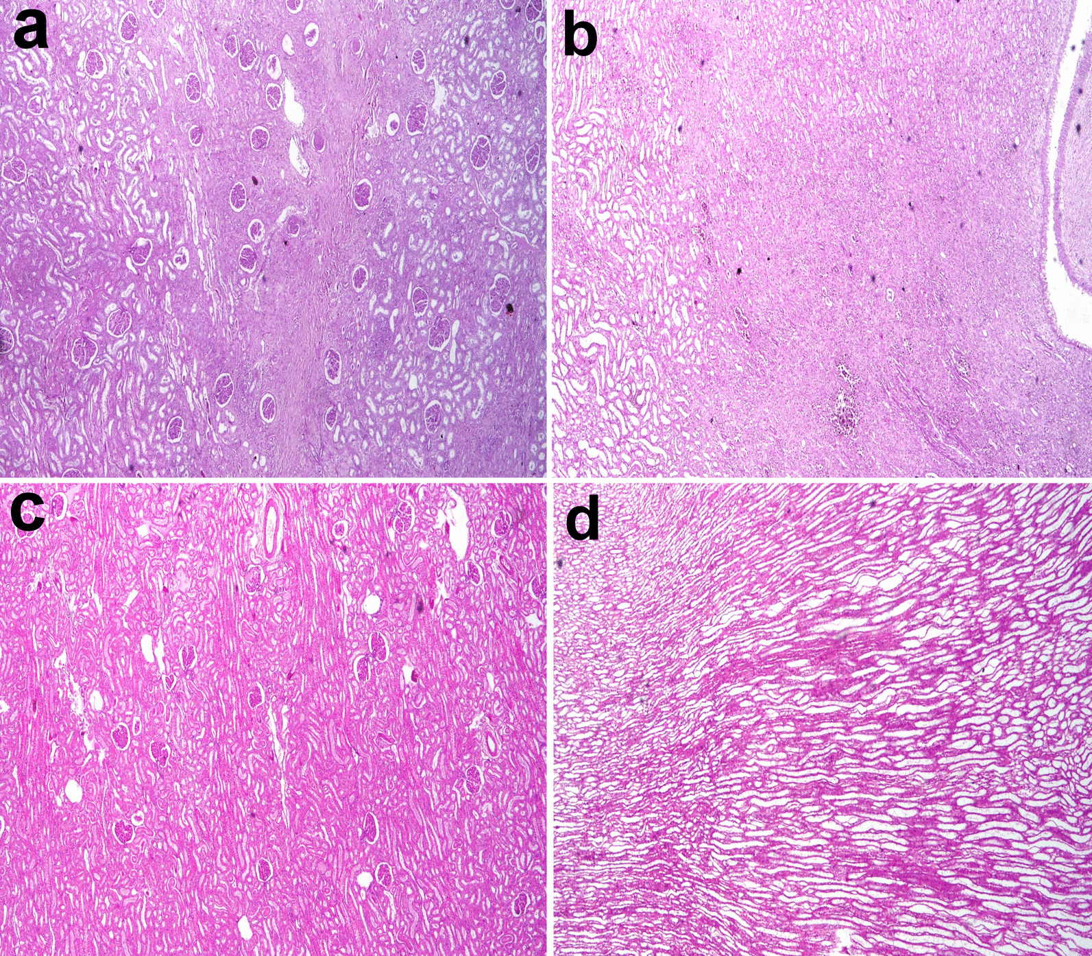
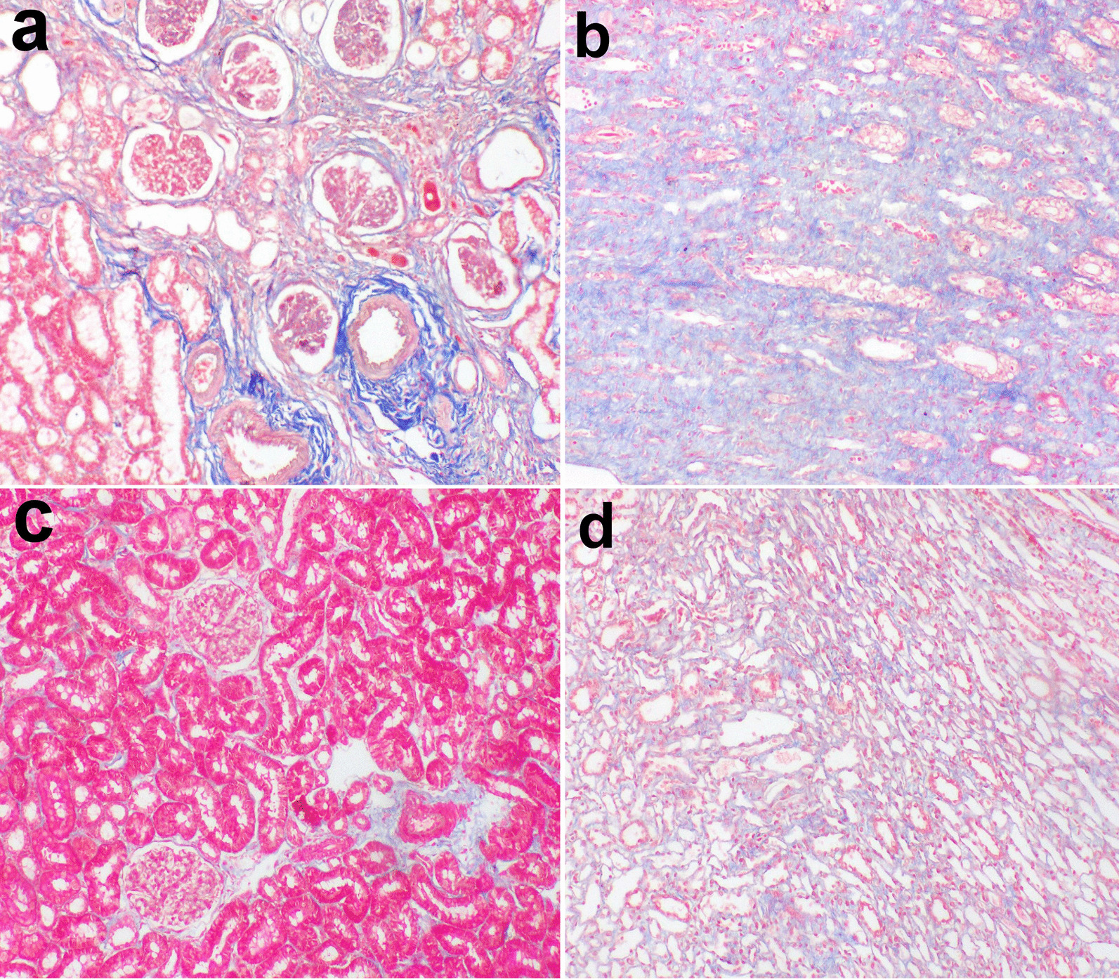
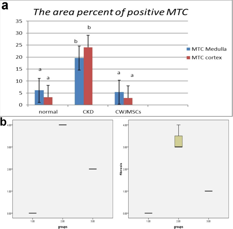
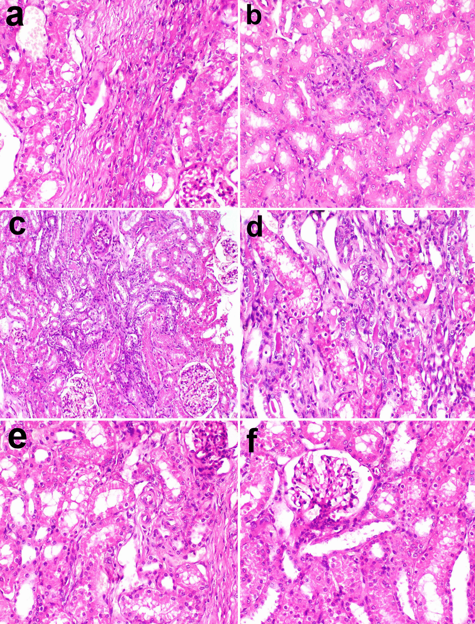
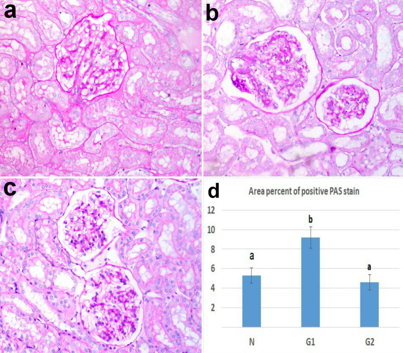
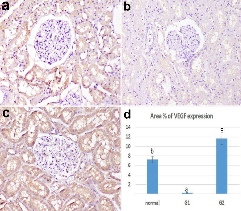
Results: The serum urea and creatinine in the treated group were significantly decreased transferring dogs in the treated group from stage 3 to stage 2 CKD according to the IRIS staging system. Histopathology of renal tissue revealed improving CKD lesions by increasing regeneration of degenerated tubules, maintaining the integrity of glomeruli. New vascularization with blood vessels remodeling were common findings. Periodic acid Schiff stain of renal tissue showed the integrity of renal tubules and thickness of the glomerular basement membrane. Fibrosis of cortex and medulla was lower in the treated group than in the CKD model as monitored by Mallory's trichrome stain (MTC). NGAL and KIM-1 genes expression were decreased while VEGF and EGF genes expression were increased indicating renal tissue repair.

Conclusions: CWJ-MSCs have a therapeutic potential in the CKD model induced in dogs.

Keywords: Camel stem cells; Chronic kidney disease; Histopathology; Kim-1; NGAL.


Conflict of interest statement

The authors declare that they have no competing interests.

Figures

Fig. 1
Fig. 1
a Spindle-shaped camel Wharton’s jelly MSCs at two weeks of culture. b MSCS of camel characterization by FACS. b A: CD105 (97%) & B: CD90 (96.5%) respectively
Fig. 2
Fig. 2
a KIM-1gene & NGAL gene expression in all experimental groups. b EGFR gene expression in all experimental groups. Columns bearing different lowercase letters are considered significant at P value < 0.05
Fig. 3
Fig. 3
Kidney of a dog showing severe diffuse interstitial fibrosis in the (a) cortex and (b) medulla in G1. c mild histological changes in the cortex and d focal fibrosis in the medulla in G2 (H and E stain X40)
Fig. 4
Fig. 4
cortex and medulla of CKD model stained by MTC (a,b) and of treated cases with CWJMSCs (c, d)
Fig. 5
Fig. 5
a the area percent of positive MTC in tissue sections of all experimental groups. Columns bearing different lowercase letters are considered significant at P value < 0.05. b Boxplot of glomerular lesion scores and fibrosis in different groups (1 = Normal group, 2 = G1, 3 = G2. The boxes are the interquartile range (IQR). The medians are the thick middle lines. The maximum and minimum values are represented by the thin horizontal lines at the top and bottom
Fig. 6
Fig. 6
photomicrographs of kidney treated with CMSC. a Sheets of stem cells embedded in a strip of connective tissue at the injection site, b the stem cells scattered in the interstitial tissue between renal tubules, c focal area of regenerated tubules showing basophilia and nuclear crowding. Note the regenerated glomerulus (arrow), d focal area of remodeling showing newly formed blood vessels, scattered stem cells, regenerated tubules, and other degenerated ones. Note the denuded renal tubule and invaded with stem cells (arrow), e note the newly formed blood vessels in the cortex (arrow), f hyperplasia of juxtaglomerular cells (arrow)
Fig. 7
Fig. 7
kidney tissue stained by PAS a control b CKD model c treated with CWJMSCs. Note the distinct brush border in (c). d the area percent of positive PAS in tissue sections of all experimental groups. Columns bearing different lowercase letters are considered significant at P value < 0.05
Fig. 8
Fig. 8
the expression of VEGF in the kidney tissue, a control, b CK model, c treated with CWJMSCs. d The area percent of VEGF expression in immunohistochemically stained sections in all experimental groups. Columns bearing different lowercase letters are considered significant at P value < 0.05

References

    1. Isaka Y. Epidermal growth factor as a prognostic biomarker in chronic kidney diseases. Ann Transl Med. 2016;4(Suppl 1):S62. doi: 10.21037/atm.2016.10.64. - DOI - PMC - PubMed
    1. Kaneko T, Shimizu A, Mii A, Fujita E, Fujino T, Kunugi S, Du X, Akimoto T, Tsuruoka S, Ohashi R, Masuda Y, Iino Y, Katayama Y, Fukuda Y. Role of matrix metalloproteinase-2 in recovery after tubular damage in acute kidney injury in mice. Nephron Exp Nephrol. 2012;122:23–35. doi: 10.1159/000346569. - DOI - PubMed
    1. Benigni A, Morigi M, Remuzzi G. Kidney regeneration. Lancet. 2010;375:1310–1317. doi: 10.1016/S0140-6736(10)60237-1. - DOI - PubMed
    1. Eirin A, Lerman LO. Mesenchymal stem cell treatment for chronic renal failure. Stem Cell Res Ther. 2014;5:83. doi: 10.1186/scrt472. - DOI - PMC - PubMed
    1. Kuppe C, Kramann R. Role of mesenchymal stem cells in kidney injury and fibrosis. Curr Opin Nephrol Hypertens. 2016;25:372–377. doi: 10.1097/MNH.0000000000000230. - DOI - PubMed

Publication types