Experimental study of the quantification of indocyanine green fluorescence in ischemic and non-ischemic anastomoses, using the SERGREEN software program
- PMID: 35908045
- PMCID: PMC9338976
- DOI: 10.1038/s41598-022-17395-6
Experimental study of the quantification of indocyanine green fluorescence in ischemic and non-ischemic anastomoses, using the SERGREEN software program
Abstract
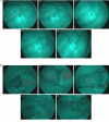
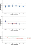
Tissue ischemia is a key risk factor in anastomotic leak (AL). Indocyanine green (ICG) is widely used in colorectal surgery to define the segments with the best vascularization. In an experimental model, we present a new system for quantifying ICG fluorescence intensity, the SERGREEN software. Controlled experimental study with eight pigs. In the initial control stage, ICG fluorescence intensity was analyzed at the level of two anastomoses, in the right and in the left colon. Control images of the two segments were taken after ICG administration. The images were processed with the SERGREEN program. Then, in the experimental ischemia stage, the inferior mesenteric artery was sectioned at the level of the anastomosis of the left colon. Fifteen minutes after the section, sequential images of the two anastomoses were taken every 30 min for the following 2 h. At the control stage, the mean scores were 134.2 (95% CI 116.3-152.2) for the right colon and 147 (95% CI 134.7-159.3) for the left colon (p = 0.174) (Scale RGB-Red, Green, Blue). The right colon remained stable throughout the experiment. In the left colon, intensity fell by 47.9 points with respect to the pre-ischemia value (p < 0.01). After the first post-ischemia determination, the values of the ischemic left colon remained stable throughout the experiment. The relative decrease in ICG fluorescence intensity of the ischemic left colon was 32.6%. The SERGREEN program quantifies ICG fluorescence intensity in normal and ischemic situations and detects differences between them. A reduction in ICG fluorescence intensity of 32.6% or more was correlated with complete tissue ischemia.
© 2022. The Author(s).
Conflict of interest statement
The authors declare no competing interests.
Figures


Similar articles
-
When should indocyanine green be assessed in colorectal surgery, and at what distance from the tissue? Quantitative measurement using the SERGREEN program.Surg Endosc. 2022 Dec;36(12):8943-8949. doi: 10.1007/s00464-022-09343-2. Epub 2022 Jun 6. Surg Endosc. 2022. PMID: 35668312
-
Meta-Analysis on the Efficacy of Indocyanine Green Fluorescence Angiography for Reduction of Anastomotic Leakage After Rectal Cancer Surgery.Am Surg. 2021 Dec;87(12):1910-1919. doi: 10.1177/0003134820982848. Epub 2020 Dec 30. Am Surg. 2021. PMID: 33377797
-
Quantitative analysis of colon perfusion pattern using indocyanine green (ICG) angiography in laparoscopic colorectal surgery.Surg Endosc. 2019 May;33(5):1640-1649. doi: 10.1007/s00464-018-6439-y. Epub 2018 Sep 10. Surg Endosc. 2019. PMID: 30203201 Free PMC article.
-
Effect of indocyanine green fluorescence angiography on preventing anastomotic leakage after colorectal surgery: a meta-analysis.Surg Today. 2021 Sep;51(9):1415-1428. doi: 10.1007/s00595-020-02195-0. Epub 2021 Jan 11. Surg Today. 2021. PMID: 33428000 Review.
-
Indocyanine green (ICG) fluorescence imaging for prevention of anastomotic leak in totally minimally invasive Ivor Lewis esophagectomy: a systematic review and meta-analysis.Dis Esophagus. 2022 Apr 19;35(4):doab056. doi: 10.1093/dote/doab056. Dis Esophagus. 2022. PMID: 34378016
Cited by
-
Indocyanine green: The guide to safer and more effective surgery.World J Gastrointest Surg. 2024 Mar 27;16(3):641-649. doi: 10.4240/wjgs.v16.i3.641. World J Gastrointest Surg. 2024. PMID: 38577071 Free PMC article.
-
Hyperspectral abdominal laparoscopy with real-time quantitative tissue oxygenation imaging: a live porcine study.Front Med Technol. 2025 Jun 5;7:1549245. doi: 10.3389/fmedt.2025.1549245. eCollection 2025. Front Med Technol. 2025. PMID: 40538619 Free PMC article.
References
-
- Golub R, Golub RW, Cantu R, Stein HD. A multivariate analysis of factors contributing to leakage of intestinal anastomoses. J. Am. Coll. Surg. 1997;184:364–372. - PubMed
MeSH terms
Substances
LinkOut - more resources
Full Text Sources

