. 2022 Jul 30;12(1):13125.
doi: 10.1038/s41598-022-17389-4.

Novel adult cortical neuron processing and screening method illustrates sex- and age-dependent effects of pharmaceutical compounds

Affiliations

Novel adult cortical neuron processing and screening method illustrates sex- and age-dependent effects of pharmaceutical compounds

Arthur Sefiani et al. Sci Rep. .

Abstract

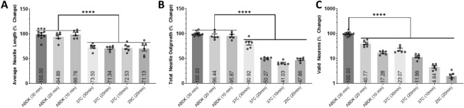
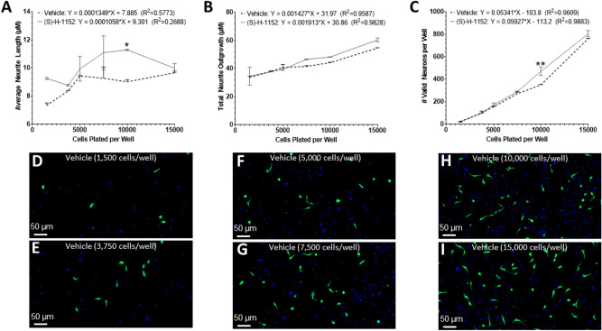
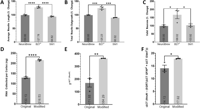
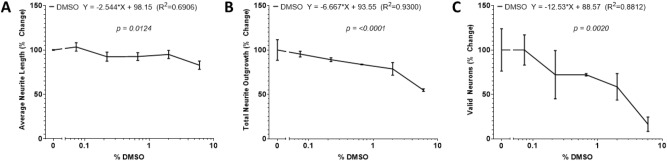
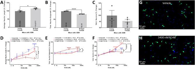
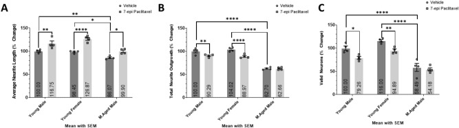
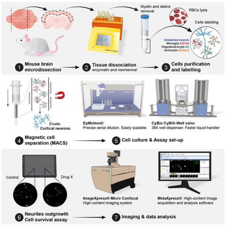
Neurodegenerative diseases and neurotraumatic injuries are typically age-associated disorders that can reduce neuron survival, neurite outgrowth, and synaptic plasticity leading to loss of cognitive capacity, executive function, and motor control. In pursuit of reducing the loss of said neurological functions, novel compounds are sought that promote neuron viability, neuritogenesis, and/or synaptic plasticity. Current high content in vitro screenings typically use cells that are iPSC-derived, embryonic, or originate from post-natal tissues; however, most patients suffering from neurodegenerative diseases and neurotrauma are of middle-age and older. The chasm in maturity between the neurons used in drug screens and those in a target population is a barrier for translational success of in vitro results. It has been historically challenging to culture adult neurons let alone conduct screenings; therefore, age-appropriate drug screenings have previously not been plausible. We have modified Miltenyi's protocol to increase neuronal yield, neuron purity, and neural viability at a reduced cost to expand our capacity to screen compounds directly in primary adult neurons. To our knowledge, we developed the first morphology-based screening system using adult cortical neurons and the first to incorporate age and sex as biological variables in a screen using adult cortical neurons. By using primary adult cortical neurons from mice that were 4 to 48 weeks old for screening pharmaceutical agents, we have demonstrated age- and sex-dependent effects on neuritogenesis and neuron survival in vitro. Utilizing age- and sex-appropriate in vitro models to find novel compounds increasing neuron survival and neurite outgrowth, made possible by our modified adult neuron processing method, will greatly increase the relevance of in vitro screening for finding neuroprotective compounds.


Conflict of interest statement

The authors declare that the research was conducted in the absence of any commercial or financial relationships that could be construed as a potential conflict of interest. A.S. and C.G.G. currently hold pending patent applications regarding the technology listed herein and are substantial owners of NeuroCreis.

Figures

Figure 1
Figure 1
The effects of Laminin coating and media supplementation and different digestion enzymes. Histograms of the (A) average neurite length, (B) total neurite outgrowth, and (C) number of valid neurons expressed as percent change relative to the control group containing no B27+ supplement or laminin coating. Primary cortical neurons isolated from young adult male mice and cultured for 2DIV. 3 wells per condition. Histograms of the (D) average neurite length, (E) total neurite outgrowth, and (F) number of valid neurons expressed as percent change relative to the MACS P&A group. Data analyzed using one-way ANOVA with Tukey’s post-hoc test comparing the mean of each condition to one another. Representative × 20 magnification images of primary cortical neurons isolated using the (G) MACS P&A and (H) 0.3 mg/mL Papain (H′) × 63 magnification) dissociation enzymes from young adult male mice and cultured for 2DIV; stained with TUBB3 (Green) and DAPI (Blue). 6 wells per condition (n = 6). *(P < 0.05), **(P < 0.01), ***(P < 0.001), ****(P < 0.0001). Graphs show mean and SEM. Scale Bar = 50 µm (D,E) or 20 µm (E′).
Figure 2
Figure 2
The effects of different dissociation protocols, timing, and temperature. Histograms of the (A) average neurite length, (B) total neurite outgrowth, and (C) number of valid neurons expressed as percent change relative to the standard gentleMACS Program 37C_ABDK_01 protocol (ABDK (30 min)). Data analyzed using one-way ANOVA with Dunnett’s post-hoc test comparing the mean of each condition to the mean of ABDK (30 min). In parentheses represents the length of the protocol whether being ran on the gentleMACS™ Octo Dissociator with Heaters (ABDK) or in a revolving apparatus incubated at 37 °C or 25 °C. Primary cortical neurons isolated from young adult male mice and cultured for 2DIV. 6 wells per condition (n = 6). *(P < 0.05), **(P < 0.01), ***(P < 0.001), ****(P < 0.0001). Graphs show mean and SEM.
Figure 3
Figure 3
The effects of cell plating density. Linear trends of the (A) average neurite length, (B) total neurite outgrowth, and (C) number of valid neurons of Vehicle and (S)-H-1152 treated primary cortical neurons isolated from young adult male mice cultured for 2DIV. The X-axis denotes the number of cells plated in each well. Representative × 20 magnification images of primary cortical neurons from young adult male mice treated with Vehicle for 2DIV and plated at (D) 1500 cells/well, (E) 3750 cells/well, (F) 5000 cells/well, (G) 7500 cells/well, (H) 10,000 cells/well, and (I) 15,000 cells/well; stained with TUBB3 (Green) and DAPI (Blue). Two-way ANOVA with Šídák’s multiple comparisons test was used to compare the means of neurons with different treatments for each respective plating density denoted by *(P < 0.05), **(P < 0.01), ***(P < 0.001), ****(P < 0.0001). Simple liner regression conducted to determine goodness of fit and if the slope differs significantly from 0. Linear regression t-test was used to compare the slope of the regression lines. 2 wells per condition (n = 2). Graphs show mean and SEM. Scale Bar = 50 µm.
Figure 4
Figure 4
The efficacy of various neuronal supplements and culture purity assessed with RNA expression analysis. Histograms of the (A) average neurite length, (B) total neurite outgrowth, and (C) number of valid neurons of primary cortical neurons treated with the respective neuronal supplement isolated from young adult male mice and cultured for 2DIV. Data is expressed as percent change relative to the B27+ supplement cohort. Data analyzed using one-way ANOVA with Dunnett’s post-hoc test comparing the mean of each condition to the mean of the B27+ supplement group. 3 wells per condition. Histograms of the (D) mass of RNA collected from each preparation, (E) the relative expression of NeuN expressed as 2−ΔCT (NeuN) and (F) the relative expression of NeuN in relation to GFAP and Glast comparing the original protocol from Miltenyi (Original) and our finalized modified protocol (Modified). − ΔCT =  − (ΔCT NeuN − (SQRT(ΔCT GFAP2 + ΔCT Glast2))). All ΔCT values are calculated as follows: ΔCT Primer = CT Primer (Sample) − CT Primer (Negative Control). Student’s T-test was used to compare the means of each cohort. RNA extracted from primary cortical neurons isolated from young adult male mice following the original and modified protocol. 3 independent samples per cohort, each analyzed in triplicate (n = 3). *(P < 0.05), **(P < 0.01), ***(P < 0.001), ****(P < 0.0001). Graphs show mean and SEM.
Figure 5
Figure 5
The effects of the drug vehicle DMSO. Linear trends of the (A) average neurite length, (B) total neurite outgrowth, and (C) number of valid neurons of primary cortical neurons isolated from young adult male mice cultured in various percentages of DMSO for 2DIV. The X-axis denotes the percentage of the neuron media comprising of DMSO. The values are expressed as percent change relative to the 0% DMSO cohort. Simple liner regression conducted to determine P value, goodness of fit, and if the slope differs significantly from 0.2 wells per condition (n = 2). Graphs show mean and SEM.
Figure 6
Figure 6
Sex and age-dependent effects of RO48. Histograms of the (A) average neurite length, (B) total neurite outgrowth, and (C) number of valid neurons extracted from the Vehicle treated group from figures (A–C), respectively, expressed as percent change relative to the young adult female (Young Female) cohort. Data analyzed using student’s T-test comparing the means of each cohort denoted by *(P < 0.05), **(P < 0.01), ***(P < 0.001), ****(P < 0.0001). Linear trends of the (D) average neurite length, (E) total neurite outgrowth, and (F) number of valid neurons of primary cortical neurons isolated from young adult male, young adult female, and middle-aged female mice cultured in various concentrations of RO48 for 2DIV. The X-axis denotes the concentration (nM) of RO48 in the media (D–F). The values are expressed as percent change relative to the Vehicle treatment group of each cohort. Representative × 20 magnification images of (G) Vehicle and (H) 3400 nM RO48 treated primary cortical neurons isolated from middle-aged female mice and cultured for 2DIV; stained with TUBB3 (Green) and DAPI (Blue). Simple liner regression was conducted to determine goodness of fit and if the slope differs significantly from 0. Linear regression t-test was used to compare the slope of the regression lines. Two-way ANOVA with Šídák’s multiple comparisons test was used to compare the means of neurons treated with 3400 nM RO48 between cohorts denoted by #(P < 0.05), ##P < 0.01), ###(P < 0.001), ####(P < 0.0001) and to compare the means of neurons treated with various concentrations of RO48 to the Vehicle treatment group of the respective cohort denoted by *(P < 0.05), **(P < 0.01), ***(P < 0.001), ****(P < 0.0001). All cohorts have equal parts Vehicle in media (0.05% DMSO). 3 wells per condition (n = 3). Graphs show mean and SEM. Scale Bar = 50 µm.
Figure 7
Figure 7
The age-dependent effects of 7-epi Paclitaxel. Histograms of the (A) average neurite length, (B) total neurite outgrowth, and (C) number of valid neurons expressed as percent change relative to the young adult male cohort with Vehicle treatment. Primary cortical neurons isolated from young adult male, young adult female, and middle-aged male mice cultured in Vehicle or 150 nM 7-epi Paclitaxel or 3DIV. Two-way ANOVA with Dunnett’s multiple comparisons test was used to compare the means of each compound to the mean of the Vehicle treated group within each respective cohort. Two-way ANOVA with Tukey’s multiple comparisons test was used to compare the mean of the Vehicle treated groups between different age cohorts. All cohorts have equal parts Vehicle in media (0.05% DMSO). 4 wells per condition (n = 4). *(P < 0.05), **(P < 0.01), ***(P < 0.001), ****(P < 0.0001). Graphs show mean and SEM.
Figure 8
Figure 8
The screening platform using primary adult neural cells. Briefly, the cortex is extracted from mice, dissociated in a dissociator, followed by the removal of debris and red blood cells. Afterwards, the glial cells are separated from neurons. All liquid handling, imaging, and analysis is done with bias and human free robots. Figure was created by Graphit Science & Art, LLC.

Similar articles

Cited by

References

    1. Kanasi E, Ayilavarapu S, Jones J. The aging population: Demographics and the biology of aging. Periodontol 2000. 2016;72:13–18. doi: 10.1111/prd.12126. - DOI - PubMed
    1. Riggs JE. The aging population: Implications for the burden of neurologic disease. Neurol. Clin. 1998;16:555–560. doi: 10.1016/S0733-8619(05)70079-7. - DOI - PubMed
    1. Statista. (Statista, statista.com, 2021).
    1. Murray CJL, et al. Disability-adjusted life years (DALYs) for 291 diseases and injuries in 21 regions, 1990–2010: A systematic analysis for the Global Burden of Disease Study 2010. The Lancet. 2012;380:2197–2223. doi: 10.1016/S0140-6736(12)61689-4. - DOI - PubMed
    1. Centers for Disease Control and Prevention. Rates of TBI-related Hospitalizations by Age Group—United States, 2001–2010, https://www.cdc.gov/traumaticbraininjury/data/rates_hosp_byage.html Accessed 21 July 2022 (2016).

Publication types

Substances