Review
. 2022 Aug:96:102189.
doi: 10.1016/j.cpr.2022.102189. Epub 2022 Jul 23.

Neural substrates of motivational dysfunction across neuropsychiatric conditions: Evidence from meta-analysis and lesion network mapping

Affiliations
Review

Neural substrates of motivational dysfunction across neuropsychiatric conditions: Evidence from meta-analysis and lesion network mapping

Chunliang Feng et al. Clin Psychol Rev. 2022 Aug.

Abstract

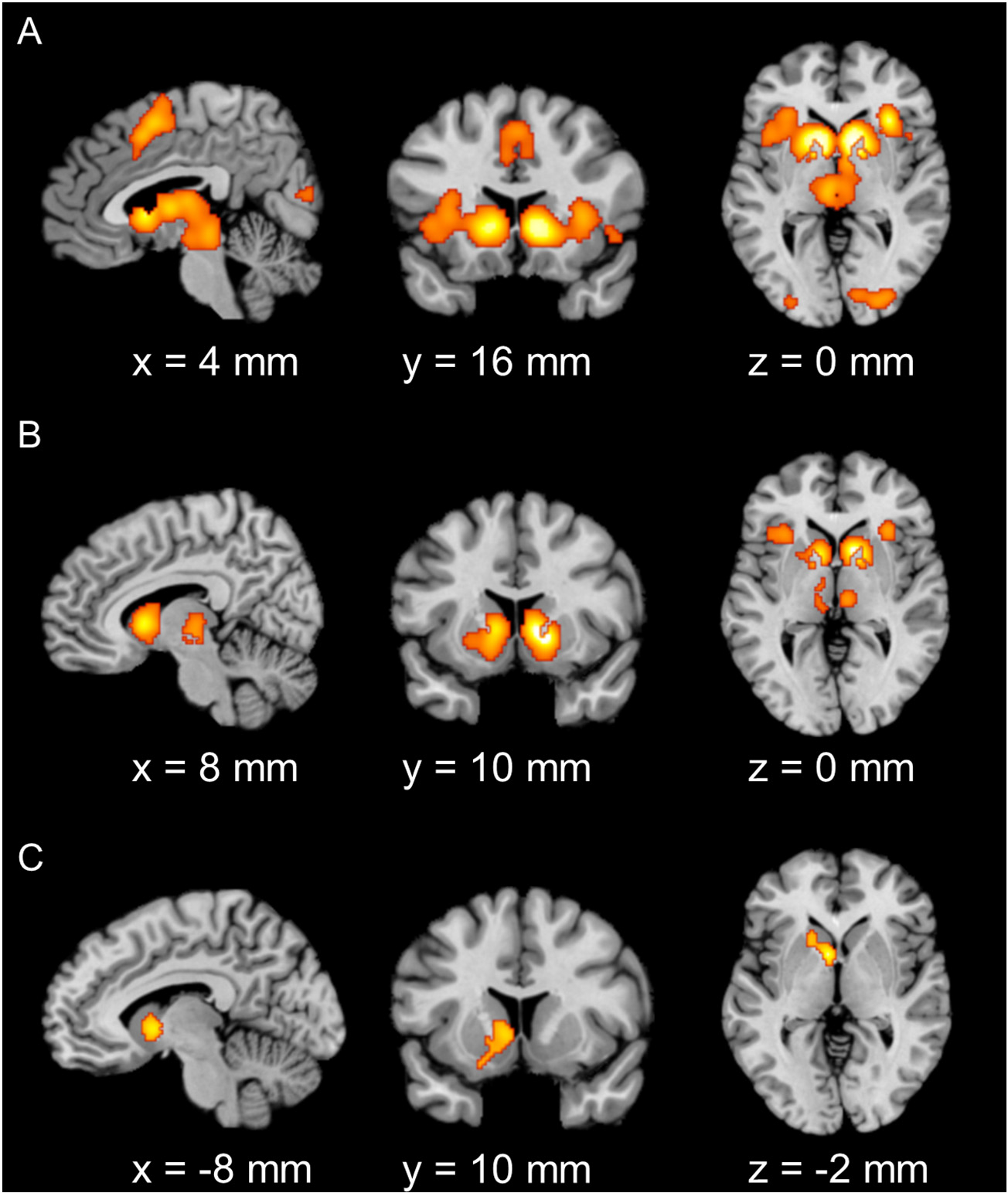
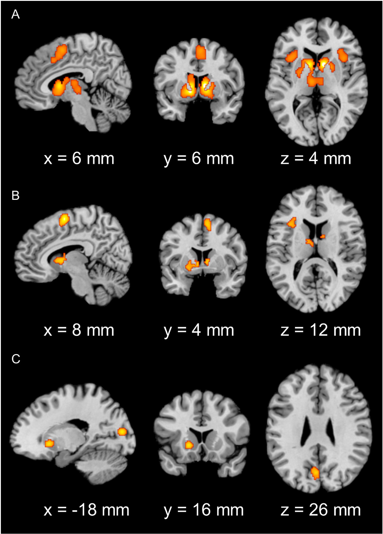
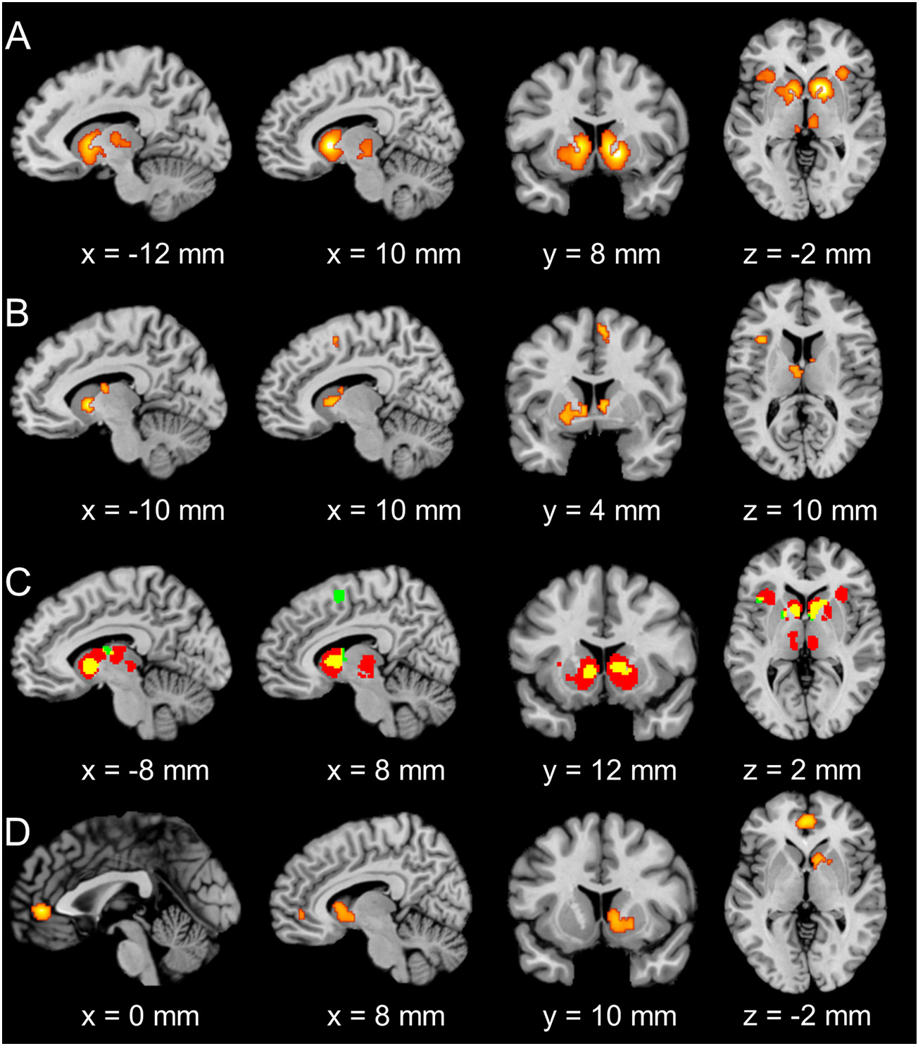
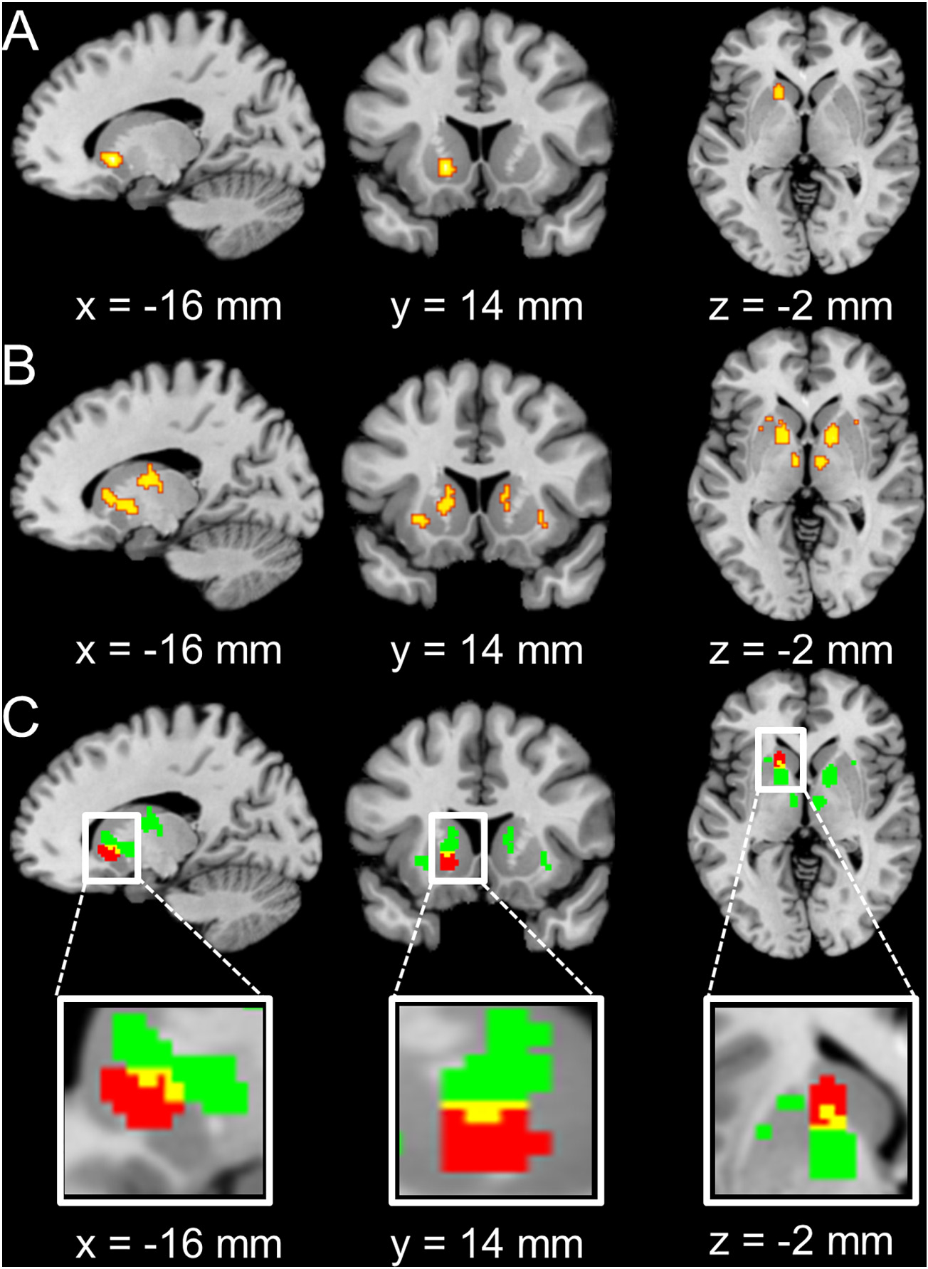
Motivational dysfunction constitutes one of the fundamental dimensions of psychopathology cutting across traditional diagnostic boundaries. However, it is unclear whether there is a common neural circuit responsible for motivational dysfunction across neuropsychiatric conditions. To address this issue, the current study combined a meta-analysis on psychiatric neuroimaging studies of reward/loss anticipation and consumption (4308 foci, 438 contrasts, 129 publications) with a lesion network mapping approach (105 lesion cases). Our meta-analysis identified transdiagnostic hypoactivation in the ventral striatum (VS) for clinical/at-risk conditions compared to controls during the anticipation of both reward and loss. Moreover, the VS subserves a key node in a distributed brain network which encompasses heterogeneous lesion locations causing motivation-related symptoms. These findings do not only provide the first meta-analytic evidence of shared neural alternations linked to anticipatory motivation-related deficits, but also shed novel light on the role of VS dysfunction in motivational impairments in terms of both network integration and psychological functions. Particularly, the current findings suggest that motivational dysfunction across neuropsychiatric conditions is rooted in disruptions of a common brain network anchored in the VS, which contributes to motivational salience processing rather than encoding positive incentive values.

Keywords: Lesion network mapping; Meta-analysis; Monetary incentive delay task; Motivation; Ventral striatum.


Conflict of interest statement

Declaration of Competing Interest

The authors are unaware of any conflicts of interest, financial or otherwise.

Figures

Fig. 1.
Fig. 1.
Significant clusters from the main meta-analysis of reward anticipation in (A) healthy controls, (B) clinical/at-risk conditions and (C) hypoactivation of clinical/at-risk conditions relative to healthy controls (cluster-level family-wise error correction, P < 0.05, with a cluster-forming threshold of P < 0.001 using 10,000 permutations). (A). Consistent maxima in the bilateral VS (extending to the amygdala, thalamus, and AI), SMA, precentral gyrus, middle occipital gyrus, and right calcarine were identified for healthy controls. (B). Consistent maxima in the bilateral VS and AI were identified for clinical/at-risk conditions. (C). Consistent maxima in the bilateral VS were found for hypoactivation of clinical/at-risk conditions relative to healthy controls.
Fig. 2.
Fig. 2.
Significant clusters from the main meta-analysis of loss anticipation in (A) healthy controls, (B) clinical/at-risk conditions, and (C) hypoactivation of clinical/at-risk conditions relative to healthy controls (cluster-level family-wise error correction, P < 0.05, with a cluster-forming threshold of P < 0.001 using 10,000 permutations). (A). Consistent maxima in the bilateral VS (extending to the amygdala, thalamus, and AI) and SMA were identified for healthy controls. (B). Consistent maxima in the bilateral VS, AI, SMA, thalamus, and precentral gyrus were identified for clinical/at-risk conditions. (C). Consistent maxima in the left VS, middle occipital gyrus, and cuneus were found for hypoactivation of clinical/at-risk conditions relative to healthy controls during loss anticipation.
Fig. 3.
Fig. 3.. Significant clusters from the main meta-analysis of reward consumption in (A) healthy controls, (B) clinical/at-risk conditions, and (C) hyperactivation of clinical/at-risk conditions relative to healthy controls (cluster-level family-wise error correction, P < 0.05, with a cluster-forming threshold of P < 0.001 using 10,000 permutations).
(A). Consistent maxima in the bilateral VS, vmPFC, and PCC were identified for healthy controls. (B). Consistent maxima in the vmPFC and right VS were identified for clinical/at-risk conditions. (C). Consistent maxima in the left IPL were found for hyperactivation of clinical/at-risk conditions relative to healthy controls during reward consumption.
Fig. 4.
Fig. 4.. The conjunction analyses for common regions for clinical/at-risk conditions and healthy controls.
(A). Consistent maxima in the bilateral VS, vmPFC, and PCC were identified for overlap of activation between clinical/at-risk conditions and healthy controls during reward anticipation. (B). Consistent maxima in the bilateral VS and vmPFC were identified for overlap of activation between clinical/at-risk conditions and healthy controls during loss anticipation. (C). Consistent maxima in the bilateral VS and vmPFC (yellow) were identified for overlap of conjunction results from A (red) and B (green). (D). Consistent maxima in the bilateral VS and vmPFC were identified for overlap of activation between clinical/at-risk conditions and healthy controls during reward consumption. (For interpretation of the references to colour in this figure legend, the reader is referred to the web version of this article.)
Fig. 5.
Fig. 5.. The conjunction analyses for common regions for clinical/at-risk conditions vs. healthy controls as well as lesion network results.
(A). Consistent maxima in the left VS were identified in the hypoactivation in clinical/at-risk conditions relative to controls during reward and loss anticipation conjunction analysis. (B). Bilateral VS and ACC were identified for the overlap of lesion-derived networks (75%). (C). The conjuction (yellow) between results from A (red) and B (green). (For interpretation of the references to colour in this figure legend, the reader is referred to the web version of this article.)

Similar articles

Cited by

References

    1. Arrondo G, Segarra N, Metastasio A, Ziauddeen H, Spencer J, Reinders NR, … Murray GK (2015). Reduction in ventral striatal activity when anticipating a reward in depression and schizophrenia: A replicated cross-diagnostic finding. Frontiers in Psychology, 6, 1280. 10.3389/fpsyg.2015.01280 - DOI - PMC - PubMed
    1. Balodis IM, Kober H, Worhunsky PD, Stevens MC, Pearlson GD, & Potenza MN (2012). Diminished frontostriatal activity during processing of monetary rewards and losses in pathological gambling. Biological Psychiatry, 71, 749–757. 10.1016/j.biopsych.2012.01.006 - DOI - PMC - PubMed
    1. Barch DM (2020). What does it mean to be transdiagnostic and how would we know? American Journal of Psychiatry, 177, 370–372. 10.1176/appi.ajp.2020.20030243 - DOI - PubMed
    1. Beck A, Schlagenhauf F, Wustenberg T, Hein J, Kienast T, Kahnt T, … Wrase J (2009). Ventral striatal activation during reward anticipation correlates with impulsivity in alcoholics. Biological Psychiatry, 66, 734–742. 10.1016/j.biopsych.2009.04.035 - DOI - PubMed
    1. Berridge KC, Robinson TE, & Aldridge JW (2009). Dissecting components of reward: “Liking”, “wanting”, and learning. Current Opinion in Pharmacology, 9, 65–73. 10.1016/j.coph.2008.12.014 - DOI - PMC - PubMed

LinkOut - more resources