. 2022 Oct:118:103381.
doi: 10.1016/j.dnarep.2022.103381. Epub 2022 Jul 26.

A redox-sensitive iron-sulfur cluster in murine FAM72A controls its ability to degrade the nuclear form of uracil-DNA glycosylase

Affiliations

A redox-sensitive iron-sulfur cluster in murine FAM72A controls its ability to degrade the nuclear form of uracil-DNA glycosylase

Jessica A Stewart et al. DNA Repair (Amst). 2022 Oct.

Abstract

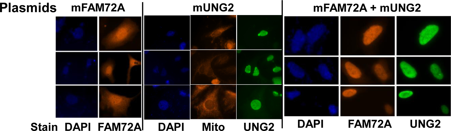
Murine FAM72A, mFAM72A, binds the nuclear form of uracil-DNA glycosylase, mUNG2, inhibits its activity and causes its degradation. In immunoprecipitation assays the human paralog, hFAM72A, binds hUNG2 and is a potential anti-cancer drug target because of its high expression in many cancers. Using purified mFAM72A, and mUNG2 proteins we show that mFAM72A binds mUNG2, and the N-terminal 25 amino acids of mUNG2 bind mFAM72A at a nanomolar dissociation constant. We also show that mFAM72A is present throughout the cells, and mUNG2 helps localize it to nuclei. Based on in silico models of mFAM72A-mUNG2 interactions, we constructed several mutants of mFAM72A and found that while they have reduced ability to deplete mUNG2, the mutations also destabilized the former protein. We confirmed that Withaferin A, a predicted lead molecule for the design of FAM72A inhibitors, binds mFAM72A with micromolar affinity but has little affinity to mUNG2. We identified two potential metal-binding sites in mFAM72A and show that one of the sites contains an Fe-S cluster. This redox-sensitive cluster is involved in the mFAM72A-mUNG2 interaction and modulates mFAM72A activity. Hydrogen peroxide treatment of cells increases mUNG2 depletion in a FAM72A-dependent fashion suggesting that mFAM72A activity is redox-sensitive.

Keywords: Class-switch recombination; Iron-sulfur cluster; Oxidative stress; Somatic hypermutation; Uracil-DNAglycosylase.


Conflict of interest statement

Conflict of interest The authors declare that there are no conflicts of interest.

Figures

Figure 1.
Figure 1.
Subcellular localization of mUNG2 and mFAM72A in HeLa cells. Fluorescence images of cells stained with DAPI (nucleus), MitoTracker Red (mitochondria) or antibodies specific for UNG or FAM72A proteins are shown. The top line shows the plasmids that were transfected into cells, while the line at the bottom shows the stain or antibody used in each image.
Figure 2.
Figure 2.
Biolayer interferometry analysis of interaction between mUNG2, mFAM72A and Withaferin A. The molecular association-dissociation curves are shown. (A) Binding of mFAM72A (immobilized) to mUNG2. The concentrations of mUNG2 are shown. (B) Binding of mFAM72A (immobilized) to 25-mer polypeptide. The concentrations of the polypeptide are shown. (C) Binding of mFAM72A (immobilized) to Withaferin A. The concentrations of the Withaferin A are shown. (D) Binding of SUMO-mUNG2 (immobilized) to Withaferin A.
Figure 3.
Figure 3.
Degradation of mUNG2 by mFAM72A and its mutants. (A) Theoretical model of mFAM72A showing the residues that were mutated. (B) Upper panel- Western blot of mUNG1 and mUNG2 proteins in HEK293T cells transfected with different pairs of plasmids. The proteins expressed by the plasmids are indicated to the left and at top of the blot. The antibodies used to visualize the proteins are indicated on the right. Lower panel- Quantification of intensities of bands from three independent blots. Mean and standard deviations are shown. EV- empty vector. (C) Western blot of mUNG2 following co-transfection of cells with mUNG2 expression plasmid with mFAM72A mutant expression plasmids. The identity of the protein bands is indicated on the right of the blot. (D) Quantification of mUNG2 levels from independent blots such as shown in part C. Mean and standard deviations are shown.
Figure 4.
Figure 4.
Role of potential metal-binding sites within FAM72A on its activity. (A) Center- The two sites, A and B, are shown within the predicted mFAM72A structure. Zoom in view of each site is shown on the side. (B and C) Western blots of cotransfection experiments using mUNG2 expression plasmid and metal-binding site mutants of mFAM72A are shown. Quantification of intensities of bands from three independent blots is presented below each blot as a bar graph. Mean and standard deviations are shown.
Figure 5.
Figure 5.. FAM72A has a redox sensitive iron-sulfur cluster.
The new pET28 mFam72a was used to purify the protein and the (A) cell pellet of mFam72a expressing E. coli was much darker in color than the mPCNA expressing cells. (B) Purified mFAM72A had a yellowish color (tube labelled 1) and when the iron-sulfur cluster was chemically reconstituted it had even darker color (tube labelled 2). (C) The UV-vis spectra the purified protein from pET28 expression system subjected to chemical reconstitution of the iron-sulfur cluster. For comparison, UV-VIS spectrum of older preparation of mFAM72A from pRSET expression system is also shown. (D) Effects of oxidation of the iron-sulfur cluster on UV- VIS absorption. The tube was opened for 10 minutes or 60 minutes prior to spectroscopy. (E) Effects of reduction of the iron-sulfur cluster on UV-VIS absorption. The protein was treated with sodium dithionite for 10 or 60 min prior to spectroscopy.
Figure 6.
Figure 6.
Effect of H2O2 treatment of cells on degradation mUNG2 promoted by mFAM72A. (A) Western blots of transfection experiments using mUNG2 expression plasmid with or without mFAM72A expression plasmid followed by treatment of cells with H2O2 at indicated concentrations. (B) Quantification of data from three independent experiments.

Similar articles

Cited by

References

    1. Heese K, The protein p17 signaling pathways in cancer, Tumour Biol, 34 (2013) 4081–4087. - PubMed
    1. Wang LT, Lin CS, Chai CY, Liu KY, Chen JY, Hsu SH, Functional interaction of Ugene and EBV infection mediates tumorigenic effects, Oncogene, 30 (2011) 2921–2932. - PubMed
    1. Rajan P, Stockley J, Sudbery IM, Fleming JT, Hedley A, Kalna G, Sims D, Ponting CP, Heger A, Robson CN, McMenemin RM, Pedley ID, Leung HY, Identification of a candidate prognostic gene signature by transcriptome analysis of matched pre- and post-treatment prostatic biopsies from patients with advanced prostate cancer, BMC Cancer, 14 (2014) 977. - PMC - PubMed
    1. Guo C, Zhang X, Fink SP, Platzer P, Wilson K, Willson JK, Wang Z, Markowitz SD, Ugene, a newly identified protein that is commonly overexpressed in cancer and binds uracil DNA glycosylase, Cancer Res, 68 (2008) 6118–6126. - PMC - PubMed
    1. Rahane CS, Kutzner A, Heese K, A cancer tissue-specific FAM72 expression profile defines a novel glioblastoma multiform (GBM) gene-mutation signature, J Neurooncol, 141 (2019) 57–70. - PubMed

LinkOut - more resources