

Pulmonary Cryptococcosis Diagnosed by a Transbronchial Lung Cryobiopsy in a Patient with Rheumatoid Arthritis
- PMID: 35908974
- PMCID: PMC10017238
- DOI: 10.2169/internalmedicine.9764-22
Pulmonary Cryptococcosis Diagnosed by a Transbronchial Lung Cryobiopsy in a Patient with Rheumatoid Arthritis
Abstract
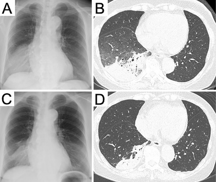
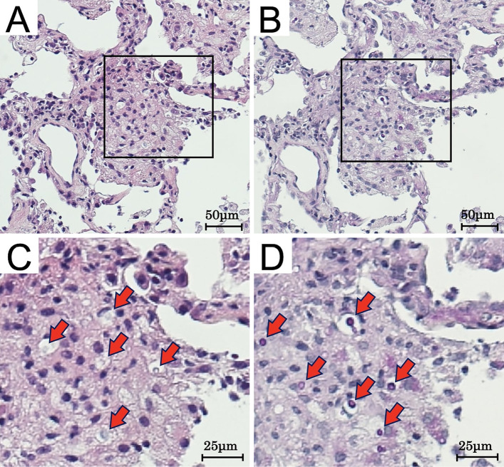
A 77-year-old woman with seronegative rheumatoid arthritis who was being treated with prednisolone (8 mg/day) and methotrexate (12 mg/week) visited our hospital with an 11-day history of a fever and dyspnea. Chest computed tomography showed infiltration in the right lower lobe. A transbronchial lung cryobiopsy (TBLC) showed cryptococcal cells, and bronchoalveolar lavage fluid later showed growth of Cryptococcus neoformans. She was treated with amphotericin B and flucytosine for about four weeks, and the pulmonary shadows improved. The treatment was then changed to fluconazole as outpatient consolidation and maintenance therapy. A rare case of pulmonary cryptococcosis diagnosed by a TBLC is reported.
Keywords: immunocompromised host; pulmonary cryptococcosis; rheumatoid arthritis; transbronchial lung cryobiopsy.
Conflict of interest statement
Figures
Similar articles
-
A rare case of Trichosporon mycotoxinivorans and Cryptococcus neoformans co-infection in lung.J Infect Chemother. 2020 Aug;26(8):838-842. doi: 10.1016/j.jiac.2020.03.002. Epub 2020 Apr 2. J Infect Chemother. 2020. PMID: 32249160
-
Isolated pulmonary cryptococcosis in an immunocompetent patient.J Bras Pneumol. 2006 Sep-Oct;32(5):476-80. J Bras Pneumol. 2006. PMID: 17268753 English, Portuguese.
-
[Case of pulmonary cryptococcosis which developed in a patient receiving abatacept therapy for rheumatoid arthritis].Nihon Kokyuki Gakkai Zasshi. 2010 Dec;48(12):980-4. Nihon Kokyuki Gakkai Zasshi. 2010. PMID: 21226309 Japanese.
-
Pulmonary cryptococcosis.Semin Respir Crit Care Med. 2011 Dec;32(6):727-34. doi: 10.1055/s-0031-1295720. Epub 2011 Dec 13. Semin Respir Crit Care Med. 2011. PMID: 22167400 Review.
-
[Cryptococcosis].Nihon Rinsho. 2008 Dec;66(12):2350-5. Nihon Rinsho. 2008. PMID: 19069104 Review. Japanese.
Cited by
-
Cryptococcosis in a patient with rheumatoid arthritis following glucocorticoid and JAK inhibitor therapy: a case description on stabilization with tocilizumab.Quant Imaging Med Surg. 2025 Apr 1;15(4):3738-3742. doi: 10.21037/qims-24-1825. Epub 2025 Mar 28. Quant Imaging Med Surg. 2025. PMID: 40235805 Free PMC article. No abstract available.
References
-
- Setianingrum F, Rautemaa-Richardson R, Denning DW. Pulmonary cryptococcosis: a review of pathobiology and clinical aspects. Med Mycol 57: 133-150, 2019. - PubMed
-
- Malabonga VM, Basti J, Kamholz SL. Utility of bronchoscopic sampling techniques for cryptococcal disease in AIDS. Chest 99: 370-372, 1991. - PubMed
-
- Zhang Y, Li N, Zhang Y, et al. . Clinical analysis of 76 patients pathologically diagnosed with pulmonary cryptococcosis. Eur Respir J 40: 1191-1200, 2012. - PubMed
-
- Chang WC, Tzao C, Hsu HH, et al. . Pulmonary cryptococcosis: comparison of clinical and radiographic characteristics in immunocompetent and immunocompromised patients. Chest 129: 333-340, 2006. - PubMed