. 2022 Aug 1;8(1):62.
doi: 10.1038/s41522-022-00324-w.

Bacterial biofilm functionalization through Bap amyloid engineering

Affiliations

Bacterial biofilm functionalization through Bap amyloid engineering

Leticia Matilla-Cuenca et al. NPJ Biofilms Microbiomes. .

Abstract

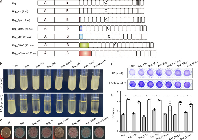
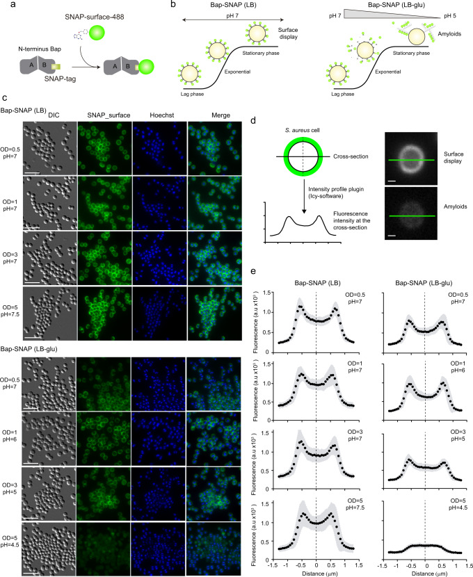
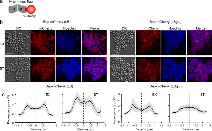
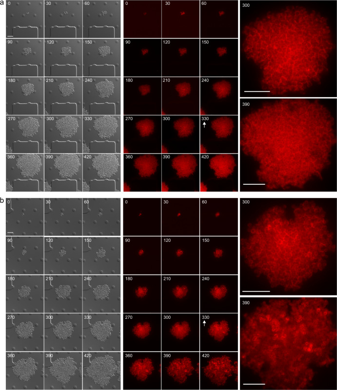
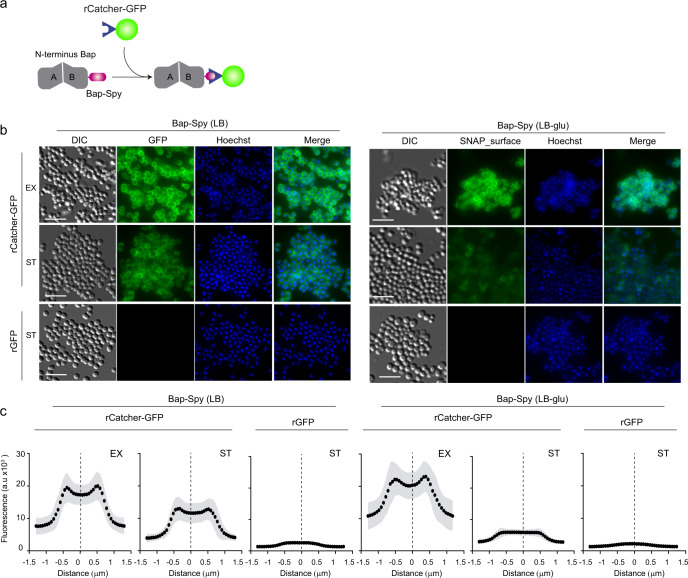
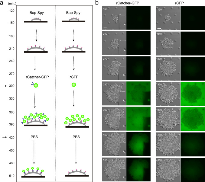
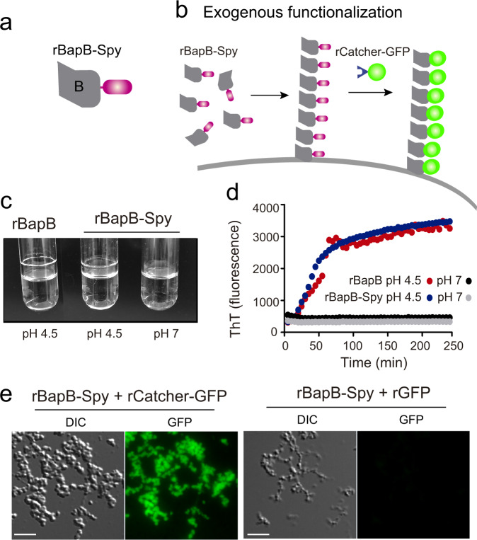
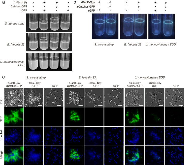
Biofilm engineering has emerged as a controllable way to fabricate living structures with programmable functionalities. The amyloidogenic proteins comprising the biofilms can be engineered to create self-assembling extracellular functionalized surfaces. In this regard, facultative amyloids, which play a dual role in biofilm formation by acting as adhesins in their native conformation and as matrix scaffolds when they polymerize into amyloid-like fibrillar structures, are interesting candidates. Here, we report the use of the facultative amyloid-like Bap protein of Staphylococcus aureus as a tool to decorate the extracellular biofilm matrix or the bacterial cell surface with a battery of functional domains or proteins. We demonstrate that the localization of the functional tags can be change by simply modulating the pH of the medium. Using Bap features, we build a tool for trapping and covalent immobilizing molecules at bacterial cell surface or at the biofilm matrix based on the SpyTag/SpyCatcher system. Finally, we show that the cell wall of several Gram-positive bacteria could be functionalized through the external addition of the recombinant engineered Bap-amyloid domain. Overall, this work shows a simple and modulable system for biofilm functionalization based on the facultative protein Bap.


Conflict of interest statement

The authors declare no competing interests.

Figures

Fig. 1
Fig. 1. Construction of engineered Bap with fusion domains.
a Collection of tags fused to the end of the amyloid domain B of Bap: His-tag (6 aa); SpyTag (13 aa); Mefp3 (48 aa); MT1 (61 aa); SNAP-tag (181 aa); mCherry (235 aa). b S. aureus expressing engineered Bap grow in suspension when they are cultured in LB media (upper panel) or form bacterial clumps in LB with glucose 0.5% (w/v) (LB-glu) overnight cultures grown under shaken conditions (200 rpm) at 37 °C (bottom panel). c Colony morphologies of S. aureus expressing engineered Bap on Congo red agar after 24 h of incubation. d Biofilms formed by S. aureus expressing engineered Bap were stained with crystal violet. For biofilm formation, bacteria were cultured in LB or LB-glu overnight at 37 °C in microtiter plates under static conditions. e Biofilms formed in LB (grey bars) or LB-glu (white bars) were quantified by solubilizing the crystal violet with alcohol-acetone and determining the absorbance at 595 nm. The error bars represent standard deviations of 3 repetitions. Statistical significance differences were determined using non-parametric one-tail Mann Whitney test: *P < 0.05.
Fig. 2
Fig. 2. SNAP-tag is an efficient tool for visualize the effect of pH on Bap-mediated multicellular behavior.
a Schematic representation of Bap-SNAP labelling mechanism. b Scheme illustrating the effect of pH on Bap-mediated multicellular behavior. When S. aureus expressing Bap-SNAP is cultured in LB the pH of the media keeps neutral along the growth curve. In this culture conditions Bap is expressed at the cell surface of the bacteria. When S. aureus expressing Bap-SNAP is cultured in LB with glucose 0.5% (w/v) (LB-glu) the pH of the media is acidified when bacteria enter in stationary phase. Bap N-terminus is processed and N-terminal fragments start to self-assemble and ultimately form amyloid fibers that mediate cell-to-cell contacts. c Immunofluorescence showing localization of Bap in S. aureus expressing Bap-SNAP grown in LB and LB-glu media at 37 °C, 200 rpm. Samples were taken at different points of the growth curve. Cells were labelled with SNAP-surface 488 substrate and Hoechst. The fluorescence of SNAP-surface 488 and Hoechst, the combination of both signals (merge panels) and the differential interference contrast (DIC) images are shown. Scale bar of panels represents 5 μm. d Schematic representation of the fluorescence intensity profile of cross-section of individual cells. The fluorescence intensity was determined using the Intensity profile plugin of Icy-software. This plugin calculates the intensity value of each pixel along a cross-section line of 3 μm that was drawn on individual cells. S. aureus Bap-SNAP individual cell grown in LB (upper right panel) and in LB-glu (lower right panel) with cross-section draw was shown. Scale bar of panels represents 0.5 μm. e Graphs correspond to the mean of the intensity profiles of cross-sections cells (n = 40). Gray shadow corresponds to standard deviation of the mean.
Fig. 3
Fig. 3. Direct fluorescent visualization of Bap-mediated multicellular behavior.
a Schematic representation of Bap-mCherry fusion. b Immunofluorescence showing localization of Bap in S. aureus expressing Bap-mCherry grown in LB and LB-glu media at 37 °C, 200 rpm until exponential phase (EX) (OD = 0.5) and stationary phase (ST) (OD = 5). The fluorescence of mCherry and Hoechst, the combination of both signals (merge panels) and the differential interference contrast (DIC) images are shown. Scale bar of panels represents 5 μm. c Graphs correspond to the mean of the intensity profiles of cross-sections cells (n~20). Gray shadow corresponds to standard deviation of the mean.
Fig. 4
Fig. 4. Real-time modulation of Bap-amyloid assembly through acidifying the growth medium.
Time-lapse fluorescence microscopy was performed to monitor mCherry expression at single-cell level in S. aureus Bap-mCherry. In order to visually compare image patterns, the histogram range of each image was modified accordingly. Bacteria were grown at 37 °C in CellAsic microfluidic plates with a continuous flow of LB for 330 min. Next, bacteria were challenged with LB (neutral pH) a or with acidified LB b for 90 min. Arrows show the point in which the media was changed. Images were taken in 30 min intervals. The mCherry fluorescence and the differential interference contrast (DIC) images are shown. Right panels show the magnification of images taken 30 min before and 60 min after media challenge. Scale bar of panels represents 5 μm.
Fig. 5
Fig. 5. SpyTag/SpyCatcher system can be used to generate a versatile tool for functionalized surface-displayed bacteria or Bap amyloid-like fibers.
a Schematic representation of Bap-Spy/rCatcher-GFP labelling mechanism. When the protein partner SpyCatcher fused to GFP is added, a covalent bond is formed between Bap-Spy and rCatcher-GFP resulting in fluorescent labeling of Bap. b Fluorescence images showing localization of Bap in S. aureus Bap-Spy grown in LB and LB-glu media at 37 °C, 200 rpm until exponential phase (EX) (OD = 0.5) and stationary phase (ST) (OD = 5). Cells were probed with purified recombinant rCatcher-GFP and purified rGFP alone (used as a control) followed by nucleic-acid stain Hoechst. The fluorescence of GFP and Hoechst, the combination of both signals (merge panels) and the differential interference contrast (DIC) images are shown. Scale bar of panels represents 5 μm. c Graphs correspond to the mean of the intensity profiles of cross-sections cells (n~30). Gray shadow corresponds to standard deviation of the mean.
Fig. 6
Fig. 6. Real time immobilization of epitopes under flow culture conditions using SpyTag/SpyCatcher system.
a Schematic diagram of the experiment. S. aureus Bap-Spy was grown in CellAsic microfluidic plates at 37 °C with a continuous flow for 300 min. Then, rCatcher-GFP (left column) and rGFP (right column) were added and the flow was maintained during 2 h with the recombinant proteins. Next, channels were washed with PBS during 90 min to remove the background fluorescence. b Images of GFP fluorescence and the differential interference contrast (DIC) at different times are shown. S. aureus Bap-Spy was grown in microfluidic plates and were incubated with rCatcher-GFP (left column) and rGFP (right column). Images were recorded every 30 min in separate fields to avoid photobleaching. Scale bar of panels represents 5 μm.
Fig. 7
Fig. 7. Recombinant rBapB-Spy protein forms aggregates under acidic conditions that can be functionalized with rCatcher-GFP.
Schematic illustration showing a rBapB-Spy and b functionalized fibers after exogenous complementation with rBapB-Spy. c rBapB-Spy aggregates under acidic conditions in a similar way of rBapB. 2 μM of purified recombinant proteins (rBapB-Spy and rBapB) were incubated in phosphate-citrate buffer at pH 4.5 and pH 7. Aggregates were only visible at acid pH. d rBapB-Spy and rBapB aggregation kinetics were monitored by following the changes in relative ThT fluorescence emission intensity. e Immunofluorescence showing rBapB-Spy aggregates probed with purified recombinant rCatcher-GFP and purified rGFP alone (used as a control). The fluorescence of GFP and the differential interference contrast (DIC) images are shown. Scale bar of panels represents 5 μm.
Fig. 8
Fig. 8. Functionalization of bacteria through exogenous complementation with rBapB-Spy.
a Exogenous complementation of biofilm negative bacteria S. aureus Δbap, E. faecalis 23 and L. monocytogenes EGD strains with rBapB-Spy coupled to purified recombinant rCatcher-GFP and purified rGFP alone (used as a control). b Fluorescence under uv-light of the rings formed by S. aureus Δbap, E. faecalis 23 and L. monocytogenes EGD cells when they are incubated with rBapB-Spy coupled to purified recombinant rCatcher-GFP and purified rGFP. c The fluorescence of GFP and Hoechst, the combination of both signals (merge panels) and the differential interference contrast (DIC) images are shown. Scale bar of panels represents 5 μm.

Similar articles

Cited by

References

    1. Shanmugam N, et al. Microbial functional amyloids serve diverse purposes for structure, adhesion and defence. Biophys. Rev. 2019;11:287–302. - PMC - PubMed
    1. Taglialegna A, Lasa I, Valle J. Amyloid structures as biofilm matrix scaffolds. J. Bacteriol. 2016;198:2579–2588. - PMC - PubMed
    1. Yarawsky AE, Johns SL, Schuck P, Herr AB. The biofilm adhesion protein Aap from Staphylococcus epidermidis forms zinc-dependent amyloid fibers. J. Biol. Chem. 2020;295:4411–4427. - PMC - PubMed
    1. Besingi RN, et al. Functional amyloids in Streptococcus mutans, their use as targets of biofilm inhibition and initial characterization of SMU_63c. Microbiol. (Read., Engl.) 2017;163:488–501. - PMC - PubMed
    1. Oli MW, et al. Functional amyloid formation by Streptococcus mutans. Microbiology. 2012;158:2903–2916. - PMC - PubMed

MeSH terms