

Computed Tomography-Based Deep Learning Nomogram Can Accurately Predict Lymph Node Metastasis in Gastric Cancer
- PMID: 35909203
- PMCID: PMC10102043
- DOI: 10.1007/s10620-022-07640-3
Computed Tomography-Based Deep Learning Nomogram Can Accurately Predict Lymph Node Metastasis in Gastric Cancer
Abstract
Background: Computed tomography is the most commonly used imaging modality for preoperative assessment of lymph node status, but the reported accuracy is unsatisfactory.
Aims: To evaluate and verify the predictive performance of computed tomography deep learning on the presurgical evaluation of lymph node metastasis in patients with gastric cancer.
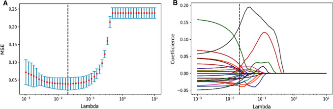
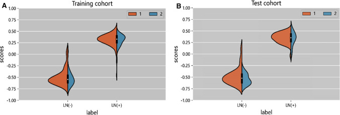
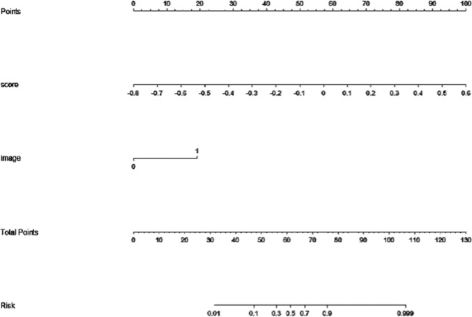
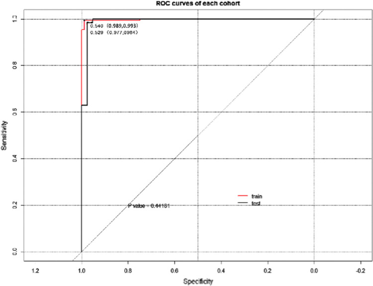
Methods: 347 patients were retrospectively selected (training cohort: 242, test cohort: 105). The enhanced computed tomography arterial phase images of gastric cancer were used for lesion segmentation, radiomics and deep learning feature extraction. Three methods were used for feature selection. Support vector machine (SVM) or random forest (RF) was used to build models. The classification performance of the models was evaluated using the area under the receiver operating characteristic curve (AUC). We also established a nomogram that included clinical predictors.
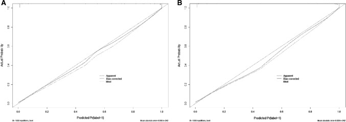
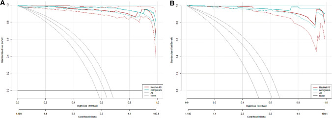
Results: The model based on ResNet50-RF showed favorable classification performance and was verified in the test cohort (AUC = 0.9803). The nomogram based on deep learning feature scores and the lymph node status reported by computed tomography showed excellent discrimination. AUC of 0.9978 was achieved in the training cohort and verified in the test cohort (AUC = 0.9914). Decision analysis curve showed the value of nomogram in clinical application.
Conclusion: The computed tomography-based deep learning nomogram can accurately and effectively evaluate lymph node metastasis in patients with gastric cancer before surgery.
Keywords: Deep learning; Gastric cancer; Lymph node metastasis; Nomogram.
© 2022. The Author(s).
Conflict of interest statement
All authors have no conflicts of interest to disclose.
Figures
Similar articles
-
Dual-energy CT-based deep learning radiomics can improve lymph node metastasis risk prediction for gastric cancer.Eur Radiol. 2020 Apr;30(4):2324-2333. doi: 10.1007/s00330-019-06621-x. Epub 2020 Jan 17. Eur Radiol. 2020. PMID: 31953668
-
CT radiomics nomogram for the preoperative prediction of lymph node metastasis in gastric cancer.Eur Radiol. 2020 Feb;30(2):976-986. doi: 10.1007/s00330-019-06398-z. Epub 2019 Aug 29. Eur Radiol. 2020. PMID: 31468157
-
Novel deep learning radiomics nomogram-based multiparametric MRI for predicting the lymph node metastasis in rectal cancer: A dual-center study.J Cancer Res Clin Oncol. 2024 Oct 9;150(10):450. doi: 10.1007/s00432-024-05986-x. J Cancer Res Clin Oncol. 2024. PMID: 39379733 Free PMC article.
-
Artificial intelligence for pre-operative lymph node staging in colorectal cancer: a systematic review and meta-analysis.BMC Cancer. 2021 Sep 26;21(1):1058. doi: 10.1186/s12885-021-08773-w. BMC Cancer. 2021. PMID: 34565338 Free PMC article.
-
State-of-the-art performance of deep learning methods for pre-operative radiologic staging of colorectal cancer lymph node metastasis: a scoping review.BMJ Open. 2024 Dec 2;14(12):e086896. doi: 10.1136/bmjopen-2024-086896. BMJ Open. 2024. PMID: 39622569 Free PMC article.
Cited by
-
Preoperative predictive model for the probability of lymph node metastasis in gastric cancer: a retrospective study.Front Oncol. 2024 Sep 27;14:1473423. doi: 10.3389/fonc.2024.1473423. eCollection 2024. Front Oncol. 2024. PMID: 39399177 Free PMC article.
-
Diagnostic performance of CT scan-based radiomics for prediction of lymph node metastasis in gastric cancer: a systematic review and meta-analysis.Front Oncol. 2023 Oct 23;13:1185663. doi: 10.3389/fonc.2023.1185663. eCollection 2023. Front Oncol. 2023. PMID: 37936604 Free PMC article.
-
Deep learning based radiomics for gastrointestinal cancer diagnosis and treatment: A minireview.World J Gastroenterol. 2022 Dec 7;28(45):6363-6379. doi: 10.3748/wjg.v28.i45.6363. World J Gastroenterol. 2022. PMID: 36533112 Free PMC article. Review.
-
Role of radiomics in predicting lymph node metastasis in gastric cancer: a systematic review.Front Med (Lausanne). 2023 Aug 16;10:1189740. doi: 10.3389/fmed.2023.1189740. eCollection 2023. Front Med (Lausanne). 2023. PMID: 37663653 Free PMC article.
-
Novel research and future prospects of artificial intelligence in cancer diagnosis and treatment.J Hematol Oncol. 2023 Nov 27;16(1):114. doi: 10.1186/s13045-023-01514-5. J Hematol Oncol. 2023. PMID: 38012673 Free PMC article. Review.
References
-
- Rice TW, Gress DM, Patil DT, Hofstetter WL, Kelsen DP, Blackstone EH. Cancer of the esophagus and esophagogastric junction-Major changes in the American Joint Committee on Cancer eighth edition cancer staging manual. CA Cancer J Clin 2017;67(4):304–317. - PubMed
Publication types
MeSH terms
LinkOut - more resources
Full Text Sources
Medical
