Review
. 2022 Oct;311(1):151-176.
doi: 10.1111/imr.13122. Epub 2022 Jul 31.

Neuroinflammation: Extinguishing a blaze of T cells

Affiliations
Review

Neuroinflammation: Extinguishing a blaze of T cells

Nail Benallegue et al. Immunol Rev. 2022 Oct.

Abstract

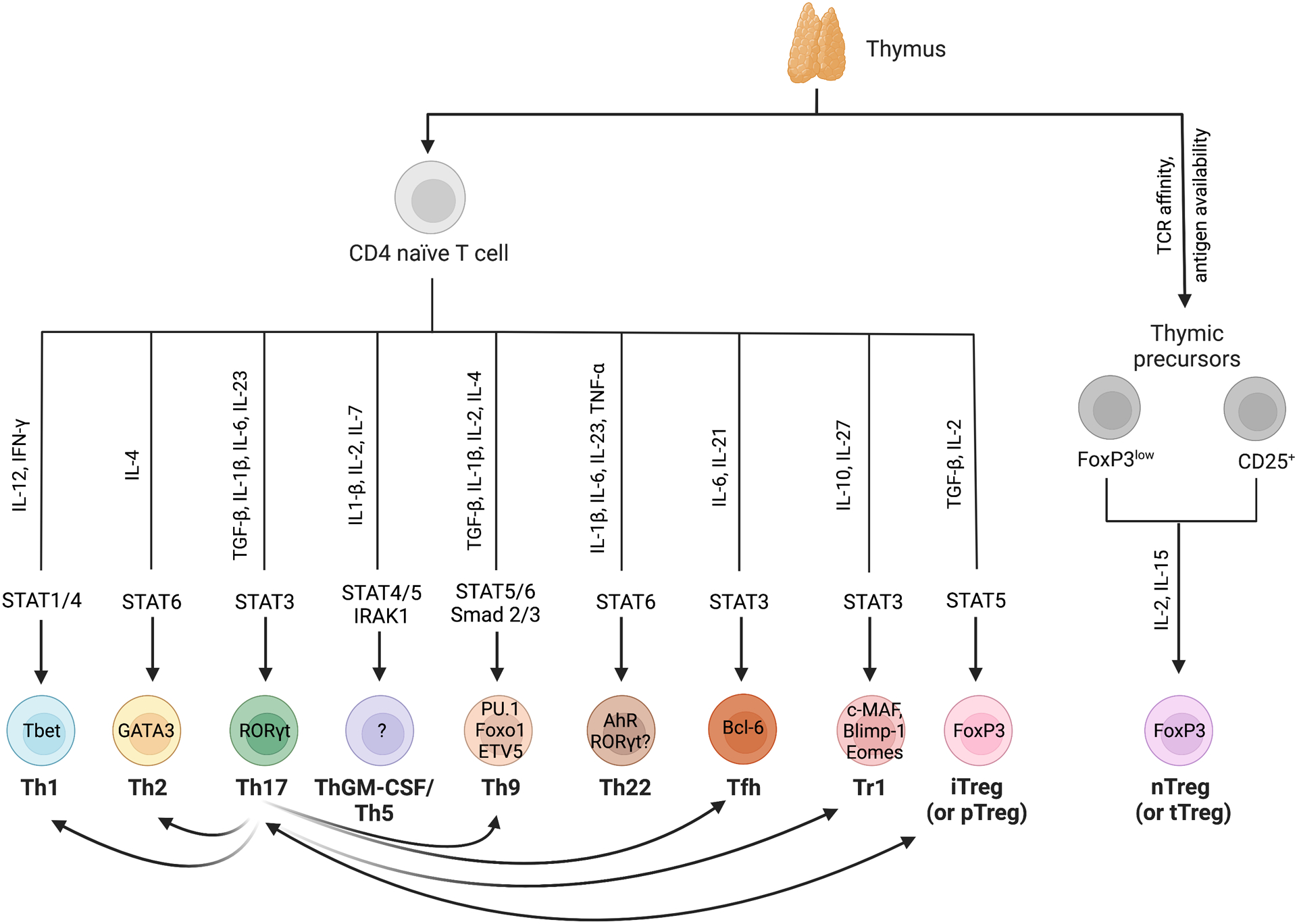
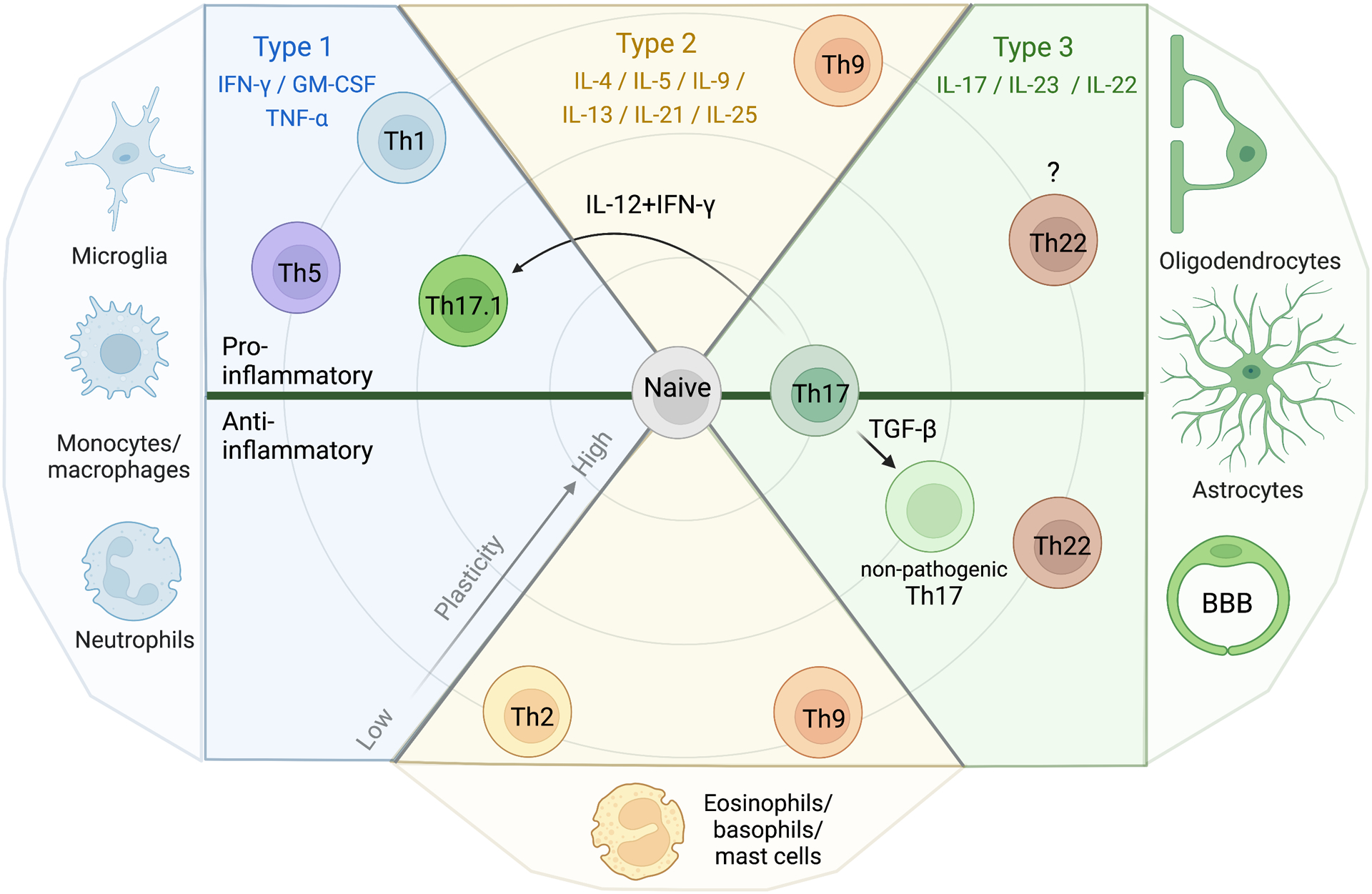
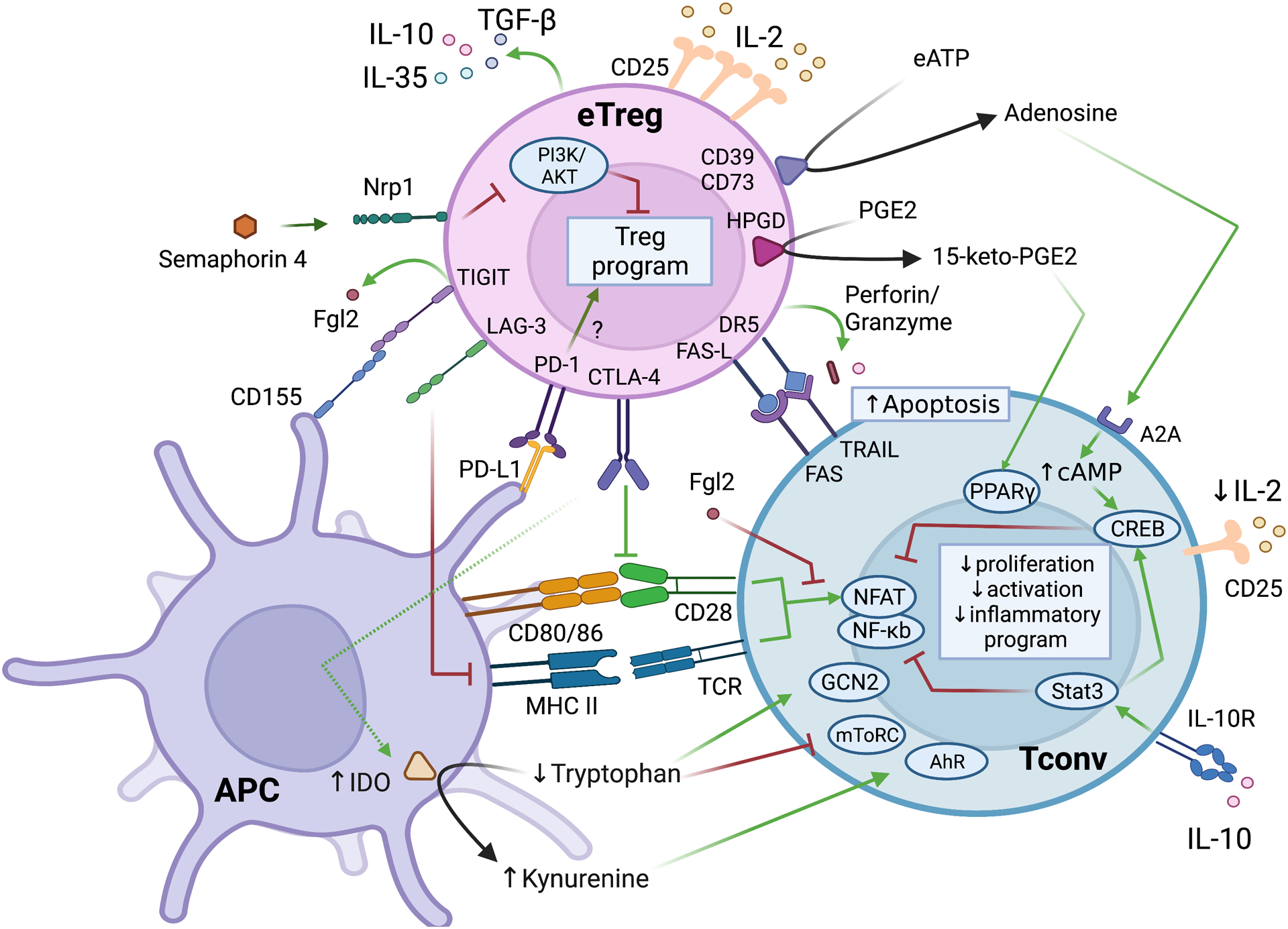
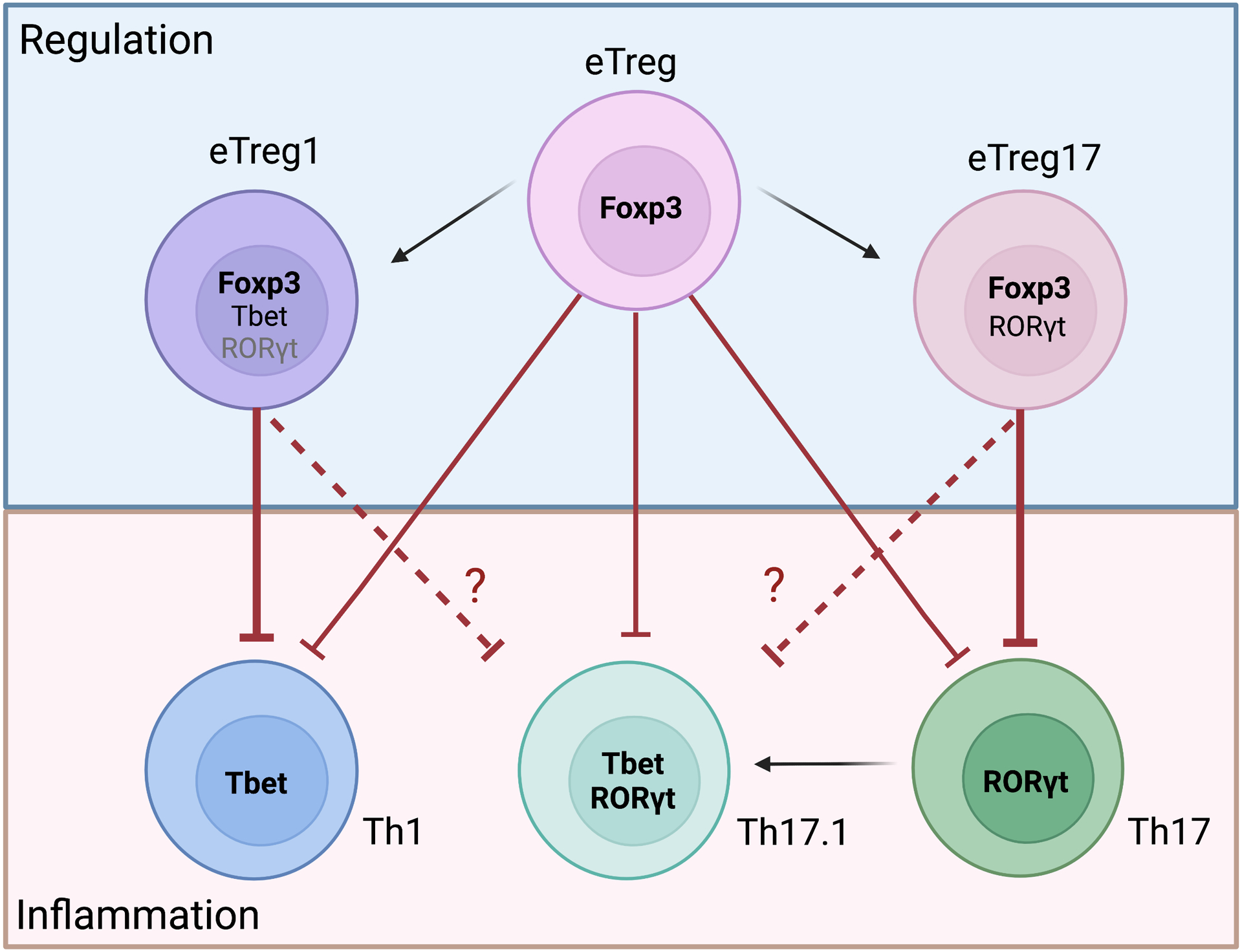
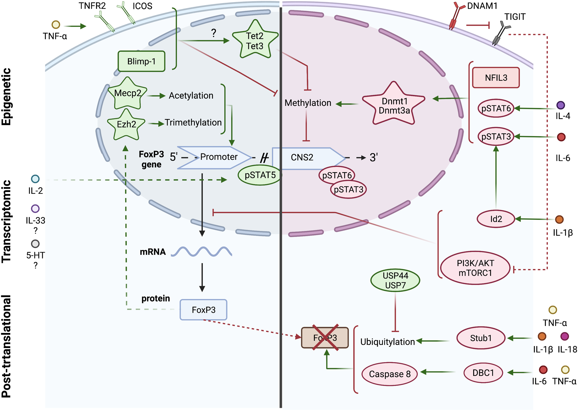
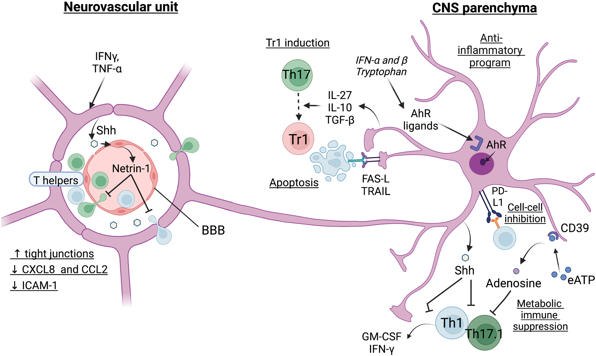
Inflammation is a biological process that dynamically alters the surrounding microenvironment, including participating immune cells. As a well-protected organ surrounded by specialized barriers and with immune privilege properties, the central nervous system (CNS) tightly regulates immune responses. Yet in neuroinflammatory conditions, pathogenic immunity can disrupt CNS structure and function. T cells in particular play a key role in promoting and restricting neuroinflammatory responses, while the inflamed CNS microenvironment can influence and reshape T cell function and identity. Still, the contraction of aberrant T cell responses within the CNS is not well understood. Using autoimmunity as a model, here we address the contribution of CD4 T helper (Th) cell subsets in promoting neuropathology and disease. To address the mechanisms antagonizing neuroinflammation, we focus on the control of the immune response by regulatory T cells (Tregs) and describe the counteracting processes that preserve their identity under inflammatory challenges. Finally, given the influence of the local microenvironment on immune regulation, we address how CNS-intrinsic signals reshape T cell function to mitigate abnormal immune T cell responses.

Keywords: T cells; Th cells; Tregs; autoimmunity; central nervous system; inflammation.


Conflict of interest statement

The authors have no conflict of interest to disclose.

Figures

Figure 1:
Figure 1:. CD4 T cell differentiation and maturation.
The thymus plays a central role in CD4 T cell maturation. After successfully passing thymic selection, CD4 naïve T cells differentiate into T helper (Th), T follicular helper (Tfh), and regulatory T cells (Tregs) upon concurrent TCR engagement and cytokine stimulation. Commitment to a given lineage is tightly regulated by selective cytokine-dependent phosphorylation of signal transducer and activator of transcription (STAT) proteins. This triggers translocation of transcription factors into the nucleus, transcription, and remodeling of chromatin accessibility of target genes. Master transcription factors exert direct or indirect control over downstream genes associated with lineage-specific phenotypes and functions. The plasticity of Th cell subsets is depicted by the curved arrows. The high plasticity of Th17 cells, inherent to RORγt modulation, makes these cells highly dynamic and capable to contextually acquire other T cell lineage-derived functions. Tregs arise from thymic precursor cells (nTregs or tTregs) or from the differentiation of CD4 naïve T cells in peripheral lymphoid organs (iTregs). iTregs also display considerable plasticity and share several elements of their transcriptional program with Th17 cells. AhR: aryl hydrocarbon receptor; Blimp-1: B lymphocyte-induced maturation protein-1; Eomes: eomesodermin; ETV5: Ets variant 5; Foxo1: forkhead box protein O1; FoxP3: forkhead box P3; GATA3: GATA binding protein 3; iTreg: inducible Treg; IFN-γ: interferon gamma; IRAK1:interleukin-1 receptor associated kinase 1; nTreg: natural Tregs; pTreg: peripheral Tregs; RORγt: retinoic-acid-receptor-related orphan nuclear receptor gamma; STAT: signal transducer and activator of transcription; Tbet: T-box expressed in T cells; TCR: T cell-receptor; Tfh: T follicular helper cells; TGF-β: transforming growth factor beta; Th: T helper cells; TNF-α: tumor necrosis factor alpha; Tr1: type 1 regulatory T cells; Treg: regulatory T cells; tTreg: thymic Tregs.
Figure 2:
Figure 2:. Novel insights into the functional classification of Th cells.
The classification of Th cells according to their cytokine production has been reconsidered. T cells can secrete a broad range of cytokines depending on environmental stimulation and state of maturation. Thus, according to their downstream cell targets, three types of Th cell responses have been proposed: the type 1 response involves modulators of monocytes/macrophages and neutrophils through GM-CSF, IFN-γ, TNF-α secretors; the type 2 response is characterized by modulators of polymorphonuclear cells like eosinophils, basophils, mast cells; and the type 3 response is mediated by IL-17 and IL-22 secretors and modulates non-immune cells like BBB endothelial cells, astrocytes and oligodendrocytes, and cells at barrier interfaces in other tissues. We emphasize the need to consider T cell flexibility when using this classification, as environmental factors can reshape T cell fate into pro- and anti-inflammatory states. Depending on the local milieu, highly plastic T cell subsets can shift the balance of the immune response.
Figure 3:
Figure 3:. Treg suppression of conventional T cells.
eTreg directly suppress Tconv cells through secretion of soluble mediators including immunosuppressive cytokines IL-10, IL-35, and TGF-β, through TIGIT-induced secretion of Fgl2, via enzymatic catabolism of soluble ATP and PGE2, and through scavenging of IL-2 in the milieu. Furthermore, eTregs induce a tolerogenic profile in APCs, triggering indirect suppression of Tconv activation, proliferation, and transcription of their pro-inflammatory factors. Indeed, eTregs induce expression of IDO in APCs, which activates the kynurenine pathway via CTLA-4 binding and results in tryptophan deprivation, reduced antigen presentation upon LAG-3 ligation, and decreased Tconv co-stimulatory signaling via CTLA-4. eTregs can also trigger death of the targeted cell upon binding to the death receptors FAS and TRAIL and by secretion of perforin and granzyme. Reinforcement of the eTreg phenotype occurs via PD-1/PD-L1 engagement, semaphorin 4 binding to Nrp1, and IL-2-mediated signaling. A2A: adenosine A2A receptor; AhR: aryl hydrocarbon receptor; APC: antigen-presenting cell; cAMP: cyclic adenosine monophosphate; CREB: C-responsive element binding; CTLA-4: cytotoxic T-lymphocyte-associated protein 4; eATP: extracellular adenosine triphosphate; eTreg : effector regulatory T cell; Fgl2: fibrinogen-like protein 2; GCN2: general control nonderepressible 2; HPGD: 15-hydroxyprostaglandin dehydrogenase; IDO: indoleamine 2,3-dioxygenase; LAG-3: lymphocyte-activation gene-3; MHC II: major histocompatibility complex II; mToRC: mammalian target of rapamycin; NFAT: nuclear factor of activated T cells; NF-κb: nuclear factor kappa-light-chain-enhancer of activated B cells; Nrp1: Neuropillin 1; PPARγ: peroxisome proliferator-activated receptor-γ; PD-1: programmed cell death protein 1; PD-L1: programmed death-ligand 1; PGE2: prostaglandin E2; STAT: signal transducer and activator of transcription; Tconv: conventional T cell; TCR: T cell-receptor; TGF-β: transforming growth factor beta; TIGIT: T cell immunoreceptor with Ig and ITIM domain; TRAIL: TNF-related apoptosis-inducing ligand.
Figure 4:
Figure 4:. Treg modulation of conventional T cells.
eTregs can acquire specialized suppressive properties according to the type of conventional T cells (Tconv) found within a given microenvironment. Thus, eTregs expressing RORγt, named eTreg17, are prone to suppress Th17 cells, whereas eTreg1 who express Tbet, efficiently regulate Th1 cells. The capacity of specialized eTregs to regulate the pathogenic Th17.1 subset has not been elucidated yet. The heterogeneity and adaptability of eTregs highlight their high degree of specialization which serves to contract inflammation under diverse conditions. eTreg: effector regulatory T cell; RORγt: retinoic-acid-receptor-related orphan nuclear receptor gamma; Tbet: T-box expressed in T cells.
Figure 5:
Figure 5:. Maintaining the regulatory T cell program during inflammation: a delicate balance.
Maintaining the suppressive phenotype of effector regulatory T cells (eTregs) is an active process which involves epigenetic, transcriptomic, and post-translational regulation of FoxP3. Demethylation of the enhancer CNS2 within the FoxP3 locus is an absolute prerequisite to initiate a stable eTreg program. External signaling catalyzes methylcytosine demethylation of CNS2, presumably via activation of the methylcytosine dioxygenase Tet2, Tet3, and Mecp2, to enable recruitment of phosphorylated STAT5 and associated-transcription factors which initiate FoxP3 transcription upon binding to the FoxP3 promoter. The required chromatin accessibility of the FoxP3 promoter is modulated by histone acetylation and trimethylation via Mecp2 and Ezh2, respectively. Conversely, inflammatory mediators promote activation of the methyltransferase Dnmt3a and Dnmt1 to inhibit CNS2 accessibility to transcription factors. STAT3 and STAT6 can directly compete with STAT5 for binding to the CNS2 locus and thus impede FoxP3 transcription. DNAM-1 competes with TIGIT for CD155 binding, resulting in the activation of the PI3K/AKT/mTORC pathway, which represses FoxP3 transcription. Similarly, IL-1β- mediated expression of Id2 blocks the transcription of FoxP3. At the protein level, pro-inflammatory cytokines can promote FoxP3 ubiquitylation and subsequent proteasomal degradation and activate a DBC1/caspase-8-mediated downregulation of the FoxP3 protein. Blimp-1: B lymphocyte-induced maturation protein-1; CNS2: conserved non-coding sequence 2; DBC1: deleted in bladder cancer protein 1; DNAM1: DNAX accessory molecule-1 (CD226); Dnmt: DNA cytosine-5-methyltransferase; Ezh2: enhancer of zeste homolog 2; ICOS: inducible T-cell costimulatory; Id2: inhibitor of DNA binding 2; Mecp2: methyl CpG binding protein 2; NFIL3: nuclear factor interleukin 3 regulated; PI3K/AKT/mTORC1: phosphoinositide 3 kinase/ protein kinase B/ mammalian target of rapamycin complex 1; Stub1: STIP1 homology and U-Box containing protein 1; Tet: Tet methylcytosine dioxygenase; TIGIT: T cell immunoreceptor with Ig and ITIM domain; TNF-α: tumor necrosis factor alpha; TNFR2: tumor necrosis factor receptor 2; USP: ubiquitin carboxyl-terminal hydrolase.
Figure 6:
Figure 6:. CNS intrinsic regulation of neuroinflammation: an astrocytic symphony orchestra.
At the CNS-barrier level, inflammation tends to disrupt the permeability of the NVU to invading T cells. In response to inflammatory mediators, activated astrocytic endfeet secrete soluble Shh which induces endothelial cell expression of netrin-1 that promotes upregulation of tight junctions to protect the endothelium, repress T cell chemoattraction and transmigration. Within the parenchyma, astrocytes are key regulators of CNS responses to inflammatory challenges. AhR signaling stimulates a switch to an anti-inflammatory profile in astrocytes. There is a multitude of mechanisms by which astrocytes regulate pathogenic T cells. Secreted Shh induces Smo-dependent signaling in the T cell compartment which imprints an anti-inflammatory program in encephalitogenic CD4 effector T cells to antagonize Th1, Th17 and ThGM-CSF phenotypes. The secretion by astrocytes of immunosuppressive cytokines IL-10 and TGF-β inhibits pathogenic T cells but also synergizes with IL-27 to initiate Th17 transdifferentiation into Tr1 cells. Astrocytes express the metabolic checkpoint molecule CD39 which enables adenosine-mediated suppression of pathogenic T cell activation and proliferation. Furthermore, direct engagement of Th cells with astrocytes triggers T cell inhibition via the PD-1/PD-L1 axis, and apoptosis upon death receptor binding. AhR: aryl hydrocarbon receptor; BBB: blood-brain-barrier; CCL2: chemokine C-C motif ligand 2; CXCL8: chemokine C-X-C ligand 8 (Interleukin 8); eATP: extracellular adenosine triphosphate; ICAM1: intercellular adhesion molecule 1; PD-L1: programmed death-ligand 1; Shh: sonic hedgehog; TGF-β: transforming growth factor beta; TNF-α: tumor necrosis factor alpha; Tr1: regulatory T cells type 1; TRAIL: TNF-related apoptosis-inducing ligand.

Similar articles

Cited by

References

    1. Walton C, King R, Rechtman L, et al. Rising prevalence of multiple sclerosis worldwide: Insights from the Atlas of MS, third edition. Mult Scler. 2020;26(14):1816–1821. - PMC - PubMed
    1. Dendrou CA, Fugger L, Friese MA. Immunopathology of multiple sclerosis. Nature reviews Immunology. 2015;15(9):545–558. - PubMed
    1. Lassmann H Pathology of inflammatory diseases of the nervous system: Human disease versus animal models. Glia. 2020;68(4):830–844. - PMC - PubMed
    1. Hayashi T, Morimoto C, Burks JS, Kerr C, Hauser SL. Dual-label immunocytochemistry of the active multiple sclerosis lesion: major histocompatibility complex and activation antigens. Annals of neurology. 1988;24(4):523–531. - PubMed
    1. Booss J, Esiri MM, Tourtellotte WW, Mason DY. Immunohistological analysis of T lymphocyte subsets in the central nervous system in chronic progressive multiple sclerosis. Journal of the neurological sciences. 1983;62(1–3):219–232. - PubMed

Publication types