. 2022 Jul 15:15:842772.
doi: 10.3389/fnmol.2022.842772. eCollection 2022.

Glutamatergic Synapse Dysfunction in Drosophila Neuromuscular Junctions Can Be Rescued by Proteostasis Modulation

Affiliations

Glutamatergic Synapse Dysfunction in Drosophila Neuromuscular Junctions Can Be Rescued by Proteostasis Modulation

Anushka Chakravorty et al. Front Mol Neurosci. .

Abstract

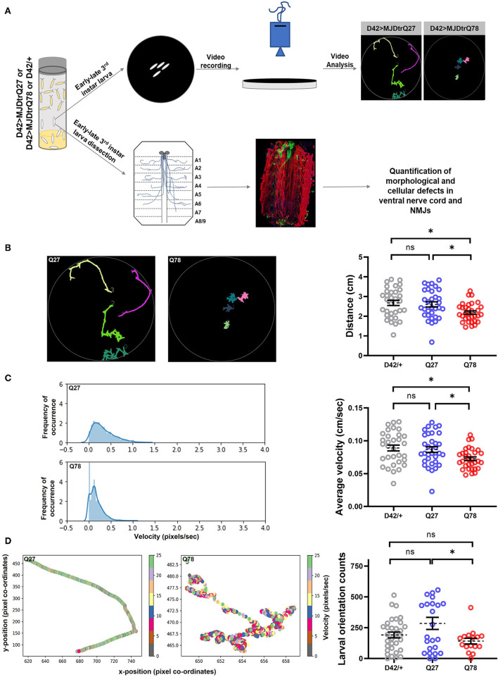
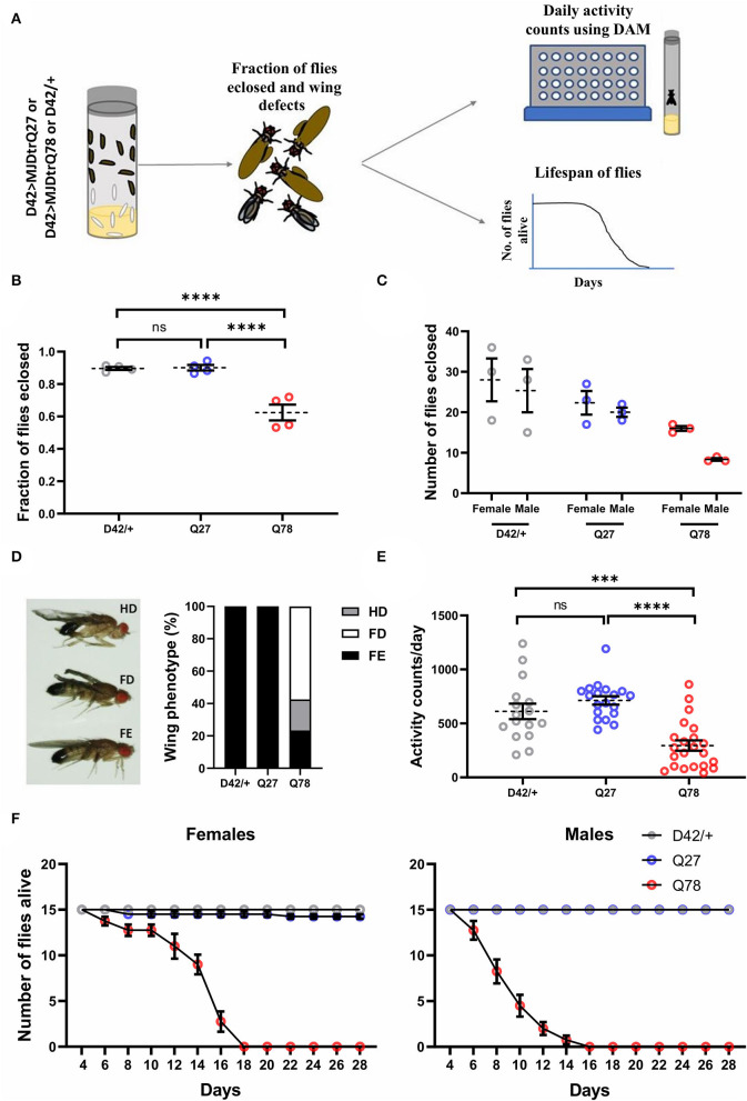
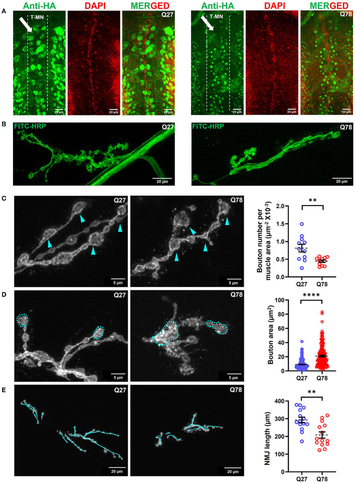
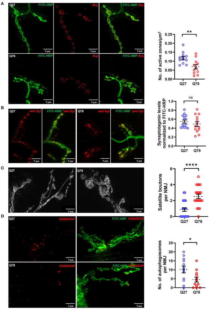
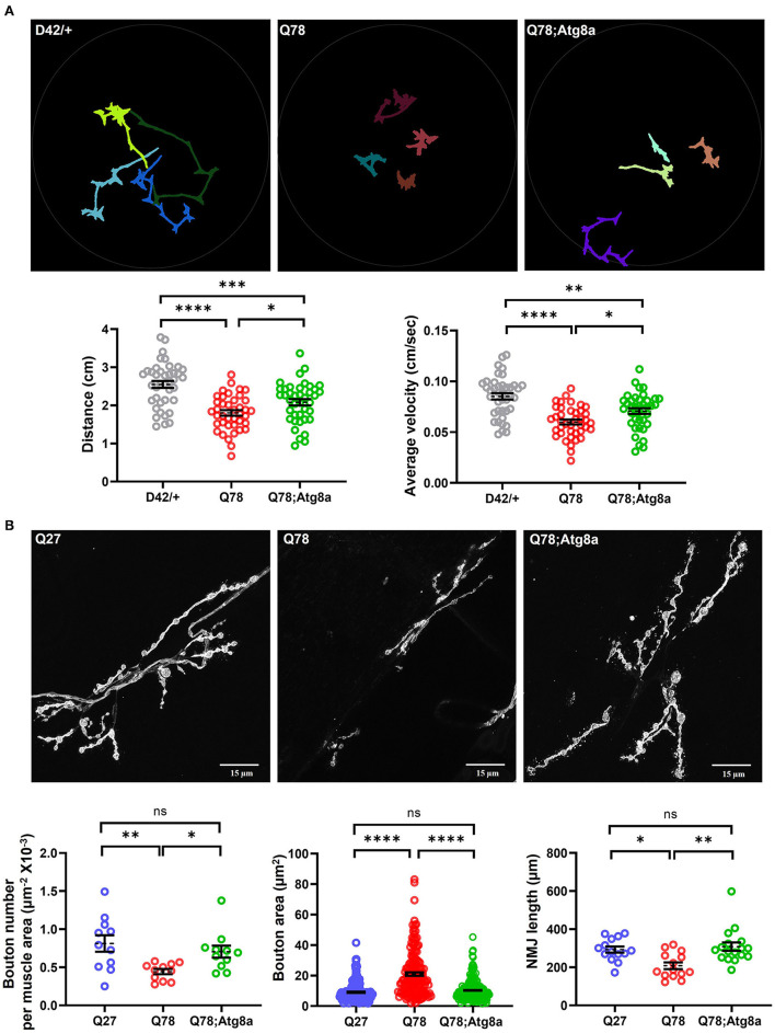
Glutamate is the major excitatory neurotransmitter in the nervous system, and the Drosophila glutamatergic neuromuscular junctions (NMJs) offer a tractable platform to understand excitatory synapse biology both in health and disease. Synaptopathies are neurodegenerative diseases that are associated with synaptic dysfunction and often display compromised proteostasis. One such rare, progressive neurodegenerative condition, Spinocerebellar Ataxia Type 3 (SCA3) or Machado-Joseph Disease (MJD), is characterized by cerebellar ataxia, Parkinsonism, and degeneration of motor neuron synapses. While the polyQ repeat mutant protein ataxin-3 is implicated in MJD, it is unclear how it leads to impaired synaptic function. In this study, we indicated that a Drosophila model of MJD recapitulates characteristics of neurodegenerative disorders marked by motor neuron dysfunction. Expression of 78 polyQ repeats of mutant ataxin-3 protein in Drosophila motor neurons resulted in behavioral defects, such as impaired locomotion in both larval and adult stages. Furthermore, defects in eclosion and lifespan were observed in adult flies. Detailed characterization of larval glutamatergic neuromuscular junctions (NMJs) revealed defects in morphological features along with compromised NMJ functioning. Autophagy, one of the key proteostasis pathways, is known to be impaired in the case of several synaptopathies. Our study reveals that overexpression of the autophagy-related protein Atg8a rescued behavioral defects. Thus, we present a model for glutamatergic synapse dysfunction that recapitulates synaptic and behavioral deficits and show that it is an amenable system for carrying out genetic and chemical biology screens to identify potential therapeutic targets for synaptopathies.

Keywords: Drosophila neuromuscular junctions; Spinocerebellar Ataxia Type 3; autophagy; glutamatergic synapse; synapse dysfunction; synaptopathy.


Conflict of interest statement

The authors declare that the research was conducted in the absence of any commercial or financial relationships that could be construed as a potential conflict of interest.

Figures

Figure 1
Figure 1
Q78 expression in motor neurons leads to larval locomotion defect. (A) A flow chart of approaches to characterize behavioral and cellular defects upon expression of mutant MJDtrQ78. Third instar larvae expressing non-pathogenic Q27 or pathogenic Q78 along with driver control D42/+ were subjected to a larval locomotion assay and simultaneously assessed for phenotypic and functional defects in NMJs. (B) Path diagrams for Q27 and Q78 larvae monitored in 1% charcoal agar plates and quantification of the total distance traversed by the driver-only control larvae (D42/+) vs. non-pathogenic Q27 and pathogenic Q78 larvae. n = 32 larvae; one-way ANOVA; post-hoc Tukey's multiple comparison test; ns, non-significant; *p < 0.05; error bars represent mean ± SEM. (C) Representative images of the velocity distribution of non-pathogenic Q27 and pathogenic Q78 larvae and quantification of the average velocity of larvae. n = 32 larvae; one-way ANOVA; post-hoc Tukey's multiple comparison test; ns, non-significant; *p < 0.05; error bar represents ± SEM. (D) A representative scatter plot showing instantaneous velocity per coordinate for non-pathogenic Q27 and pathogenic Q78 larvae. The number of times the larvae traversed linearly in the arena without reorientation/coiling was plotted as orientation counts. n = 32 larvae; one-way ANOVA; post-hoc Tukey's multiple comparison test; ns, non-significant; *p < 0.05; error bar represents mean ± SEM.
Figure 2
Figure 2
Eclosion, locomotion, and lifespan are affected in adult flies on the expression of Q78 in motor neurons. (A) A flow chart depicting phenotypes characterized in adult flies expressing non-pathogenic Q27 or pathogenic Q78 in motor neurons using the D42-Gal4 driver. A fraction of adult flies that successfully emerged from pupae, wing phenotypes, locomotion, and adult lifespan was assayed. (B) Quantification of fraction of flies eclosed. n > 280 pupae from 4 vials; 3x2 contingency test followed by Fisher's Exact test; ****p < 0.0001; ns, non-significant; error bars represent mean ± SEM. (C) Quantification of the number of male and female flies eclosed. n ≥ 70 flies. (D) Images depicting wing phenotypes of adult flies expressing pathogenic Q78 under D42-Gal4 and quantification of the percentage of wing phenotypes observed. n ≥ 47 flies; FD, full degeneration; HD, half degeneration; FE, fully expanded. (E) Quantification of activity counts per day. n ≥ 16 flies per genotype; one-way ANOVA; post-hoc Tukey's HSD; ****p < 0.0001; ***p < 0.001; ns, non-significant; error bars represent mean ± SEM. (F) Survivorship plots showing the number of flies alive across 28 days. n = 60 flies for both males and females; error bars depict mean ± SEM.
Figure 3
Figure 3
Q78 expression in motor neurons leads to multiparametric changes in the morphology of NMJs. (A) An image depicting the cell bodies of motor neurons in the ventral nerve cord (VNCs) of Drosophila larvae. An arrow depicts the alignment of the thoracic motor neuron (T-MN) cell bodies. FITC-HRP marks the neuronal membrane. HA antibody was used for staining the pathogenic or non-pathogenic ATXN3 polyQ protein. (B) Image depicting the NMJs of the third instar larva marked by FITC-HRP in Q27 and Q78-expressing larvae. The morphometric changes observed are further quantified. (C) The number of boutons per muscle area in Q27 and Q78-expressing larvae and quantification of the same. Arrowheads depict the varicosities or boutons. All quantifications were done on NMJs of muscle 6/7 of abdominal hemisegment A2. n > 11; Student's t-test; **p < 0.01. An error bar represents mean ± SEM. (D) The area of boutons in Q27 and Q78-expressing larvae and quantification of the same. Markings indicate the areas considered for quantification. All quantifications were done on NMJs of muscle 6/7 of abdominal segments A4-A6. n > 160; Student's t-test; ****p < 0.0001. An error bar represents mean ± SEM. (E) Length of NMJ branches and the main arbor in Q27 and Q78-expressing larvae and quantification of the same. Markings represent the lengths considered for quantification. All quantifications were done on NMJs of muscle 6/7 of abdominal segments A4-A6. n > 12; Student's t-test; **p < 0.01. An error bar represents mean ± SEM.
Figure 4
Figure 4
Q78 expression in larval NMJs leads to functional changes in glutamatergic synapses. (A) A representative image of active zones marked by anti-Brp in the NMJs of the third-instar larva marked by FITC-HRP in Q27 and Q78-expressing larvae. Quantification of the number of active zones normalized to the area of NMJ. All quantifications were done on NMJs of muscle 6/7 of abdominal segments A4-A6. n ≥ 12; Student's t-test; **p < 0.01. An error bar represents mean ± SEM. (B) Representative images of synaptotagmin (Syt) intensity in the third instar larval NMJ marked by FITC-HRP in Q27 and Q78-expressing larvae and quantification of Syt signal intensity in the boutons. All quantifications were done on NMJs of muscle 6/7 of abdominal segments A4-A6. n ≥ 15; Student's t-test; ns, non-significant. An error bar represents mean ± SEM. (C) Representative images depicting satellite boutons budding from the main boutons in NMJs with Q78 expression along with quantification of the number of satellite boutons per NMJ. All quantifications were done on NMJs of muscle 6/7 of abdominal segments A4-A6. n > 25; Student's t-test; ****p < 0.0001. An error bar represents mean ± SEM. (D) The figure depicts the number of autophagosomes marked by anti-GABARAP in NMJs stained by FITC-HRP in Q27- and Q78-expressing larvae. All quantifications were done on NMJs of muscle 6/7 of abdominal segments A4-A6. n > 12; Student's t-test; *p < 0.05. An error bar represents mean ± SEM.
Figure 5
Figure 5
The behavioral and morphological defects in larval glutamatergic synapses are rescued upon Atg8a overexpression. (A) Path diagrams for indicated larvae monitored in 1% charcoal agar plates and quantification of total distance traversed and average velocity of the driver-only control larvae (D42/+) vs. pathogenic Q78 and rescue Q78; Atg8a larvae. n = 40 larvae; one-way ANOVA; post-hoc Tukey's multiple comparison test; *p < 0.05; **p < 0.01; ***p < 0.001; ****p < 0.0001; ns, non-significant. Error bars represent mean ± SEM. (B) Representative images of NMJs of indicated larvae from muscle 6/7 of abdominal hemisegment A2. Quantification of the number of boutons per muscle area (n > 11 NMJs), bouton area (n > 160 boutons), and NMJ arbor length (n > 12 NMJs). Quantifications for the number of boutons per area of muscle were done on hemisegment A2. Quantifications for the bouton area and NMJ arbor length were done on NMJs of muscle 6/7 of abdominal segments A4-A6. One-way ANOVA; post-hoc Tukey's multiple comparison test; *p < 0.05, **p < 0.01, ***p < 0.001, ****p < 0.0001. ns, non-significant. Error bars represent mean ± SEM.

Similar articles

Cited by

References

    1. Alhindi A., Boehm I., Chaytow H. (2021). Small junction, big problems: neuromuscular junction pathology in mouse models of amyotrophic lateral sclerosis (ALS). J. Anat. 10.1111/joa.13463. [Epub ahead of print]. - DOI - PMC - PubMed
    1. Alves S., Régulier E., Nascimento-Ferreira I., Hassig R., Dufour N., Koeppen A., et al. . (2008). Striatal and nigral pathology in a lentiviral rat model of Machado-Joseph disease. Hum. Mol. Genet. 17, 2071–2083. 10.1093/hmg/ddn106 - DOI - PubMed
    1. Andres-Alonso M., Kreutz M. R., Karpova A. (2021). Autophagy and the endolysosomal system in presynaptic function. Cell. Mol. Life Sci. 78, 2621–2639. 10.1007/s00018-020-03722-5 - DOI - PMC - PubMed
    1. Ashkenazi A., Bento C. F., Ricketts T., Vicinanza M., Siddiqi F., Pavel M., et al. . (2017). Polyglutamine tracts regulate beclin 1-dependent autophagy. Nature 545, 108–111. 10.1038/nature22078 - DOI - PMC - PubMed
    1. Bae J. R., Kim S. H. (2017). Synapses in neurodegenerative diseases. BMB Rep. 50, 237–246. 10.5483/BMBRep.2017.50.5.038 - DOI - PMC - PubMed

LinkOut - more resources