. 2022 Jul 13:15:920221.
doi: 10.3389/fnmol.2022.920221. eCollection 2022.

Multi-mtDNA Variants May Be a Factor Contributing to Mitochondrial Function Variety in the Skin-Derived Fibroblasts of Leber's Hereditary Optic Neuropathy Patients

Affiliations

Multi-mtDNA Variants May Be a Factor Contributing to Mitochondrial Function Variety in the Skin-Derived Fibroblasts of Leber's Hereditary Optic Neuropathy Patients

Shun Yao et al. Front Mol Neurosci. .

Abstract

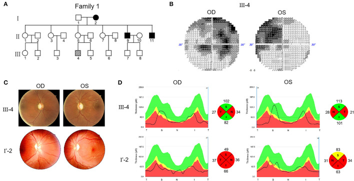
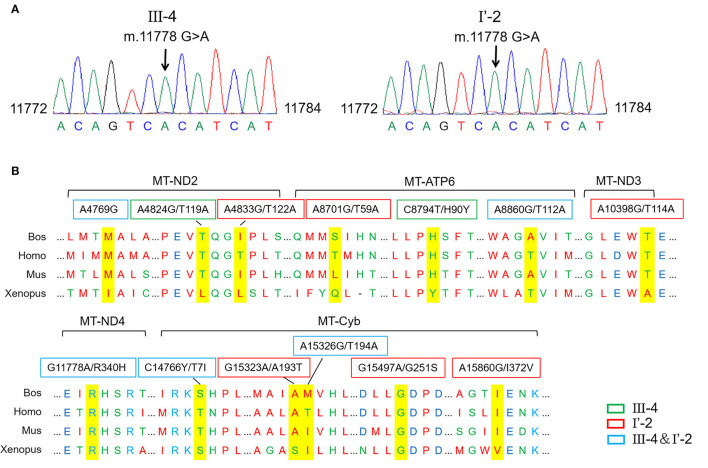
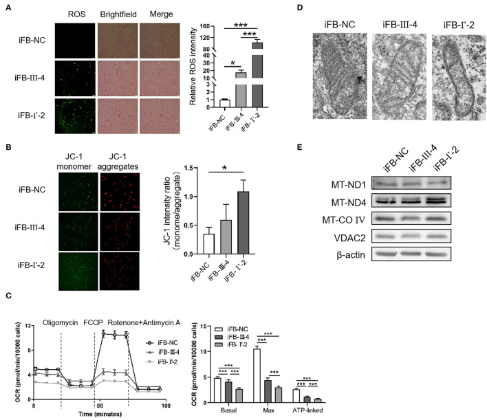
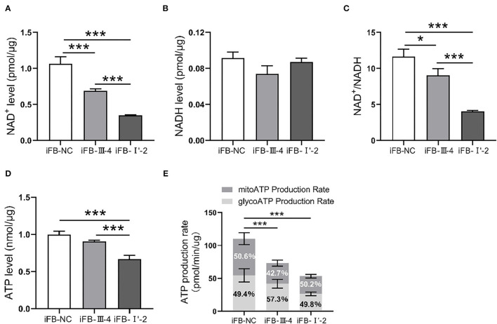
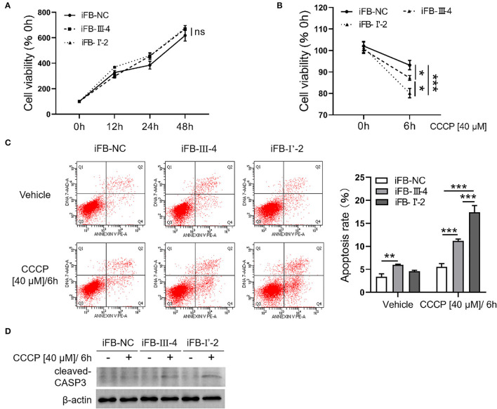
Heterogeneity is a major feature of Leber's hereditary optic neuropathy (LHON) and has a significant impact on the manifestation and diagnosis of the disease. This study explored whether multiple variations in mitochondrial genes were associated with the heterogeneity, mainly phenotypic heterogeneity. Ophthalmic examinations were conducted in two probands with LHON with G11778A and multiple mitochondrial DNA gene (mtDNA) variants. Skin fibroblast cell lines were generated from patients and age- and sex-matched controls. ROS levels, mitochondrial membrane potential, cell energy respiration, and metabolic functions were measured. Flow cytometry and cell viability tests were performed to evaluate the cell apoptosis levels and fate. We found that cells with more mtDNA variants had higher ROS levels, lower mitochondrial membrane potential, and weaker respiratory function. Flow cytometry and cell viability testing showed that multiple mtDNA variants are associated with different levels of cell viability and apoptosis. In conclusion, we found that skin-derived fibroblast cells from G11778A LHON patients could be used as models for LHON research. Multi-mtDNA variants contribute to mitochondrial function variety, which may be associated with heterogeneity in patients with LHON.

Keywords: Leber's hereditary optic neuropathy (LHON); cell viability; heterogeneity; mitochondria dysfunction; mtDNA variants.


Conflict of interest statement

The authors declare that the research was conducted in the absence of any commercial or financial relationships that could be construed as a potential conflict of interest.

Figures

Figure 1
Figure 1
Pedigree and features of probands with Leber's hereditary optic neuropathy (LHON). (A) Pedigree of two probands. Arrow denotes the proband. (B) Visual field test results of proband III-4. (C) Fundus photographs of two probands. (D) RNFL assay of two probands after swept-source optical coherence tomography (SS-OCT) detection.
Figure 2
Figure 2
Different mitochondrial DNA gene (mtDNA) variations of two probands. (A) The mutation m.11778G>A was confirmed in probands by the Sanger sequencing. (B) Conservative analysis of mutant amino acids in four species. Bos, Bostaurus; Homo, Homo sapiens; Mus, Musmusculus; Xenopus, Xenopuslaevis.
Figure 3
Figure 3
Mitochondria damaged severely in immortal fibroblast (iFB)-LHON cells with more variants. (A) ROS level was detection in different fibroblast cell lines: iFB-NC, iFB-III-4, and iFB-I'-2. *p ≤ 0.05, ***p ≤ 0.001. (B) JC-1 monome and aggregate were pictured. The ratio of fluorescence intensity between monomer and aggregate characterized mitochondrial membrane potential. *p ≤ 0.05. (C) oxygen consumption rate (OCR) value of three fibroblast cells was detected using seahorse. The basal (OCR value = OCR value at the beginning, meaning the level of energy metabolism rate in basal state), max (OCR value = OCR value at the highest point after FCCP treatment, meaning the maximum respiration rate that the cell can achieve) and ATP-linked (OCR value = OCR value decreased after oligomycin treatment, meaning the respiration part used for ATP synthesis) OCR values were calculated. ***p ≤ 0.001. (D) Mitochondrial morphology in iFB-NC, iFB-III-4, and iFB-I'-2 cells was photographed by electron microscope. (E) The protein levels of MT-ND1, MT-ND4, MT-CO IV, and VDAC2 were detected by Western blot. The β-actin was used as reference.
Figure 4
Figure 4
The iFB-LHON cells with more variants have lower energy synthesis capacity. (A,B) NAD+ and NADH level in mitochondria were tested in fibroblast cells. ***p ≤ 0.001. (C) The ratio of NAD+/NADH was calculated. *p ≤ 0.05, ***p ≤ 0.001. (D) Fibroblast cells were collected and ATP level in mitochondria was examined in both cells. ***p ≤ 0.001. (E) ATP production rates in fibroblast cells were detected by seahorse XF96 analysis. The mitoATP production rate refers to the ATP production rate related to mitochondrial oxidative phosphorylation (expressed as pmol ATP/min). The glycoATP production rate associated with the conversion of glucose to lactic acid in the glycolysis pathway (also expressed as pmol ATP/min). ***p ≤ 0.001.
Figure 5
Figure 5
Fibroblast cells with more mtDNA variants were more sensitive to stimuli. (A) Fibroblast cells were cultured for 48 h. The cell viability was detected in 0, 12, 24, and 48 h, respectively, using CCK8. (B) Cells were treated with CCCP [40 μM] for 6 h and cell viability was measured before and after treatment. *p ≤ 0.05, ***p ≤ 0.001. (C) CCCP [40 μM/6 h] treatment was performed in fibroblast cells and apoptosis level was detected using flow cytometry. **p ≤ 0.01, ***p ≤ 0.001. (D) Fibroblasts cells were collected after CCCP [40 μM/6 h] treatment and cleaved-CASP3 was detected using western blotting.

Similar articles

Cited by

References

    1. Agarwal S., Muqit M. M. K. (2021). PTEN-induced kinase 1 (PINK1) and Parkin: Unlocking a mitochondrial quality control pathway linked to Parkinson's disease. Curr. Opin. Neurobiol. 72, 111–119. 10.1016/j.conb.2021.09.005 - DOI - PubMed
    1. Agip A. A., Blaza J. N., Bridges H. R., Viscomi C., Rawson S., Muench S. P., et al. . (2018). Cryo-EM structures of complex I from mouse heart mitochondria in two biochemically defined states. Nat. Struct. Mol. Biol. 25, 548–556. 10.1038/s41594-018-0073-1 - DOI - PMC - PubMed
    1. Almasieh M., Catrinescu M. M., Binan L., Costantino S., Levin L. A. (2017). Axonal Degeneration in retinal ganglion cells is associated with a membrane polarity-sensitive redox process. J. Neurosci. 37, 3824–3839. 10.1523/JNEUROSCI.3882-16.2017 - DOI - PMC - PubMed
    1. Amore G., Romagnoli M., Carbonelli M., Barboni P., Carelli V., La Morgia C. (2021). Therapeutic options in hereditary optic neuropathies. Drugs 81, 57–86. 10.1007/s40265-020-01428-3 - DOI - PMC - PubMed
    1. Brandon M. C., Lott M. T., Nguyen K. C., Spolim S., Navathe S. B., Baldi P., et al. . (2005). MITOMAP: a human mitochondrial genome database−2004 update. Nucleic Acids Res 33, D611–D613. 10.1093/nar/gki079 - DOI - PMC - PubMed

LinkOut - more resources