. 2022 Feb 19:3:100117.
doi: 10.1016/j.crmicr.2022.100117. eCollection 2022.

Phenotype to genotype in Neurospora crassa: Association of the scumbo phenotype with mutations in the gene encoding ceramide C9-methyltransferase

Affiliations

Phenotype to genotype in Neurospora crassa: Association of the scumbo phenotype with mutations in the gene encoding ceramide C9-methyltransferase

Erin L Bredeweg et al. Curr Res Microb Sci. .

Abstract

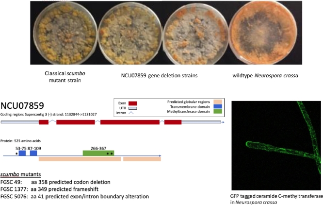
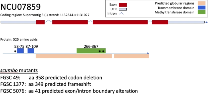
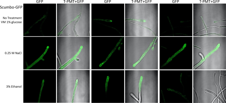
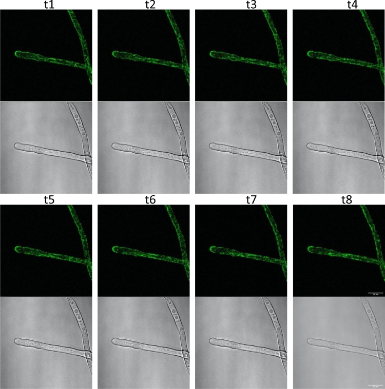
Using a legacy of genetic mutants of Neurospora crassa, paired with resequencing efforts through JGI, we have identified the gene responsible for the 'scumbo' mutant. This early morphological mutant was described as "Irregular flat, spreading growth with knobby protrusions and abnormal conidiation, but no free conidia. Mycelium usually appears yellowish rather than orange. Female fertile." (Perkins, Radford et al. 2000). Our further investigation has found new insights into the identity and associated functions of scumbo as a ceramide C9 methyltransferase, previously annotated as "similar to cyclopropane-fatty-acyl-phospholipidsynthase", encoded by the gene NCU07859. This enzyme performs a fungal-specific methyl modification of glycosyl-ceramides and has implications for membrane homeostasis and hyphal polarity in filamentous fungi.

Keywords: Ceramide C9-methyltransferase; Fast-forward genetics; Fungal genetics; Fungal genomics; Fungi; Morphology; Neurospora; Scumbo.


Conflict of interest statement

The authors declare that they have no known competing financial interests or personal relationships that could have appeared to influence the work reported in this paper.

Figures

Image, graphical abstract
Graphical abstract
Fig. 1
Fig. 1
Gene model with exon structures and placement of scumbo mutant features in FGSCNeurospora strains. Secondary structure and SMART/pfam domains were predicted by eukaryotic linear motif (Kumar, Gouw et al. 2019). NCU07859 is 525 amino acids long and features two transmembrane domains (spanning amino acids 53-75 and 87-109) and a methyltransferase domain (amino acids 266-367) identified from SMART/Pfam domain families. Resequencing of FGSC49 found a codon deletion in the methyltransferase domain. Resequencing of FGSC1377 found a frameshift in the methyltransferase domain. Resequencing of FGSC5076 found an altered exon/intron boundary at amino acid 41 upstream of the transmembrane domain. Each mutation location is marked by asterisk.
Fig. 2
Fig. 2
Generation and testing of scumbo homokaryons susceptibility to hygromycin B. To establish the segregation of the scumbo phenotype, we crossed FGSC13992, identified as a heterokaryon NCU07859 from the knockout collection, to OR74A (FGSC2489). FGSC13992 was used as the maternal strain. Ascospores were collected and germinated by heat shock, isolated to individual slants, and tested for mating type using spot plates. XEB23.1 is mat A, XEB23.2 is mat a. To test susceptibility to hygromycin B, we first grew hyphal tissue on small agar plates, cutting 3 mm squares as a means of equal inoculum. Plates were cultured for 7 days before imaging.
Fig. 3
Fig. 3
Plate growth with osmotic stressors. To assay growth on stress conditions, conidia or hyphal fragments were grown on plates containing VM for 24-36 hours. 3 mm agar blocks were cut and placed on the center of each plate and sealed using micropore surgical tape. Plates were imaged after 5 days of growth. VM:Vogel's medium with 1.5% sucrose.
Fig. 4
Fig. 4
Microscopy of Scumbo-GFP. A strain with NCU07859 transcriptionally fused with a C-terminal GFP tag, imaged under different conditions. Conidia were cultured in 2 mL of media with the indicated supplements for 16 hours with no agitation at room temperature. Tissue was then transferred to a microscope slide with 5-10 μl of media and covered with a coverslip for image capture on a Leica 710 confocal microscope. Three representative paired images are shown, normalized to a common GFP range using Fiji. At the left of each pair is the GFP channel, right is the overlay of GFP with photomultiplier tube (T-PMT) transmission.
Fig. 5
Fig. 5
Scumbo-GFP in a time-series. Images were taken of the same hyphae of Scumbo-GFP, showing accumulation at the end of the hyphae's Spitzenkörper, or collection of vesicles at the hyphal tip. Scumbo signal fluctuates along the endomembrane system. Each timepoint represents a capture over approximately 2 minutes by scanning confocal microscope. Scale bar is 10 μm in t8.

Similar articles

Cited by

References

    1. Abramsky T., Tatum E. Differential inhibition of branching enzyme in a morphological mutant and in wild type Neurospora - Influence of carbon source in the growth medium. Biochimica Et Biophysica Acta. 1976;421(1):106–114. - PubMed
    1. Akhberdi O., Zhang Q., Wang H., Li Y., Chen L., Wang D., Yu X., Wei D., Zhu X. Roles of phospholipid methyltransferases in pycnidia development, stress tolerance and secondary metabolism in the taxol-producing fungus Pestalotiopsis microspore. Microbiological Research. 2018;210:33–42. - PubMed
    1. Baker S.E. Selection to sequence: opportunities in fungal genomics. Environmental Microbiology. 2009;11(12):2955–2958. - PubMed
    1. Baker S.E. Protein hyperproduction in fungi by design. Applied microbiology Biotechnology. 2018;102(20):8621–8628. - PMC - PubMed
    1. Baker S.E., Bredeweg E.L. Comparative genomics, resequencing and fast forward genetics in Aspergillus and Penicillium. Aspergillus and Penicillium in the Post-genomic Era. 2016:17–26.