Case Reports
. 2022 Jul 22:14:2235-2241.
doi: 10.2147/CMAR.S366771. eCollection 2022.

An Ovarian Large-Cell Neuroendocrine Carcinoma Accompanied by Clear Cell Carcinoma with Specific High Level of AFP: Case Report and Review of the Literature

Affiliations
Case Reports

An Ovarian Large-Cell Neuroendocrine Carcinoma Accompanied by Clear Cell Carcinoma with Specific High Level of AFP: Case Report and Review of the Literature

Jian Qiu et al. Cancer Manag Res. .

Abstract

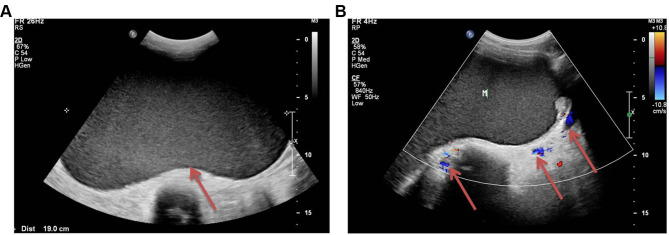
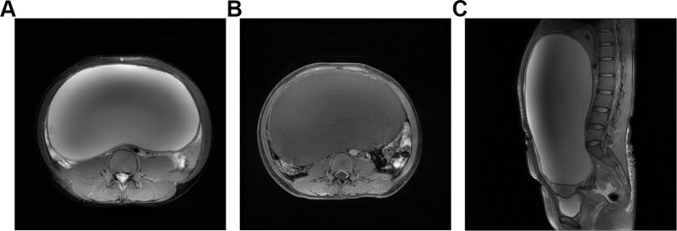
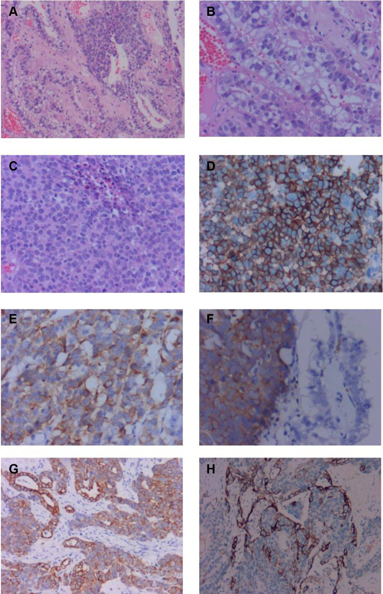
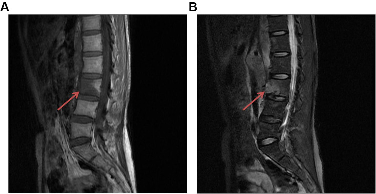
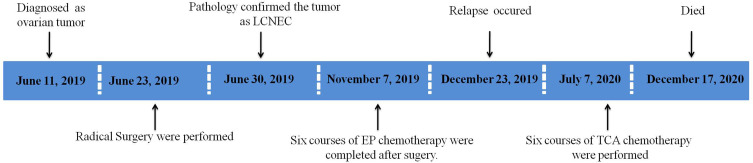
Large cell neuroendocrine carcinoma (LCNEC) is a rare histological subtype of ovarian cancer. A few cases have been reported in the literature with extreme invasiveness and a poor prognosis. However, there still have not been accepted criteria for diagnosis and treatment of LCNEC. Here we report an unmarried 37 year-old woman who was diagnosed with LCNEC associated with clear cell carcinoma and the tumor index was manifested with a specific increase of AFP. The case received six courses of etoposide and carboplatin chemotherapy as an adjuvant therapy after primary curative surgery. However, she relapsed within 6 months after surgery and metastasized rapidly to distant organs despite combined chemotherapy of paclitaxel, cisplatin, and bevacizumab, and died 18 months after primary surgery. This is the first reported case of LCNEC manifested with a specific increase of AFP and characteristically metastasized to the spine as recurrence. Reviewing our case as well as previously reported cases, LCNEC present with aggressive malignancy and vulnerable to distant metastasis through a hematogenous approach, we conjectured that adding Bevacizumab in primary chemotherapy may be beneficial to extend disease-free survival. But so far there is no recommendation of this regimen for treatment of LCNEC in current guidelines. Further research is needed to confirm this view so as to find the best treatment of LCNEC and improve the prognosis of these patients.

Keywords: large cell neuroendocrine carcinoma; ovarian tumors.


Conflict of interest statement

The authors declare that they have no conflicts of interest in relation to this work.

Figures

Figure 1
Figure 1
Abdominal ultrasound scan showed there was a huge mass in the pelvic and abdominal cavity. (A) The ultrasound showed a huge cystic mass in the pelvic and abdominal cavity (Arrow); (B) A few blood flow signals were found around the mass (Arrow).
Figure 2
Figure 2
Pelvic MRI enhancement scan of the huge mass in the sagittal plane and axial plane. (A) MRI of the mass with increased signal on T2W in the axial plane. (B) MRI of the mass with decreased signal on T1W in the axial plane. (C) MRI of the mass with increased signal on T2W in the sagittal plane.
Figure 3
Figure 3
Pathological and immunohistochemical findings of this case. (A) Microscopic finding (hematoxylin-eosin [HE] staining): Junction area of clear cell carcinoma and large cell neuroendocrine carcinoma (original magnification, ×10). (B) Area of clear cell carcinoma (original magnification, ×20). (C) Area of large cell neuroendocrine carcinoma, poorly-differentiated large cells (original magnification, ×20). (DF) Immunohistochemical studies of this case for representative neuroendocrine markers: CD56 (D: ×20), chromogranin A (E: ×20) and synaptophysin (F: ×20). Immunohistochemical test also found CK and CK20 were positive (G and H×10).
Figure 4
Figure 4
Lumbar MRI enhancement scan showed destruction of L3 vertebra. (A) Destruction of L3 vertebra caused by tumor invasion with decreased signal on T1W in the sagittal plane (Arrow). (B) Destruction of L3 vertebra caused by tumor invasion with increased signal on T2W in the sagittal plane (Arrow).
Figure 5
Figure 5
Timeline of the process of diagnosis and treatments of this case.

Similar articles

Cited by

References

    1. Eichhorn JH, Young RH. Neuroendocrine tumors of the genital tract. Am J Clin Pathol. 2001;115:S94–S112. doi:10.1309/64CW-WKGK-49EF-BYD1 - DOI - PubMed
    1. Chenevert J, Bessette P, Plante M, Tetu B, Dube V. Mixed ovarian large cell neuroendocrine carcinoma, mucinous adenocarcinoma, and teratoma: a report of two cases and review of the literature. Pathol Res Pract. 2009;205(9):657–661. doi:10.1016/j.prp.2009.01.013 - DOI - PubMed
    1. Oshita T, Yamazaki T, Akimoto Y, et al. Clinical features of ovarian large-cell neuroendocrine carcinoma: four case reports and review of the literature. Exp Ther Med. 2011;2(6):1083–1090. doi:10.3892/etm.2011.325 - DOI - PMC - PubMed
    1. Gardner GJ, Reidy-Lagunes D, Gehrig PA. Neuroendocrine tumors of the gynecologic tract: a Society of Gynecologic Oncology (SGO) clinical document. Gynecol Oncol. 2011;122(1):190–198. doi:10.1016/j.ygyno.2011.04.011 - DOI - PubMed
    1. Hirasawa T. Ovarian neuroendocrine carcinoma associated with mucinous carcinoma and teratoma. Nihon Rinsho. 2004;62(5):973–978. - PubMed

Publication types