

IL-33/ST2 Activation Is involved in Ro60-Regulated Photosensitivity in Cutaneous Lupus Erythematosus
- PMID: 35909659
- PMCID: PMC9328989
- DOI: 10.1155/2022/4955761
IL-33/ST2 Activation Is involved in Ro60-Regulated Photosensitivity in Cutaneous Lupus Erythematosus
Abstract
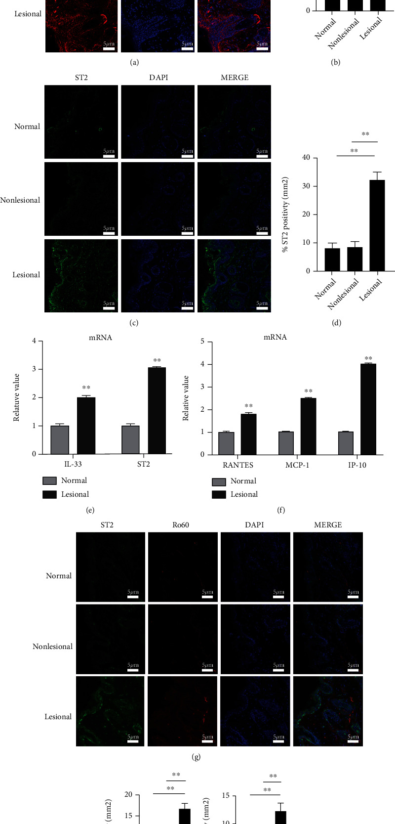
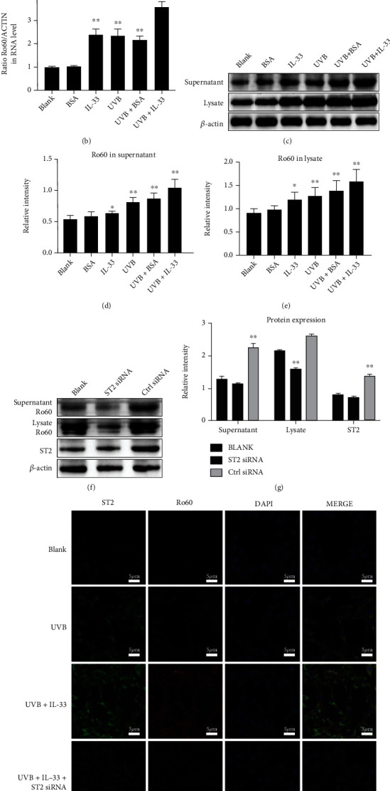
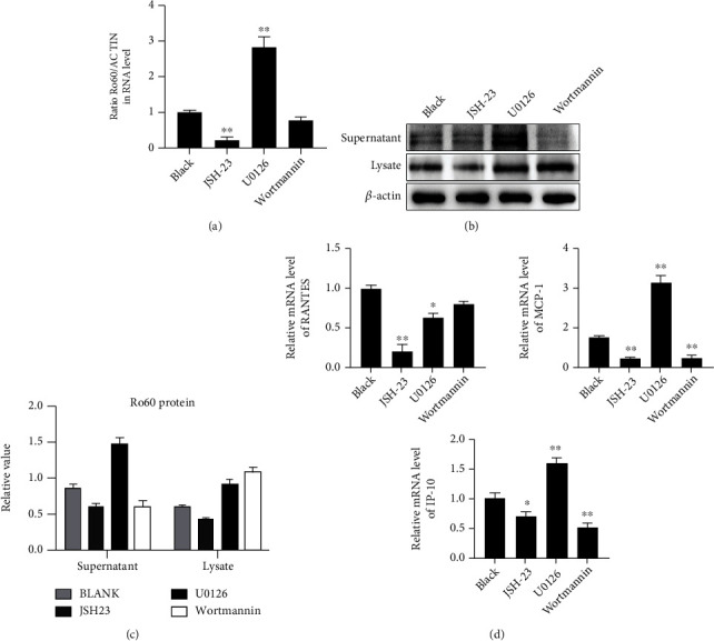
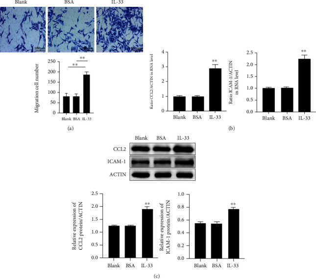
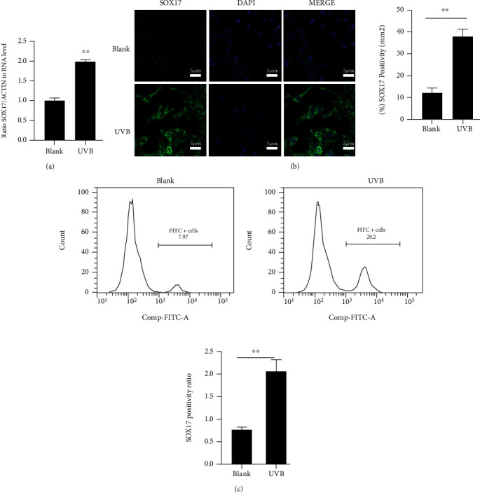
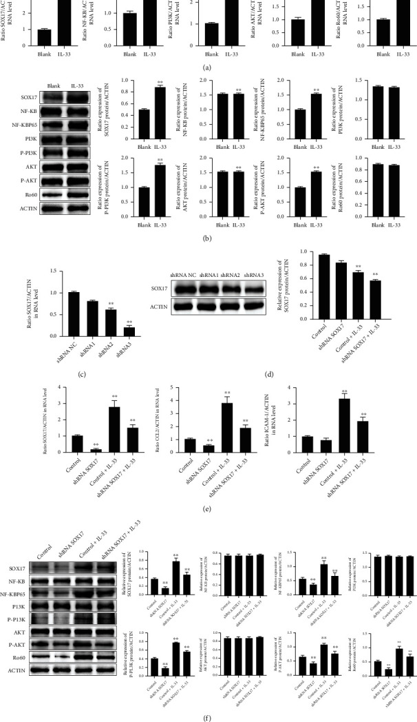
Interleukin- (IL-) 33 contributes to various inflammatory processes. IL-33/ST2 activation participates in systemic lupus erythematous via binding to the receptor of Suppression of Tumorigenicity 2 protein (ST2). However, whether IL-33/ST2 interferes with the nosogenesis of cutaneous lupus erythematosus (CLE) has not been reported so far. Herein, we proposed to disclose the impacts on IL-33/ST2 activation and Ro60 on CLE and their potential implications in the photosensitization of CLE cells. IL-33, ST2, and Ro60 in CLE patients' skin lesions were detected. Murine keratinocytes stimulated with or without IL-33 were irradiated by ultraviolet B (UVB), and the levels of Ro60 and inflammation markers were determined. Keratinocytes were cocultured with J774.2 macrophages and stimulated with IL-33 for analysis of chemostasis. The results identified that IL-33, ST2, and downstream inflammation markers were significantly upregulated in CLE lesions with Ro60 overexpression. Additionally, IL-33 treatment promoted the upregulation of Ro60 induced by UVB treatment in murine keratinocytes. Moreover, IL-33 stimulates keratinocytes to induce macrophage migration via enhancing the generation of the chemokine (C-C motif) ligands 17 and 22. Meanwhile, the silencing of ST2 or nuclear factor-kappa B (NF-κB) suppression abolished IL-33-induced upregulation of Ro60 in keratinocytes. Similarly, the inhibition of SOX17 expression was followed by downregulation of Ro60 in keratinocytes following IL-33 stimulation. In addition, UVB irradiation upregulated SOX17 in keratinocytes. Conclusively, the IL-33/ST2 axis interferes with Ro60-regulated photosensitization via activating the NF-κB- and PI3K/Akt- and SOX17-related pathways.
Copyright © 2022 Yitian Song et al.
Conflict of interest statement
The authors declare that they have no conflicts of interest.
Figures
Similar articles
-
ST2L promotes VEGFA-mediated angiogenesis in gastric cancer by activating TRAF6/PI3K/Akt/NF-κB pathway via IL-33.Sci Rep. 2024 Nov 2;14(1):26393. doi: 10.1038/s41598-024-76763-6. Sci Rep. 2024. PMID: 39488565 Free PMC article.
-
TWEAK/Fn14 Activation Participates in Ro52-Mediated Photosensitization in Cutaneous Lupus Erythematosus.Front Immunol. 2017 May 31;8:651. doi: 10.3389/fimmu.2017.00651. eCollection 2017. Front Immunol. 2017. PMID: 28620393 Free PMC article.
-
Photosensitivity and type I IFN responses in cutaneous lupus are driven by epidermal-derived interferon kappa.Ann Rheum Dis. 2018 Nov;77(11):1653-1664. doi: 10.1136/annrheumdis-2018-213197. Epub 2018 Jul 18. Ann Rheum Dis. 2018. PMID: 30021804 Free PMC article.
-
Ethnic differences in immunogenetic features and photosensitivity of cutaneous lupus erythematosus.Arch Dermatol Res. 2009 Jan;301(1):111-5. doi: 10.1007/s00403-008-0897-3. Epub 2008 Sep 17. Arch Dermatol Res. 2009. PMID: 18797890 Review.
-
The role of cytokines in the pathogenesis of cutaneous lupus erythematosus.Cytokine. 2015 Jun;73(2):326-34. doi: 10.1016/j.cyto.2015.01.031. Epub 2015 Mar 9. Cytokine. 2015. PMID: 25767072 Review.
Cited by
-
Knowledge, attitudes, and practices toward biologics among systemic lupus erythematosus patients: a cross-sectional study.Front Public Health. 2025 Mar 11;13:1445576. doi: 10.3389/fpubh.2025.1445576. eCollection 2025. Front Public Health. 2025. PMID: 40135161 Free PMC article.
-
Topical miRNA Delivery via Elastic Liposomal Formulation: A Promising Genetic Therapy for Cutaneous Lupus Erythematosus (CLE).Int J Mol Sci. 2025 Mar 14;26(6):2641. doi: 10.3390/ijms26062641. Int J Mol Sci. 2025. PMID: 40141283 Free PMC article.
-
sST2 Levels Show No Association with Helicobacter pylori Infection in Asymptomatic Patients: Implications for Biomarker Research.Dig Dis Sci. 2023 Aug;68(8):3293-3299. doi: 10.1007/s10620-023-08005-0. Epub 2023 Jun 20. Dig Dis Sci. 2023. PMID: 37338618 Free PMC article.
References
MeSH terms
Substances
LinkOut - more resources
Full Text Sources
Molecular Biology Databases
