. 2022 Jul 21:2022:6206883.
doi: 10.1155/2022/6206883. eCollection 2022.

Stem Cells from Human Exfoliated Deciduous Teeth Attenuate Atopic Dermatitis Symptoms in Mice through Modulating Immune Balance and Skin Barrier Function

Affiliations

Stem Cells from Human Exfoliated Deciduous Teeth Attenuate Atopic Dermatitis Symptoms in Mice through Modulating Immune Balance and Skin Barrier Function

Hao Xiong et al. Mediators Inflamm. .

Abstract

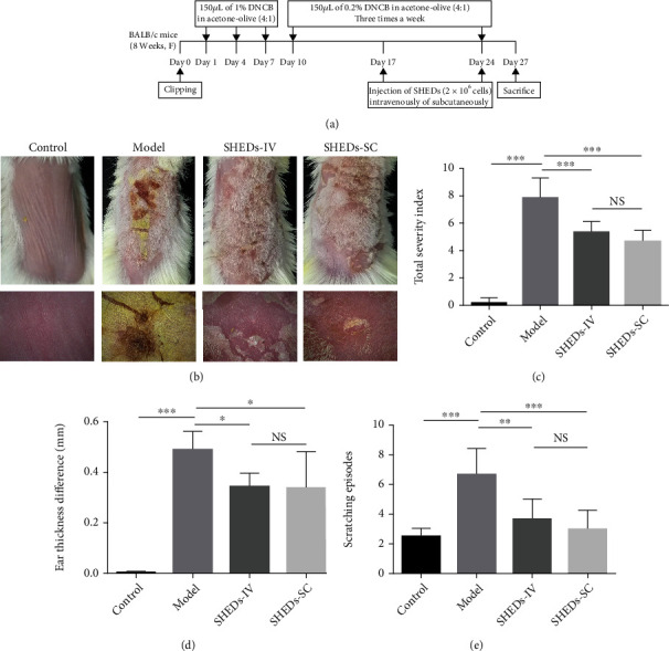
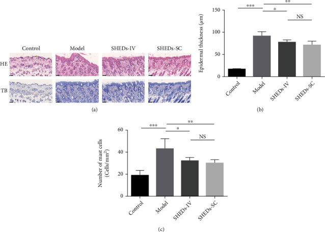
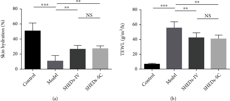
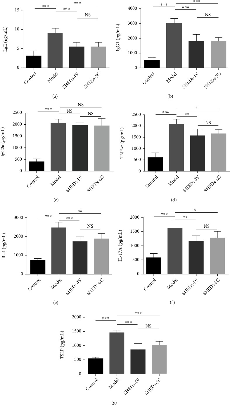
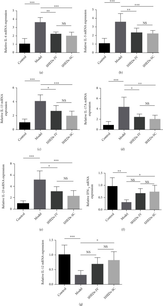
Atopic dermatitis (AD) is a chronic skin inflammatory disease associated with immune abnormalities and disrupted skin barrier function. Mesenchymal stem cells (MSCs) have been suggested as an alternative therapeutic option in AD. Stem cells from human exfoliated deciduous teeth (SHEDs) are a unique postnatal stem cell population with high immunomodulatory properties. The aim of this study was to explore the effects of SHEDs on AD in the BALB/c mouse model induced by 2,4-dinitrochlorobenzene (DNCB). SHEDs were administrated intravenously or subcutaneously, and clinical severity, histopathological findings, skin barrier function, and organ indexes were evaluated. Skin tissue cytokine mRNA levels and serum cytokine protein levels were further analysed. SHED administration significantly alleviated AD clinical severity, including dermatitis scores, ear thickness, scratching behaviour, and infiltration of mast cells. In addition, disrupted skin barrier function and enlarged spleens were restored by SHED administration. Further, SHED treatment reduced the levels of IgE, IgG1, and thymic stromal lymphopoietin (TSLP) in the serum and the modulated expression of Th1-, Th2-, and Th17-associated cytokines in skin lesions. In conclusion, SHEDs attenuated AD-like skin lesions in mice by modulating the immune balance and skin barrier function. SHEDs could be a potential new treatment agent for AD.


Conflict of interest statement

The authors declare no conflict of interest.

Figures

Figure 1
Figure 1
Experimental procedure and effect of SHEDs on the clinical features of AD-like symptoms induced by DNCB in BALB/c mice at the time of sacrifice. (a) Schematic diagram of the experimental procedure for the induction of AD lesions and SHED administration. (b) Cutaneous manifestations in mice in each group. (c) Clinical skin severity scores of AD-like skin lesions in BALB/c mice of each group. The total score is the sum of individual scores determined based on the symptoms of erythema/haemorrhage, scarring/dryness, edema, and excoriation/erosion. (d) Ear thickness difference in BALB/c mice of each group. (e) Scratching behaviour difference in BALB/c mice of each group. All the data are expressed as mean ± SD (n = 6). Three repeated times of each experiment were conducted.
Figure 2
Figure 2
Effect of SHEDs on cutaneous histopathological observations. (a) Representative histologic findings of cutaneous tissue sections stained with hematoxylin and eosin (HE) or toluidine blue (TB) (magnification 200x; scale bar = 100 μm). (b) The thicknesses of the epidermis in BALB/c mice of each group. (c) The number of infiltrated mast cells in 1 mm2 of skin lesion in BALB/c mice of each group. All the data are expressed as mean ± SD (n = 6). Three repeated times of each experiment were conducted.
Figure 3
Figure 3
Effect of SHEDs on skin barrier. (a) Skin hydration in BALB/c mice of each group. (b) Transepidermal water loss (TEWL) in BALB/c mice of each group. All the data are expressed as mean ± SD (n = 6). Three repeated times of each experiment were conducted.
Figure 4
Figure 4
Effect of SHEDs on AD-related immunoglobulin and cytokine expression in serum and skin tissue. (a–c) The immunoglobulin levels of IgE, IgG1, and IgG2a in serum. (d–g) The cytokine levels of TNF-α, IL-4, IL-17A, and TSLP in serum. All the data are expressed as mean ± SD (n = 6). Three repeated times of each experiment were conducted.
Figure 5
Figure 5
Effect of SHEDs on AD-related cytokine expression in skin tissue. (a–e) The relative mRNA levels of Th2- and Th17-mediated cytokines including IL-4, IL-5, IL-13, IL-17A, and IL-23. (f and g) The relative mRNA levels of Th1-mediated cytokines including IFN-γ and IL-12. All the data are expressed as mean ± SD (n = 6). Three repeated times of each experiment were conducted.

Similar articles

Cited by

References

    1. Stander S. Atopic dermatitis. The New England Journal of Medicine . 2021;384(12):1136–1143. doi: 10.1056/NEJMra2023911. - DOI - PubMed
    1. Langan S. M., Irvine A. D., Weidinger S. Atopic dermatitis. Lancet . 2020;396(10247):345–360. doi: 10.1016/S0140-6736(20)31286-1. - DOI - PubMed
    1. Sroka-Tomaszewska J., Trzeciak M. Molecular mechanisms of atopic dermatitis pathogenesis. International Journal of Molecular Sciences . 2021;22(8):p. 4130. doi: 10.3390/ijms22084130. - DOI - PMC - PubMed
    1. Yang X., Kambe N., Takimoto-Ito R., Kabashima K. Advances in the pathophysiology of atopic dermatitis revealed by novel therapeutics and clinical trials. Pharmacology & Therapeutics . 2021;224:p. 107830. doi: 10.1016/j.pharmthera.2021.107830. - DOI - PubMed
    1. Mirershadi F., Ahmadi M., Rezabakhsh A., Rajabi H., Rahbarghazi R., Keyhanmanesh R. Unraveling the therapeutic effects of mesenchymal stem cells in asthma. Stem Cell Research & Therapy . 2020;11(1):p. 400. doi: 10.1186/s13287-020-01921-2. - DOI - PMC - PubMed

LinkOut - more resources