

Proteome Informatics in Tibetan Sheep (Ovis aries) Testes Suggest the Crucial Proteins Related to Development and Functionality
- PMID: 35909681
- PMCID: PMC9334778
- DOI: 10.3389/fvets.2022.923789
Proteome Informatics in Tibetan Sheep (Ovis aries) Testes Suggest the Crucial Proteins Related to Development and Functionality
Abstract
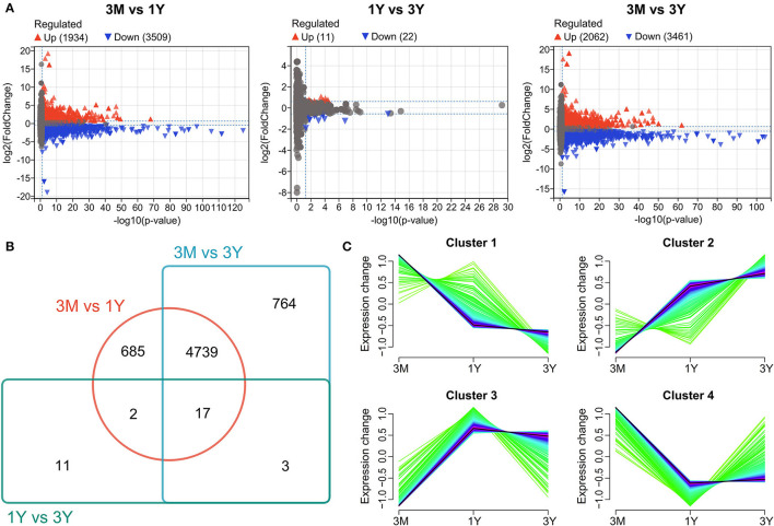
Testis has an indispensable function in male reproduction of domestic animals. Tibetan sheep (Ovis aries) is a locally adapted breed of sheep raised in the Qinghai-Tibet Plateau, with outsized roles in providing the livelihood for millions of residents. Nevertheless, less is known on how protein expression and their functional roles in developmental testes of such breed limit their use in breeding efforts. In this study, we obtained comprehensive protein profiles from testes of Tibetan sheep at three developmental stages (including pre-puberty, post-puberty, and adulthood) using data-independent acquisition-based proteomic strategy to quantitatively identify the differentially abundant proteins (DAPs) associated with testicular development and function and to unravel the molecular basis of spermatogenesis. A total of 6,221 proteins were differentially expressed in an age-dependent manner. The reliability of the gene expression abundance was corroborated by quantitative PCR and targeted parallel reaction monitoring. These DAPs were significantly enriched to biological processes concerning spermatid development and sperm deformation, mitosis, glycolytic process, cell-cell/extracellular matrix (ECM) junctions, cell proliferation, apoptosis, and migration and to the pathways including, developmental process and sexual reproduction-related (such as VEGF, estrogen, insulin, GnRH, Hippo, PI3K-Akt, mTOR, MAPK, and AMPK), and testicular cell events-related pathways (such as tight/gap/adherens junctions, ECM-receptor interaction, regulation of actin cytoskeleton, glycolysis, cell cycle, and meiosis). Based on these bioinformatics analysis, we constructed four protein-protein interaction network, among which the proteins are involved in mitosis, meiosis, spermiogenesis, and testicular microenvironment, respectively. Altogether, these bioinformatics-based sequencing results suggest that many protein-coding genes were expressed in a development-dependent manner in Tibetan sheep testes to contribute to the testicular cell development and their surrounding microenvironment remodeling at various stages of spermatogenesis. These findings have important implications for further understanding of the mechanisms underlying spermatogenesis in sheep and even other plateau-adapted animals.
Keywords: Sertoli cell; data-independent acquisition; germ cell; sheep; spermatogenesis; targeted proteomics; testis.
Copyright © 2022 Li, Wang, Luo, An, Li, Su, Shi, Chen, Zhang and Ma.
Conflict of interest statement
The authors declare that the research was conducted in the absence of any commercial or financial relationships that could be construed as a potential conflictof interest.
Figures


Similar articles
-
Identification and functional characterization of developmental-stage-dependent piRNAs in Tibetan sheep testes.J Anim Sci. 2023 Jan 3;101:skad189. doi: 10.1093/jas/skad189. J Anim Sci. 2023. PMID: 37282774 Free PMC article.
-
The regulatory role of BMP4 in testicular Sertoli cells of Tibetan sheep.J Anim Sci. 2023 Jan 3;101:skac393. doi: 10.1093/jas/skac393. J Anim Sci. 2023. PMID: 36440761 Free PMC article.
-
Integration analysis of transcriptome and metabolome revealed the potential mechanism of spermatogenesis in Tibetan sheep (Ovis aries) at extreme high altitude.Genomics. 2024 Nov;116(6):110949. doi: 10.1016/j.ygeno.2024.110949. Epub 2024 Oct 9. Genomics. 2024. PMID: 39389270
-
Nitric oxide and cyclic nucleotides: their roles in junction dynamics and spermatogenesis.Oxid Med Cell Longev. 2008 Oct-Dec;1(1):25-32. doi: 10.4161/oxim.1.1.6856. Oxid Med Cell Longev. 2008. PMID: 19794905 Free PMC article. Review.
-
Multiple signaling pathways in Sertoli cells: recent findings in spermatogenesis.Cell Death Dis. 2019 Jul 17;10(8):541. doi: 10.1038/s41419-019-1782-z. Cell Death Dis. 2019. PMID: 31316051 Free PMC article. Review.
Cited by
-
Quantitative Proteomic Analysis Reveals Key Proteins Involved in Testicular Development of Yaks.Int J Mol Sci. 2024 Aug 2;25(15):8433. doi: 10.3390/ijms25158433. Int J Mol Sci. 2024. PMID: 39126002 Free PMC article.
-
Semen Quality, Testicular Cell Apoptosis, and Transcriptome Analysis Following Mild Scrotal Heat Stress in Wugu-Hu Crossbred and Hu Rams.Animals (Basel). 2025 Mar 3;15(5):724. doi: 10.3390/ani15050724. Animals (Basel). 2025. PMID: 40076007 Free PMC article.
-
Identification of postnatal development dependent genes and proteins in porcine epididymis.BMC Genomics. 2023 Dec 4;24(1):729. doi: 10.1186/s12864-023-09827-y. BMC Genomics. 2023. PMID: 38049726 Free PMC article.
References
LinkOut - more resources
Full Text Sources
Miscellaneous