. 2022 Jul 15:13:932793.
doi: 10.3389/fpls.2022.932793. eCollection 2022.

Highly Overexpressed AtC3H18 Impairs Microgametogenesis via Promoting the Continuous Assembly of mRNP Granules

Affiliations

Highly Overexpressed AtC3H18 Impairs Microgametogenesis via Promoting the Continuous Assembly of mRNP Granules

Liai Xu et al. Front Plant Sci. .

Abstract

Plant CCCH zinc-finger proteins form a large family of regulatory proteins function in many aspects of plant growth, development and environmental responses. Despite increasing reports indicate that many CCCH zinc-finger proteins exhibit similar subcellular localization of being localized in cytoplasmic foci, the underlying molecular mechanism and the connection between this specific localization pattern and protein functions remain largely elusive. Here, we identified another cytoplasmic foci-localized CCCH zinc-finger protein, AtC3H18, in Arabidopsis thaliana. AtC3H18 is predominantly expressed in developing pollen during microgametogenesis. Although atc3h18 mutants did not show any abnormal phenotype, possibly due to redundant gene(s), aberrant AtC3H18 expression levels caused by overexpression resulted in the assembly of AtC3H18-positive granules in a dose-dependent manner, which in turn led to male sterility phenotype, highlighting the importance of fine-tuned AtC3H18 expression. Further analyzes demonstrated that AtC3H18-positive granules are messenger ribonucleoprotein (mRNP) granules, since they can exhibit liquid-like physical properties, and are associated with another two mRNP granules known as processing bodies (PBs) and stress granules (SGs), reservoirs of translationally inhibited mRNAs. Moreover, the assembly of AtC3H18-positive granules depends on mRNA availability. Combined with our previous findings on the AtC3H18 homologous genes in Brassica campestris, we concluded that appropriate expression level of AtC3H18 during microgametogenesis is essential for normal pollen development, and we also speculated that AtC3H18 may act as a key component of mRNP granules to modulate pollen mRNAs by regulating the assembly/disassembly of mRNP granules, thereby affecting pollen development.

Keywords: Arabidopsis thaliana; AtC3H18; mRNP granules; pollen development; processing bodies (PBs); stress granules (SGs).


Conflict of interest statement

The authors declare that the research was conducted in the absence of any commercial or financial relationships that could be construed as a potential conflict of interest. The handling editor declared a past collaboration with one of the authors, LH.

Figures

FIGURE 1
FIGURE 1
Temporal and spatial expression pattern of AtC3H18. (A) Structure of AtC3H18 encoded protein. CCCH, CCCH zinc-finger motif; LOTUS, Limkain, Oskar, and TUdor-containing proteins 5 and 7; RRM, RNA-recognition motif; LCR, low complexity region; NES, Nuclear export signal. (B) Relative expression analysis showed that AtC3H18 is highly expressed in pollen. The data was downloaded from Arabidopsis eFP Browser. Values are mean ± SD. R, root; Ste, stem; VeR, vegetative rosette; CaL, cauline leaf; Fl, flower stage 12; Si, seeds stage 5 w/siliques; Car, carpels at Fl; Pe, petals at Fl; Se, sepals at Fl; Sta, stamens at Fl; MP, mature pollen. GUS staining of ProAtC3H18:GUS transgenic plants in inflorescence (C), floral buds (D), sepal (E), petal (F), mature anther (G), mature unfertilized pistil (H), wild-type pistil hand-pollinated with ProAtC3H18:GUS pollen (I), ProAtC3H18:GUS pistil hand-pollinated with wild-type pollen (J), mature pollen grains (K), germinated pollen (L), 7-d-old seedling (M), cauline leaf (N), stem (O) and silique (P). (Q) GFP fluorescence intensity of ProAtC3H18:GFP transgenic anthers. (R) GFP fluorescence signal in germinated pollen. EMS, MMS, LMS, early, mid, late microspore; EBP, LBP, early, late bicellular pollen; TCP, tricellular pollen; MPG, mature pollen grain. Bars = 2 mm in (C), 200 μm at stage 9 and stage 10 in (D), (E) to (J), 500 μm at stage 11 to stage 13 in (D), 20 μm in (K) and (L), 1 mm in (M) to (P), 100 μm in (Q), and 10 μm in (R).
FIGURE 2
FIGURE 2
Overexpression of AtC3H18 results in three types of pollen phenotypes. (A) Alexander staining of a representative anther from a T1 line of AtC3H18 overexpression transgenic plants. (B) Alexander staining of anthers and scanning electron microscopy (SEM) of pollen grains produced by three types of AtC3H18 overexpression transgenic plants named Type-I, Type-II and Type-III, respectively. (C) qRT-PCR analysis of AtC3H18 in inflorescences of WT, Type-I, Type-II, and Type-III transgenic plants. TUB4 was used as the normalization control, and the expression of AtC3H18 in WT was set as 1. Values are mean ± SD. (D) Seed number per silique. Error bars represent SD, n = 30. WT, wild-type; Type-I/II/III, Type-I/II/III ProAtC3H18:AtC3H18-GFP transgenic plants; ProAtC3H18:GFP, ProAtC3H18:GFP control line; ♀Type-II/III X ♂. WT, emasculated flowers of Type-II/III plants cross-pollinated with wild-type pollen. Asterisks on columns indicate statistically significant differences from the WT calculated using Student’s t-test: *, P ≤ 0.05; **, P ≤ 0.01; and ***, P ≤ 0.001. (E) Representative images of fully development siliques. Bar = 2 mm.
FIGURE 3
FIGURE 3
Semi-thin transverse sections of AtC3H18 overexpression transgenic anthers. Semi-thin sections of anthers from the wild-type (A–F), Type-I (G–L), Type-II (M–R) and Type-III (S–X) of ProAtC3H18:AtC3H18-GFP transgenic plants at anther development stage 5 (A,G,M,S), stage 7 (B,H,N,T), stage 8 (C,I,O,U), stage9/10 (D,J,P,V), stage 11 (E,K,Q,W), and stage 12/13 (F,L,R,X). Asterisks in (V) indicate microspores with less cytoplasm. MMC, microspore mother cell; Msp, microspore; P, pollen; dP, degraded pollen; T, tapetum; Tds, tetrads. Bars = 25 μm.
FIGURE 4
FIGURE 4
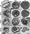
Transmission electron microscopy (TEM) of different types of AtC3H18 overexpression transgenic pollen. Ultrastructure of microspores at different developmental stages from the wild-type (A–C), Type-I (D–F), Type-II (G–I), and Type-III (J–L) of ProAtC3H18:AtC3H18-GFP transgenic plants. Late-UNM, late uninucleate microspore; BCP, bicellular pollen; TCP, tricellular pollen; N, nucleus; V, Vacuole; VN, vegetative nucleus. Bars = 5 μm.
FIGURE 5
FIGURE 5
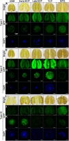
Observation of pollen nucleus development and AtC3H18-GFP fusion protein in anthers and pollen grains of different types of AtC3H18 overexpression transgenic plants. Anthers and pollen grains from different developmental stages of Type-I (A–D), Type-II (E–H), and Type-III (I–L) of ProAtC3H18:AtC3H18-GFP transgenic plants were displayed. GFP images of pollen showed the formation of AtC3H18-positive granules (C,G,K). DAPI staining labeled pollen nuclei (D,H,L). Images in (C,D,G,H,K,L) were merged from multiple cutting planes. UNM, uninucleate microspores; Early/Late-BCP, early/late-bicellular pollen; TCP, immature tricellular pollen; MPG, mature pollen grain. Bars = 100 μm in Bright and GFP images of anthers, 5 μm in GFP and DAPI staining images of pollen.
FIGURE 6
FIGURE 6
The elevated expression of AtC3H18 can promote the formation of AtC3H18-positive granules in pollen. (A) Anthers from T1 lines of AtC3H18 overexpression transgenic plants usually contain wild-type pollen, Type-I, Type-II and Type-III transgenic pollen, simultaneously. Bars = 20 μm. (B–F) The scatter plots showed the relationships between the number of AtC3H18-positive granules and the fluorescence intensity of transgenic pollen produced by the T3 lines of AtC3H18 overexpression transgenic plants, named OE#3-2-11 (B), OE#9-2-2 (C), OE#9-2-4 (D), OE#21-1-3 (E), and OE#21-1-4 (F), respectively. Each point represented an individual pollen, and at least 60 pollen were analyzed for each plants. Lines in OE#3-2-11 (B), OE#9-2-4 (D) and OE#21-1-4 (F) depicted the degree of linear regression. (G) A heat shock of 37°C for 30 min promotes the formation of large AtC3H18-positive granules. Bars = 5 μm.
FIGURE 7
FIGURE 7
AtC3H18 can co-localize with PB and SG marker proteins during heat stress. Co-localization images of free-GFP and GFP-AtC3H18 fusion protein with RFP-DCP2 (A,B,E,F) or RFP-PABP8 (C,D,G,H) in N. benthamiana leaf epidermal cells at room temperature (RT, A–D) and after heat treatment at 42°C for 90 min (E–H), respectively. Pictures represent epifluorescence (GFP and RFP) and merged images (Merge). All the images shown here are the magnified images of the areas depicted by the white frames in Supplementary Figure 7. All Images were merged from multiple cutting planes. Bars = 25 μm. (I–L), Quantitative analysis of co-localization.
FIGURE 8
FIGURE 8
The formation process and the highly dynamic nature of AtC3H18-positive granules. (A) The highly dynamic formation of AtC3H18-positive granules during heat stress. Pictures represent white field images (Bright), epifluorescence (GFP-AtC3H18 and RFP-DCP2), merged images (Merge), and magnified images in Merge [Merge (zoom)]. Bars = 25 μm. (B) Shapes of AtC3H18-positive granules in N. benthamiana leaf epidermal cells during heat treatment. Bars = 5 μm. (C) FRAP images showed fluorescence recovery of two representative AtC3H18-positive granules at different time points in vivo. Red arrowhead marks photobleaching events, red circles represent the bleaching areas. Bars = 5 μm. (D) FRAP recovery curves of two representative AtC3H18-positive granules in (C). Red arrowheads mark photobleaching events. (E) Montages of AtC3H18-positive granules deforming and fusing during heat treatment. The next two rows of images are the enlargements of the first row. Bars = 20 μm in the first line and 5 μm in the second and third lines. All Images were captured from a single cutting plane.
FIGURE 9
FIGURE 9
LOTUS, RRM and N-terminus are responsible for targeting AtC3H18 to the cytoplasmic foci, and mRNAs are required for the formation of AtC3H18-poisitive granules. (A) Schematic diagram of AtC3H18 protein structure and the constructs used for the subcellular localization analyzes of truncated AtC3H18. (B) The localization of CCCH zinc finger motif, LOTUS domain and RRM domain in N. benthamiana leaf epidermal cells at room temperature (RT) and after heat stress. (C) The localization of N-Terminus (N-Ter, amino acids 1 to 158) and C-Terminus (C-Ter, amino acids 291 to 317 and 389 to 536). The localization of AtC3H18 (D) and the quantification of granules (E) in young roots of ProAtC3H18:AtC3H18-GFP transgenic seedlings after heat treatment and after 3 h recovery. The localization of AtC3H18 (F) and the quantification of granules (G) in young roots of ProAtC3H18:AtC3H18-GFP transgenic seedlings after heat treatment with or without CHX treatment. Pictures represent at least three independent analyzes. (E,G), n = 5 biological repeats, mean ± SD. ***P < 0.001. All Images were merged from multiple cutting planes. Bars = 25 μm.

Similar articles

Cited by

References

    1. Aizer A., Kalo A., Kafri P., Shraga A., Ben-Yishay R., Jacob A., et al. (2014). Quantifying mRNA targeting to P-bodies in living human cells reveals their dual role in mRNA decay and storage. J. Cell Sci. 127 4443–4456. 10.1242/jcs.152975 - DOI - PubMed
    1. Alberti S., Gladfelter A., Mittag T. (2019). Considerations and Challenges in Studying Liquid-Liquid Phase Separation and Biomolecular Condensates. Cell 176 419–434. 10.1016/j.cell.2018.12.035 - DOI - PMC - PubMed
    1. Anantharaman V., Zhang D., Aravind L. (2010). OST-HTH: a novel predicted RNA-binding domain. Biol. Direct 5:13. 10.1186/1745-6150-5-13 - DOI - PMC - PubMed
    1. Anderson P., Kedersha N. (2006). RNA granules. J. Cell Biol. 172 803–808. - PMC - PubMed
    1. Anderson P., Kedersha N. (2008). Stress granules: the Tao of RNA triage. Trends Biochem. Sci. 33 141–150. 10.1016/j.tibs.2007.12.003 - DOI - PubMed

LinkOut - more resources