. 2022 Jul 14:13:942261.
doi: 10.3389/fphar.2022.942261. eCollection 2022.

Exogenous proline enhances susceptibility of NSCLC to cisplatin via metabolic reprogramming and PLK1-mediated cell cycle arrest

Affiliations

Exogenous proline enhances susceptibility of NSCLC to cisplatin via metabolic reprogramming and PLK1-mediated cell cycle arrest

Bingjie Han et al. Front Pharmacol. .

Abstract

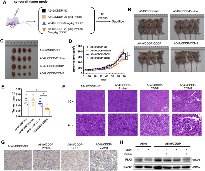
The occurrence of cisplatin resistance is still the main factor limiting the therapeutic effect of non-small cell lung cancer (NSCLC). It is urgent to elucidate the resistance mechanism and develop novel treatment strategies. Targeted metabolomics was first performed to detect amino acids' content in cisplatin-resistant cancer cells considering the relationship between tumour metabolic rearrangement and chemotherapy resistance and chemotherapy resistance. We discovered that levels of most amino acids were significantly downregulated, whereas exogenous supplementation of proline could enhance the sensitivity of NSCLC cells to cisplatin, evidenced by inhibited cell viability and tumour growth in vitro and xenograft models. In addition, the combined treatment of proline and cisplatin suppressed ATP production through disruption of the TCA cycle and oxidative phosphorylation. Furthermore, transcriptomic analysis identified the cell cycle as the top enriched pathway in co-therapy cells, accompanied by significant down-regulation of PLK1, a serine/threonine-protein kinase. Mechanistic studies revealed that PLK1 inhibitor (BI2536) and CDDP have synergistic inhibitory effects on NSCLC cells, and cells transfected with lentivirus expressing shPLK1 showed significantly increased toxicity to cisplatin. Inhibition of PLK1 inactivated AMPK, a primary regulator of cellular energy homeostasis, ultimately leading to cell cycle arrest via FOXO3A-FOXM1 axis mediated transcriptional inhibition in cisplatin-resistant cells. In conclusion, our study demonstrates that exogenous proline exerts an adjuvant therapeutic effect on cisplatin resistance, and PLK1 may be considered an attractive target for the clinical treatment of cisplatin resistance in NSCLC.

Keywords: NSCLC; cisplatin resistance; metabolic reprogramming; plk1; proline.


Conflict of interest statement

The authors declare that the research was conducted in the absence of any commercial or financial relationships that could be construed as a potential conflict of interest.

Figures

FIGURE 1
FIGURE 1
The amino acid targeted metabolic profiling reveals significantly altered metabolites in A549 cells with cisplatin resistance. (A) Heatmap of differential amino acids quantified in A549 and A549/CDDP cells. (B) Boxplots of the relative concentration distribution of representative differential amino acids, including glutamate, methionine, tyrosine, isoleucine, leucine, valine, tryptophan, phenylalanine, and proline. *p < 0.05, **p < 0.01, ***p < 0.001, ****p < 0.0001.
FIGURE 2
FIGURE 2
Proline supplementation significantly enhanced the sensitivity of cancer cells to cisplatin. (A) The viability of control or proline-treated A549 and A549/CDDP cells exposed to different concentrations of cisplatin for 72 h was analyzed by MTT assay. (B) EdU assay for the effect of 20 mM proline alone or combined with 10 μM CDDP on the proliferation of A549 and A549/CDDP cells after 48 h in vitro. (C) The abundance of ATP in A549 and A549/CDDP cells treated with 20 mM proline, 10 μM CDDP, and their combination for 72 h. (D) The cell cycle distribution of A549 (up) and A549/CDDP (down) cells after 20 mM proline, 5 μM CDDP, and the combination treatment for 48 h was detected by flow cytometry. (E,F) Quantification analysis of cell cycle arrest in (D). (G,H) Analysis of apoptosis in A549 and A549/CDDP cells exposed to 20 mM proline, 5 μM CDDP, and their combination for 72 h by flow cytometry. EdU: 5-ethynyl-2′-deoxyuridine. *p < 0.05, **p < 0.01, ***p < 0.001.
FIGURE 3
FIGURE 3
Transcriptomic profiling reveals the alteration of gene transcription levels in A549 and A549/CDDP cells upon 20 mM proline, 10 μM CDDP, and their combination treatment. (A) PCA analysis of transcriptomic profiles obtained from A549 and A549/CDDP cells exposed to different treatments. Each dot represents a biological repeat. (B) Venn plot shows the overlap of significantly differential genes of four groups (group 1/2/3/4) in A549 and A549/CDDP cells. (C) GO analysis was performed on 108 significantly differential genes corresponding to (B). (D,E) KEGG enrichment pathway analysis of upregulated and downregulated mRNAs in (B). (F) Relative transcription levels of PLK1 in A549 and A549/CDDP cells were detected by qRT-PCR. (G) Effects of proline, CDDP, and their combination on protein levels of p-PLK1 and PLK1. (H) The bands in (G) were quantified and presented as the mean ± SEM of three independent experiments. Statistical significance was determined by a two-tailed, paired Student’s t-test. *p < 0.05, **p < 0.01, ***p < 0.001.
FIGURE 4
FIGURE 4
Combination of proline and cisplatin results in NSCLC growth inhibition in vivo. (A) The xenograft tumour model investigates the effects of 20 g/kg proline, 3 mg/kg cisplatin and their combined therapy on cisplatin-resistant A549 cells. (B) Representative imaging of mice 10 weeks after injection of A549/CDDP cells upon different treatments. (C) Combined treatment of proline and CDDP effectively inhibited A549/CDDP cell’s subcutaneous tumour growth in nude mice. (D) The tumour volume was monitored every 3 days to generate the tumour growth curves. (E) The tumours were extracted and weighed. (F) H&E-stained tumour sections. (G) Sections of tumours were stained with PLK1 antibody by immunohistochemical analysis. (H) Detection of protein expression levels of PLK1 in various groups by western blotting. The data are the means ± SEMs of three independent experiments. *p < 0.05.
FIGURE 5
FIGURE 5
The knockdown of PLK1 enhances the efficacy of CDDP by inducing cell cycle arrest. (A) CI heatmaps of A549 (up) and A549/CDDP (down) cells were treated with BI2536 and CDDP at the indicated doses. (B) MTT assays were performed to assess cell viability of A549 and A549/CDDP cells stably transfected with PLKO.5 and PLK1 shRNA with or without 5 μM cisplatin treatment for 24, 48, and 72 h. (C) The cell cycle distribution in NC shRNA- and PLK1 shRNA-transfected cells treated with or without 5 μM cisplatin was detected by flow cytometry.
FIGURE 6
FIGURE 6
The expression levels of markers involved in cell cycle arrest induced by PLK1 inhibition. (A) The levels of AMPK, AKT, p-AKT, FoxO3A, p-FoxO3A, FoxM1, P53, and CCNB1 in the parental and cisplatin-resistant A549 cells infected with PLK1 siRNA. (B) The shNC- or shPLK1-transfected A549 and A549/CDDP cells were incubated with 5 μM CDDP to detect the expression levels of PLK1-related proteins. (C) Working model showing the underlying mechanism of proline enhanced cisplatin sensitivity in NSCLC cells.

References

    1. An Y., Wang B., Wang X., Dong G., Jia J., Yang Q., et al. (2020). SIRT1 inhibits chemoresistance and cancer stemness of gastric cancer by initiating an AMPK/FOXO3 positive feedback loop. Cell Death Dis. 11 (2), 115. 10.1038/s41419-020-2308-4 - DOI - PMC - PubMed
    1. Bivona T. G., Doebele R. C. (2016). A framework for understanding and targeting residual disease in oncogene-driven solid cancers. Nat. Med. 22 (5), 472–478. 10.1038/nm.4091 - DOI - PMC - PubMed
    1. Boroughs L. K., DeBerardinis R. J. (2015). Metabolic pathways promoting cancer cell survival and growth. Nat. Cell Biol. 17 (4), 351–359. 10.1038/ncb3124 - DOI - PMC - PubMed
    1. Bray F., Ferlay J., Soerjomataram I., Siegel R. L., Torre L. A., Jemal A., et al. (2018). Global cancer statistics 2018: GLOBOCAN estimates of incidence and mortality worldwide for 36 cancers in 185 countries. Ca. Cancer J. Clin. 68 (6), 394–424. 10.3322/caac.21492 - DOI - PubMed
    1. Butler M., van der Meer L. T., van Leeuwen F. N. (2021). Amino acid depletion therapies: starving cancer cells to death. Trends Endocrinol. Metab. 32 (6), 367–381. 10.1016/j.tem.2021.03.003 - DOI - PubMed