

Discovery of Leishmania Druggable Serine Proteases by Activity-Based Protein Profiling
- PMID: 35910377
- PMCID: PMC9335491
- DOI: 10.3389/fphar.2022.929493
Discovery of Leishmania Druggable Serine Proteases by Activity-Based Protein Profiling
Abstract
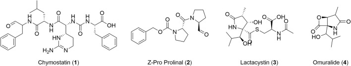
Leishmaniasis are a group of diseases caused by parasitic protozoa of the genus Leishmania. Current treatments are limited by difficult administration, high cost, poor efficacy, toxicity, and growing resistance. New agents, with new mechanisms of action, are urgently needed to treat the disease. Although extensively studied in other organisms, serine proteases (SPs) have not been widely explored as antileishmanial drug targets. Herein, we report for the first time an activity-based protein profiling (ABPP) strategy to investigate new therapeutic targets within the SPs of the Leishmania parasites. Active-site directed fluorophosphonate probes (rhodamine and biotin-conjugated) were used for the detection and identification of active Leishmania serine hydrolases (SHs). Significant differences were observed in the SHs expression levels throughout the Leishmania life cycle and between different Leishmania species. Using iTRAQ-labelling-based quantitative proteomic mass spectrometry, we identified two targetable SPs in Leishmania mexicana: carboxypeptidase LmxM.18.0450 and prolyl oligopeptidase LmxM.36.6750. Druggability was ascertained by selective inhibition using the commercial serine protease inhibitors chymostatin, lactacystin and ZPP, which represent templates for future anti-leishmanial drug discovery programs. Collectively, the use of ABPP method complements existing genetic methods for target identification and validation in Leishmania.
Keywords: ABPP; Leishmania; fluorophosphonate; proteomics; serine protease inhibitors. Discovery of Leishmania druggable serine proteases; serine proteases.
Copyright © 2022 Porta, Isern, Kalesh and Steel.
Conflict of interest statement
The authors declare that the research was conducted in the absence of any commercial or financial relationships that could be construed as a potential conflict of interest.
Figures


Similar articles
-
Profiling Serine Hydrolases in the Leishmania Host-Pathogen Interactome Using Cell-Permeable Activity-Based Fluorophosphonate Probes.Chembiochem. 2025 May 27;26(10):e202500160. doi: 10.1002/cbic.202500160. Epub 2025 May 14. Chembiochem. 2025. PMID: 40146885 Free PMC article.
-
Pretreatment with serine protease inhibitors impairs Leishmania amazonensis survival on macrophages.Parasit Vectors. 2025 Jan 23;18(1):23. doi: 10.1186/s13071-024-06630-w. Parasit Vectors. 2025. PMID: 39849543 Free PMC article.
-
Protease inhibitors in potential drug development for Leishmaniasis.Indian J Biochem Biophys. 2013 Oct;50(5):363-76. Indian J Biochem Biophys. 2013. PMID: 24772958 Review.
-
In vitro anti-leishmanial efficacy of potato tuber extract (PTEx): leishmanial serine protease(s) as putative target.Exp Parasitol. 2014 Nov;146:11-9. doi: 10.1016/j.exppara.2014.08.009. Epub 2014 Aug 14. Exp Parasitol. 2014. PMID: 25128800
-
Understanding serine proteases implications on Leishmania spp lifecycle.Exp Parasitol. 2018 Jan;184:67-81. doi: 10.1016/j.exppara.2017.11.008. Epub 2017 Nov 22. Exp Parasitol. 2018. PMID: 29175018 Review.
Cited by
-
Clickable Probes for Pathogen Proteasomes: Synthesis and Applications.ACS Omega. 2024 Aug 2;9(32):34829-34840. doi: 10.1021/acsomega.4c04316. eCollection 2024 Aug 13. ACS Omega. 2024. PMID: 39157084 Free PMC article.
-
Profiling Serine Hydrolases in the Leishmania Host-Pathogen Interactome Using Cell-Permeable Activity-Based Fluorophosphonate Probes.Chembiochem. 2025 May 27;26(10):e202500160. doi: 10.1002/cbic.202500160. Epub 2025 May 14. Chembiochem. 2025. PMID: 40146885 Free PMC article.
-
Activity-based protein profiling: A graphical review.Curr Res Pharmacol Drug Discov. 2023 Aug 24;5:100164. doi: 10.1016/j.crphar.2023.100164. eCollection 2023. Curr Res Pharmacol Drug Discov. 2023. PMID: 37692766 Free PMC article. Review.
-
Microplate-Based Enzymatic Activity Assay Protocol Powered by Activity-Based Probes.Methods Mol Biol. 2025;2921:119-137. doi: 10.1007/978-1-0716-4502-4_6. Methods Mol Biol. 2025. PMID: 40515987
-
Drug Discovery for Cutaneous Leishmaniasis: A Review of Developments in the Past 15 Years.Microorganisms. 2023 Nov 23;11(12):2845. doi: 10.3390/microorganisms11122845. Microorganisms. 2023. PMID: 38137989 Free PMC article. Review.
References
Grants and funding
LinkOut - more resources
Full Text Sources