

An Assay Combining Droplet Digital PCR With Propidium Monoazide Treatment for the Accurate Detection of Live Cells of Vibrio vulnificus in Plasma Samples
- PMID: 35910629
- PMCID: PMC9335127
- DOI: 10.3389/fmicb.2022.927285
An Assay Combining Droplet Digital PCR With Propidium Monoazide Treatment for the Accurate Detection of Live Cells of Vibrio vulnificus in Plasma Samples
Abstract
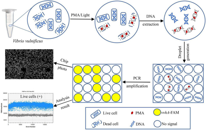
Vibrio vulnificus (V. vulnificus) is one of the most common pathogenic Vibrio species to humans; therefore, the establishment of timely and credible detection methods has become an urgent requirement for V. vulnificus illness surveillance. In this study, an assay combining droplet digital PCR (ddPCR) with propidium monoazide (PMA) treatment was developed for detecting V. vulnificus. The primers/probes targeting the V. vulnificus hemolysin A (vvhA) gene, amplification procedures, and PMA processing conditions involved in the assay were optimized. Then, we analyzed the specificity, sensitivity, and ability to detect live cell DNA while testing the performance of PMA-ddPCR in clinical samples. The optimal concentrations of primers and probes were 1.0 and 0.3 μM, respectively. The annealing temperature achieving the highest accuracy in ddPCR assay was 60°C. With an initial V. vulnificus cell concentration of 108 CFU/mL (colony-forming units per milliliter), the optimal strategy to distinguish live cells from dead cells was to treat samples with 100 μM PMA for 15 min in the dark and expose them to LED light with an output wavelength of 465 nm for 10 min. The specificity of the PMA-ddPCR assay was tested on 27 strains, including seven V. vulnificus strains and 20 other bacterial strains. Only the seven V. vulnificus strains were observed with positive signals in specificity analysis. Comparative experiments on the detection ability of PMA-ddPCR and PMA-qPCR in pure cultures and plasma samples were performed. The limit of detection (LOD) and the limit of quantitation (LOQ) in pure culture solutions of V. vulnificus were 29.33 and 53.64 CFU/mL in PMA-ddPCR, respectively. For artificially clinical sample tests in PMA-ddPCR, V. vulnificus could be detected at concentrations as low as 65.20 CFU/mL. The sensitivity of the PMA-ddPCR assay was 15- to 40-fold more sensitive than the PMA-qPCR in this study. The PMA-ddPCR assay we developed provides a new insight to accurately detect live cells of V. vulnificus in clinical samples, which is of great significance to enhance public health safety and security capability and improve the emergency response level for V. vulnificus infection.
Keywords: Vibrio vulnificus; accurate detection; clinical; droplet digital PCR; propidium monoazide; vvhA gene.
Copyright © 2022 Hu, Fu, Zhang, Pan, Xia, Zhu and Guo.
Conflict of interest statement
JX is employed by Pilot Gene Technologies (Hangzhou) Co., Ltd. The remaining authors declare that the research was conducted in the absence of any commercial or financial relationships that could be construed as a potential conflict of interest.
Figures


Similar articles
-
Absolute quantification of viable Vibrio cholerae in seawater samples using multiplex droplet digital PCR combined with propidium monoazide.Front Microbiol. 2023 Jun 9;14:1149981. doi: 10.3389/fmicb.2023.1149981. eCollection 2023. Front Microbiol. 2023. PMID: 37362935 Free PMC article.
-
Enumeration of Viable Non-Culturable Vibrio cholerae Using Droplet Digital PCR Combined With Propidium Monoazide Treatment.Front Cell Infect Microbiol. 2021 Nov 2;11:753078. doi: 10.3389/fcimb.2021.753078. eCollection 2021. Front Cell Infect Microbiol. 2021. PMID: 34796126 Free PMC article.
-
Real-time PCR detection of Vibrio vulnificus in oysters: comparison of oligonucleotide primers and probes targeting vvhA.Appl Environ Microbiol. 2005 Oct;71(10):5702-9. doi: 10.1128/AEM.71.10.5702-5709.2005. Appl Environ Microbiol. 2005. PMID: 16204478 Free PMC article.
-
Development of a sensitive and specific qPCR assay in conjunction with propidium monoazide for enhanced detection of live Salmonella spp. in food.BMC Microbiol. 2013 Dec 1;13:273. doi: 10.1186/1471-2180-13-273. BMC Microbiol. 2013. PMID: 24289661 Free PMC article.
-
A Propidium Monoazide (PMAxx)-Droplet Digital PCR (ddPCR) for the Detection of Viable Burkholderia cepacia Complex in Nuclease-Free Water and Antiseptics.Microorganisms. 2022 Apr 30;10(5):943. doi: 10.3390/microorganisms10050943. Microorganisms. 2022. PMID: 35630385 Free PMC article.
Cited by
-
Evaluation of the diagnostic performance of an immunochromatographic test for Chlamydia trachomatis.Pract Lab Med. 2024 May 27;40:e00412. doi: 10.1016/j.plabm.2024.e00412. eCollection 2024 May. Pract Lab Med. 2024. PMID: 38867761 Free PMC article.
-
A Novel RAA Combined Test Strip Method Based on Dual Gene Targets for Pathogenic Vibrio vulnificus in Aquatic Products.Foods. 2023 Sep 28;12(19):3605. doi: 10.3390/foods12193605. Foods. 2023. PMID: 37835259 Free PMC article.
-
Public health aspects of Vibrio spp. related to the consumption of seafood in the EU.EFSA J. 2024 Jul 23;22(7):e8896. doi: 10.2903/j.efsa.2024.8896. eCollection 2024 Jul. EFSA J. 2024. PMID: 39045511 Free PMC article.
References
-
- Álvarez-Contreras A. K., Quiñones-Ramírez E. I., Vázquez-Salinas C. (2021). Prevalence, detection of virulence genes and antimicrobial susceptibility of pathogen Vibrio species isolated from different types of seafood samples at “La Nueva Viga” market in Mexico City. Antonie. Van. Leeuwenhoek. 114 1417–1429. 10.1007/s10482-021-01591-x - DOI - PubMed
LinkOut - more resources
Full Text Sources