. 2022 Jul 18;12(12):5596-5614.
doi: 10.7150/thno.73650. eCollection 2022.

Turmeric-derived nanovesicles as novel nanobiologics for targeted therapy of ulcerative colitis

Affiliations

Turmeric-derived nanovesicles as novel nanobiologics for targeted therapy of ulcerative colitis

Caifang Gao et al. Theranostics. .

Abstract

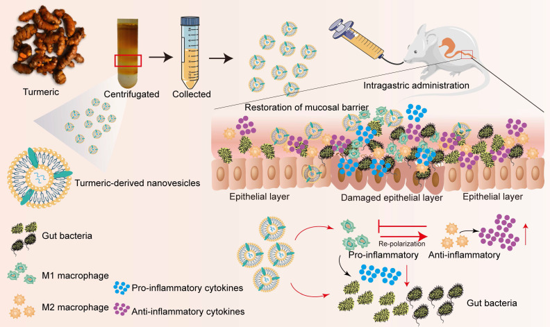
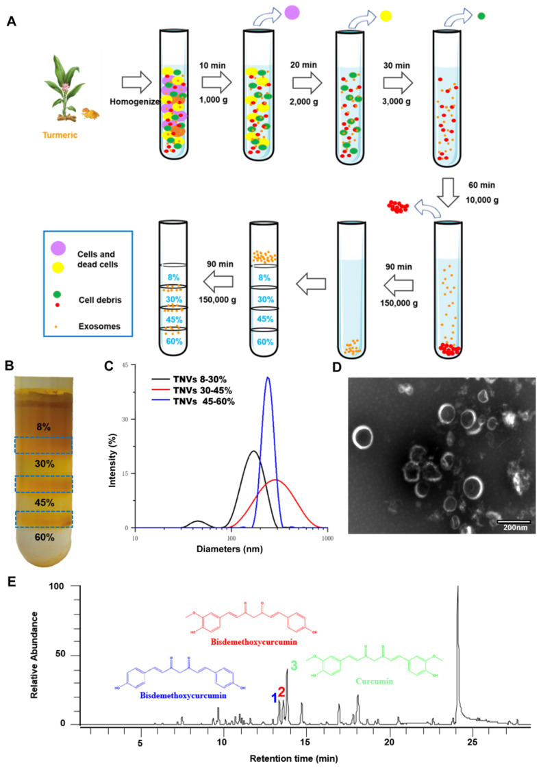
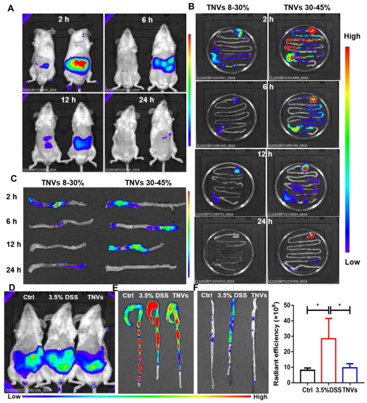
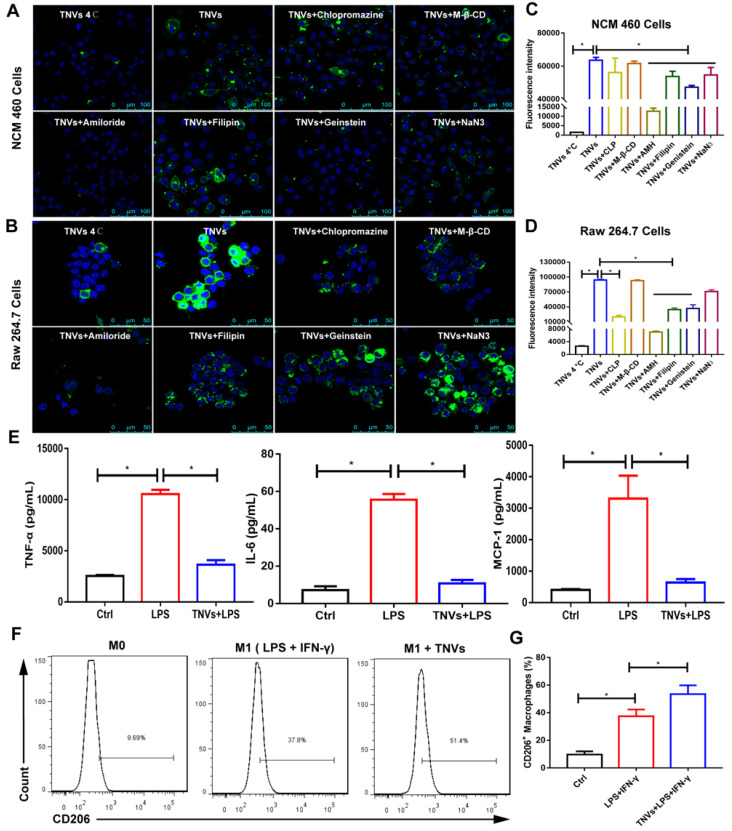
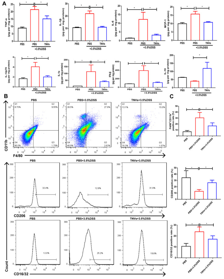
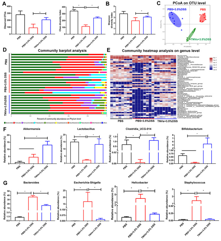
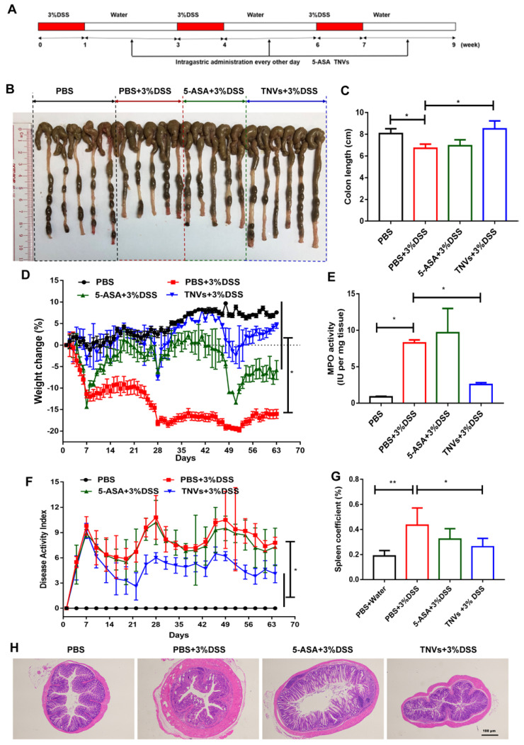
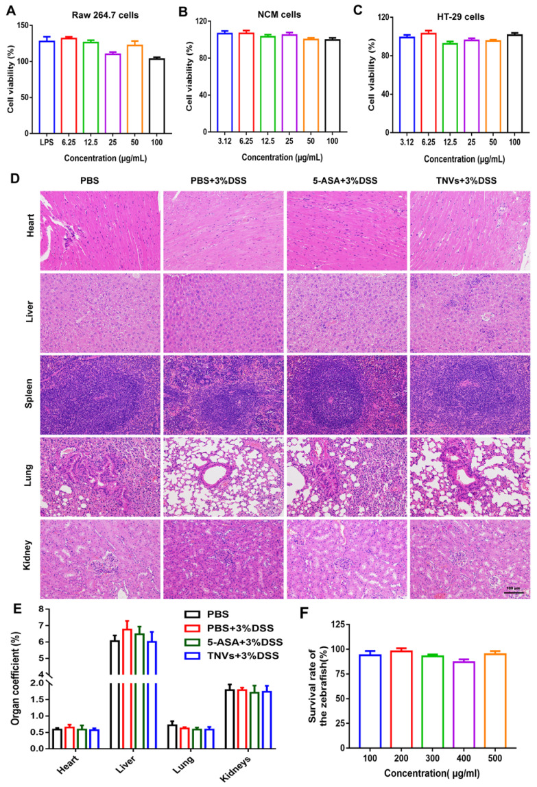
Rationale: Ulcerative colitis (UC), a typical kind of inflammatory bowel disease (IBD), is an idiopathic chronic intestinal inflammation. Conventional therapeutic strategies mainly focus on the rebalance of pro-inflammation and anti-inflammation cytokines, whereas targeting damaged intestinal barriers, imbalanced intestinal microbiota and dysregulated mucosal immune responses in UC remain a big challenge. The objective of this study was to develop turmeric-derived nanovesicles (TNVs) for alleviation of colitis and explore the underlying mechanisms. Methods: TNVs were isolated and purified through differential centrifugation. The targeted ability was evaluated on the dextran sulfate sodium (DSS)-induced mouse model by IVIS imaging system. The anti-inflammation efficacy was studied in lipopolysaccharide (LPS)-induced macrophages and DSS-induced acute and chronic colitic mouse model. In addition, the influence of TNVs on the intestinal microbiota was investigated via 16S rRNA microbiome sequence and the condition of macrophage polarization after TNVs treatment was analyzed by flow cytometry. Results: TNVs were isolated and characterized as nano-size spheroids. The IVIS imaging experiment indicated that orally administrated TNVs could accumulate in the inflamed colon sites and exhibited superior anti-inflammatory activity both in vitro and in vivo. The 16S rRNA sequencing suggested the important role of TNVs in the regulation of gut microbiota. Further, TNVs could promote the transformation of M1 phenotype to M2 macrophages and restore the damaged intestinal epithelium barrier to exert the anti-colitis efficacy. Conclusion: Collectively, oral administration of TNVs exhibited excellent anti-inflammatory efficacy through restoring the damaged intestinal barrier, regulating the gut microbiota and reshaping the macrophage phenotype. This study sheds light on the application of natural exosome-like nanovesicles for the treatment of UC.

Keywords: Fresh herbs; Gut microbiota; Macrophage polarization; Turmeric-derived nanovesicles; Ulcerative colitis.


Conflict of interest statement

Competing Interests: The authors have declared that no competing interest exists.

Figures

Scheme 1
Scheme 1
Schematic illustration of the preparation procedure and anti-colitic efficacy of TNVs. TNVs exert potent anti-inflammation activity via restoring the damaged intestinal barrier, modulating the intestinal microbiota and altering the aberrant mucosa immune environment, especially reshaping the macrophage polarization condition.
Figure 1
Figure 1
Isolation and characterization of turmeric-derived nanovesicles (TNVs). A. The isolation and purification scheme. B. The representative image of purified TNVs after sucrose gradient centrifugation. C. The size distribution of TNVs from various bands between different sucrose concentration. D. The transmission electron microscopy of TNVs between 30-45% sucrose solution. E. The LC-MS spectrum of curcuminoids in TNVs.
Figure 2
Figure 2
In vivo and ex vivo distribution of DiR-labeled TNVs. A. Whole body imaging of the mice at 2, 6, 12 and 24 h after oral administration of TNVs originated from 8%-30% (left) and 30%-45% (right) band. B. The distribution of DiR-labeled TNVs in the gastrointestinal tract at different time points. C. The distribution of DiR-labeled TNVs in the distal colons at different time point. D. Whole body imaging of the mice (healthy, DSS-induced and TNVs treated) at 6 h with DiR-labeled TNVs treatment. E-F. The accumulation and radiant efficiency statistics of DiR-labeled TNVs in the feces-contained and no-feces colon dissected from mice at different condition. Data are shown as mean ± SD, n = 3. Significance as *P < 0.05.
Figure 3
Figure 3
In vitro cell internalization mechanisms, anti-inflammatory properties and the effect on the macrophage polarization of TNVs. A-B. Representative fluorescence images of cellular uptake of DiO-labeled TNVs in NCM 460 cells and Raw 264.7 macrophages after co-incubation for 6 h and additional different inhibitors treatment 1 h. C-D. Fluorescence intensity of cellular uptake of DiO-labeled TNVs in NCM 460 cells and Raw 264.7 macrophages determined using flow cytometry. E. In vitro anti-inflammatory activities of TNVs on Raw 264.7 macrophages. The concentrations of pro-inflammatory cytokines (TNF-α, IL-6, and MCP-1) were quantified using ELISA assay. F-G. Flowcytometry analysis of CD206 expression in Raw 264.7 macrophages. Data are shown as mean ± SD, n = 3. Significance as *P < 0.05.
Figure 4
Figure 4
TNVs alleviated colitis-related symptoms in DSS-induced acute colitic mice. A. The flow chart represents the establishment of mice colitis model and drug treatment design. B-C. Representative images of colons and average colon length from different groups. D. Body weight change expressed as the percentage of the day-zero weight. E. DAI, F. Colonic MPO activity, G. Representative histological sections of distal colons stained with H&E (4× and 10× magnification). Data shown as means ± SD, n = 8. The principle of determining disease activity index (DAI) is the summation of the body weight loss (0- <1%, 1- 1~5%, 2- 5~10%, 3- 10~20%, 4- > 20%), stool consistency (0- normal, 2- loose, 4- diarrhea) and rectal bleeding (0- normal, 2- positive finding, 4- gross bleeding).
Figure 5
Figure 5
TNVs mitigated intestinal inflammation responses and reshaped macrophage differentiation in colonic lamina propria. A. Cytokine productions of TNF-α, IL-1β, IL-6, MCP-1, IL-12p70, IL-1α, IFN-β and IL-10 in colon tissues. B-C. The FACS analysis of F4/80+CD11b+macrophage and the positive rate of CD16/32 and CD206 gated on F4/80+CD11b+ macrophages from colonic lamina propria in different groups. Data are shown as means ± SD; n = 5. Significance as *P < 0.05.
Figure 6
Figure 6
TNVs regulated the composition of intestinal microbiota. A-B. Estimation of microbial community observed operational taxonomic units (OTU) richness and α-diversity (chao and shannon index). C. PCoA plot illustrating the intestinal microbiota β-diversity. D. Relative abundance of intestinal microbiota. Phylum-level taxonomy are presented as a percentage of total sequences. E. Heatmap of the relative abundance of genus-level taxonomy for each mouse. F-G. Relative abundance of representative beneficial and harmful taxa. Data are shown as means ± SD; n = 8. Significance as *P < 0.05.
Figure 7
Figure 7
TNVs extenuated colitis-related symptoms in DSS-induced chronic colitic mice. A. The flow chart represents the establishment of mice colitis model and drug treatment design. B-C. Representative images of colons and average colon length from different groups. D. Body weight change expressed as the percentage of the day-zero weight. E. Colonic MPO activity, F. DAI, G. Spleen coefficient, and H. histological sections of distal colons stained with H&E (4× magnification). Data shown as means ± SD, n = 5. Significance as *P < 0.05.
Figure 8
Figure 8
In vitro and in vivo biosafety evaluation of TNVs. A-C. Viabilities of Raw 264.7, NCM 460 and HT-29 cells after incubation with TNVs at different concentrations for 24 h, n = 6. D. H&E staining of main organs slices from mice H&E staining of main organs slices from mice treated with TNVs. E. The organs coefficients, n = 5, and F. The survival rate of zebrafish after treatment with TNVs at different concentrations for 24 h, n = 15. Data shown as means ± SD.

Similar articles

Cited by

References

    1. Xavier RJ, Podolsky DK. Unravelling the pathogenesis of inflammatory bowel disease. Nature. 2007;448:427–34. - PubMed
    1. Kaplan GG, Ng SC. Understanding and preventing the global increase of inflammatory bowel disease. Gastroenterology. 2017;152:313–321.e2. - PubMed
    1. Hanauer SB. Update on the etiology, pathogenesis and diagnosis of ulcerative colitis. Nat Clin Pract Gastroenterol Hepatol. 2004;1:26–31. - PubMed
    1. Neurath MF. Current and emerging therapeutic targets for IBD. Nat Rev Gastroenterol Hepatol. 2017;14:269–278. - PubMed
    1. Dubinsky MC. Azathioprine, 6-mercaptopurine in inflammatory bowel disease: pharmacology, efficacy, and safety. Clin Exp Gastroenterol Hepatol. 2004;2:731–743. - PubMed

Publication types