A Fatal Case Coronavirus Disease 2019 - Associated Acute Hemorrhagic Necrotizing Encephalopathy
- PMID: 35910821
- PMCID: PMC9336601
- DOI: 10.4103/jgid.jgid_185_20
A Fatal Case Coronavirus Disease 2019 - Associated Acute Hemorrhagic Necrotizing Encephalopathy
Abstract
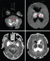
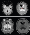
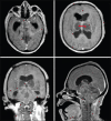
Coronavirus disease 2019 (COVID-19) has been reported in association with a variety of brain imaging findings such as acute hemorrhagic necrotizing encephalopathy. To the best of our knowledge, we are reporting a second case of acute necrotizing hemorrhagic encephalopathy associated with COVID-19, which was fatal in a few hours in a 56-year-old male without a specific history. We claim that this case is important because this case shows that the unconscious patients are potentially infected by severe acute respiratory syndrome coronavirus 2 (SARS-CoV-2) and might cause the horizontal infection. In order to end the pandemic of SARS-CoV-2 diseases, the diagnosis of the disease must be prompt and not overlook any findings. We think that diffusion magnetic resonance imaging is a promising and useful sequence to evaluate the changes in brain tissue in the acute necrotizing encephalopathy.
Keywords: Acute hemorrhagic necrotizing encephalopathy; COVID-19; SARS-CoV-2; death.
Copyright: © 2022 Journal of Global Infectious Diseases.
Conflict of interest statement
There are no conflicts of interest.
Figures


Similar articles
-
Clinical and morphological features of SARS-COV-2 associated acute hemorrhagic necrotizing encephalopathy: case report.Egypt J Neurol Psychiatr Neurosurg. 2021;57(1):158. doi: 10.1186/s41983-021-00413-1. Epub 2021 Nov 27. Egypt J Neurol Psychiatr Neurosurg. 2021. PMID: 34866892 Free PMC article.
-
A fatal case of COVID-19-associated acute necrotizing encephalopathy.Eur J Neurol. 2021 Nov;28(11):3870-3872. doi: 10.1111/ene.14966. Eur J Neurol. 2021. PMID: 34655265 Free PMC article.
-
COVID-19-Associated Acute Asymmetric Hemorrhagic Necrotizing Encephalopathy: A Case Report.Neurohospitalist. 2022 Apr;12(2):371-376. doi: 10.1177/19418744211055360. Neurohospitalist. 2022. PMID: 35401914 Free PMC article.
-
Novel coronavirus disease 2019 (COVID-19) and neurodegenerative disorders.Dermatol Ther. 2020 Jul;33(4):e13591. doi: 10.1111/dth.13591. Epub 2020 May 26. Dermatol Ther. 2020. PMID: 32412679 Free PMC article. Review.
-
Acute hemorrhagic leukoencephalitis in a COVID-19 patient-a case report with literature review.Neuroradiology. 2021 May;63(5):653-661. doi: 10.1007/s00234-021-02667-1. Epub 2021 Feb 11. Neuroradiology. 2021. PMID: 33575849 Free PMC article. Review.
Cited by
-
Defining the Clinicoradiologic Syndrome of SARS-CoV-2 Acute Necrotizing Encephalopathy: A Systematic Review and 3 New Pediatric Cases.Neurol Neuroimmunol Neuroinflamm. 2024 Jan;11(1):e200186. doi: 10.1212/NXI.0000000000200186. Epub 2023 Dec 7. Neurol Neuroimmunol Neuroinflamm. 2024. PMID: 38086061 Free PMC article.
-
Acute necrotizing encephalopathy associated with COVID-19: case series and systematic review.J Neurol. 2023 Nov;270(11):5171-5181. doi: 10.1007/s00415-023-11915-8. Epub 2023 Sep 11. J Neurol. 2023. PMID: 37695531 Review.
-
Acute Necrotizing Encephalopathy in Adult Patients With COVID-19: A Systematic Review of Case Reports and Case Series.J Clin Neurol. 2023 Nov;19(6):597-611. doi: 10.3988/jcn.2022.0431. Epub 2023 Jul 13. J Clin Neurol. 2023. PMID: 37455513 Free PMC article.
References
-
- Porto L, Lanferman H, Möller-Hartmann W, Jacobi G, Zanella F. Acute necrotising encephalopathy of childhood after exanthema subitum outside Japan or Taiwan. Neuroradiology. 1999;41:732–4. - PubMed
Publication types
LinkOut - more resources
Full Text Sources
Miscellaneous
