The Establishment of a Mouse Model for Degenerative Kyphoscoliosis Based on Senescence-Accelerated Mouse Prone 8
- PMID: 35910839
- PMCID: PMC9329026
- DOI: 10.1155/2022/7378403
The Establishment of a Mouse Model for Degenerative Kyphoscoliosis Based on Senescence-Accelerated Mouse Prone 8
Retraction in
-
Retracted: The Establishment of a Mouse Model for Degenerative Kyphoscoliosis Based on Senescence-Accelerated Mouse Prone 8.Oxid Med Cell Longev. 2023 Sep 27;2023:9753462. doi: 10.1155/2023/9753462. eCollection 2023. Oxid Med Cell Longev. 2023. PMID: 37810564 Free PMC article.
Abstract
Objective: Degenerative kyphoscoliosis (DKS) is a complex spinal deformity associated with degeneration of bones, muscles, discs, and facet joints. The aim of this study was to establish an animal model of degenerative scoliosis that recapitulates key pathological features of DKS and to validate the degenerative changes in senescence-accelerated mouse prone 8 (SAMP8) mice.
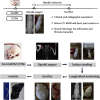
Methods: Thirty male mice were divided into 2 groups: 10 bipedal C57BL/6J mice were used as the control group, and 20 bipedal SAMP8 mice were used as the experimental group. Mice were bipedalized under general anesthesia. The incidence of scoliosis and bone quality was determined using radiographs and in vivo micro-CT images 4, 8, and 12 weeks after surgery, respectively. Histomorphological studies of muscle samples were performed after sacrifice at 12 weeks after surgery.
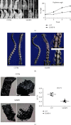
Results: On the 12th week, the incidence rates of kyphosis in C57BL/6J and SAMP8 groups were 50% and 100%, respectively. Overall, the incidence and angle of kyphosis were significantly higher in the bipedal SAMP8 group compared to the C57BL/6J group (44.7°± 6.2° vs. 84.3°± 10.3°, P<0.001). Based on 3D reconstruction of the entire spine, degeneration of the intervertebral disc was observed in bipedal SAMP8 mice, including the reduction of disc height and the formation of vertebral osteophytes. The bone volume ratio (BV/TV) was significantly suppressed in the bipedal SAMP8 group compared with the bipedal C57BL/6J group. In addition, HE staining and Mason staining of the paraspinal muscle tissue showed chronic inflammation and fibrosis in the muscles of the bipedal SAMP8 group.
Conclusions: The SAMP8 mouse model can be taken as a clinically relevant model of DKS, and accelerated aging of the musculoskeletal system promotes the development of kyphosis.
Copyright © 2022 Zongshan Hu et al.
Conflict of interest statement
The authors declare that they have no conflicts of interest.
Figures


Similar articles
-
Development and Characterization of a Novel Bipedal Standing Mouse Model of Intervertebral Disc and Facet Joint Degeneration.Clin Orthop Relat Res. 2019 Jun;477(6):1492-1504. doi: 10.1097/CORR.0000000000000712. Clin Orthop Relat Res. 2019. PMID: 31094848 Free PMC article.
-
Paraspinal myopathy-induced intervertebral disc degeneration and thoracolumbar kyphosis in TSC1mKO mice model-a preliminary study.Spine J. 2022 Mar;22(3):483-494. doi: 10.1016/j.spinee.2021.09.003. Epub 2021 Oct 13. Spine J. 2022. PMID: 34653636
-
Long-term whole-body vibration induces degeneration of intervertebral disc and facet joint in a bipedal mouse model.Front Bioeng Biotechnol. 2023 Mar 17;11:1069568. doi: 10.3389/fbioe.2023.1069568. eCollection 2023. Front Bioeng Biotechnol. 2023. PMID: 37008038 Free PMC article.
-
The senescence-accelerated prone mouse (SAMP8): a model of age-related cognitive decline with relevance to alterations of the gene expression and protein abnormalities in Alzheimer's disease.Exp Gerontol. 2005 Oct;40(10):774-83. doi: 10.1016/j.exger.2005.05.007. Epub 2005 Jul 18. Exp Gerontol. 2005. PMID: 16026957 Review.
-
Surgical treatment for kyphoscoliosis in Cohen syndrome.Nagoya J Med Sci. 2013 Aug;75(3-4):279-86. Nagoya J Med Sci. 2013. PMID: 24640185 Free PMC article. Review.
Cited by
-
Retracted: The Establishment of a Mouse Model for Degenerative Kyphoscoliosis Based on Senescence-Accelerated Mouse Prone 8.Oxid Med Cell Longev. 2023 Sep 27;2023:9753462. doi: 10.1155/2023/9753462. eCollection 2023. Oxid Med Cell Longev. 2023. PMID: 37810564 Free PMC article.
-
Establishment of a mouse model of ovarian oxidative stress induced by hydrogen peroxide.Front Vet Sci. 2024 Nov 6;11:1484388. doi: 10.3389/fvets.2024.1484388. eCollection 2024. Front Vet Sci. 2024. PMID: 39568483 Free PMC article.
References
-
- García-Ramos C. L., Obil-Chavarría C. A., Molina-Choez D. D., Reyes-Sánchez A. Epidemiological and radiological profile of patients with degenerative scoliosis: 20 year experience at a referral institute. Acta Ortopédica Mexicana . 2018;32(2):60–64. - PubMed
-
- Wang N. G., Wang Y. P., Qiu G. X., et al. Radiological evaluation of intervertebral angles on short-segment fusion of degenerative lumbar scoliosis. Zhonghua Wai Ke Za Zhi . 2010;48(7):506–510. - PubMed
Publication types
MeSH terms
LinkOut - more resources
Full Text Sources
Medical

