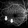
Retinal Vasculitis in a Patient With Mixed Connective Tissue Disease Following COVID-19 Infection: Correlation or Coincidence?
- PMID: 35911323
- PMCID: PMC9328413
- DOI: 10.7759/cureus.26365
Retinal Vasculitis in a Patient With Mixed Connective Tissue Disease Following COVID-19 Infection: Correlation or Coincidence?
Abstract
We report the systemic and ophthalmic findings in a female patient with mixed connective tissue disease (MCTD) who subsequently developed retinal vasculitis following coronavirus disease 2019 (COVID-19) reinfection. The patient was a known case of MCTD maintained in remission on immunosuppressive treatment. She subsequently developed retinal vasculitis with areas of capillary non-perfusion in the right eye. This was a finding not seen previously. She was started on an enhanced immunosuppressive regimen along with scatter laser photocoagulation. COVID-19 has been reported to lead to the development of autoimmune disease, both de novo as well as the worsening of pre-existing disease. The onset of retinal vasculitis may potentially be due to a post-COVID-19 exacerbation of her pre-existing MCTD. Physicians should be aware of this possibility and screen for the same.
Keywords: covid-19; fundoscopy; mixed connective tissue disease; retinal vasculitis; systemic inflammatory and autoimmune disease.
Copyright © 2022, Mehta et al.
Conflict of interest statement
The authors have declared that no competing interests exist.
Figures


Similar articles
-
Frosted Branch Angiitis in the Setting of Active COVID-19 Infection and Underlying Mixed Connective Tissue Disease.Cureus. 2023 Mar 28;15(3):e36819. doi: 10.7759/cureus.36819. eCollection 2023 Mar. Cureus. 2023. PMID: 36998920 Free PMC article.
-
Retinal vasculitis and vitreous hemorrhage associated with mixed connective tissue disease: retinal vasculitis in MCTD.Int Ophthalmol. 2005 Aug-Oct;26(4-5):159-61. doi: 10.1007/s10792-006-9015-8. Epub 2007 Jan 3. Int Ophthalmol. 2005. PMID: 17200794
-
Retinal vasculopathy associated with mixed connective tissue disease.Ocul Immunol Inflamm. 2010 Jan;18(1):13-5. doi: 10.3109/09273940903402629. Ocul Immunol Inflamm. 2010. PMID: 20128643
-
[Retinal vasculitis and systemic diseases].Rev Med Interne. 2018 Sep;39(9):721-727. doi: 10.1016/j.revmed.2018.04.013. Epub 2018 Jun 20. Rev Med Interne. 2018. PMID: 29933971 Review. French.
-
[Autoimmune hepatitis associated with mixed connective tissue disease: report of a case and a review of the literature].Nihon Rinsho Meneki Gakkai Kaishi. 2001 Apr;24(2):75-80. doi: 10.2177/jsci.24.75. Nihon Rinsho Meneki Gakkai Kaishi. 2001. PMID: 11411090 Review. Japanese.
Cited by
-
Frosted Branch Angiitis in the Setting of Active COVID-19 Infection and Underlying Mixed Connective Tissue Disease.Cureus. 2023 Mar 28;15(3):e36819. doi: 10.7759/cureus.36819. eCollection 2023 Mar. Cureus. 2023. PMID: 36998920 Free PMC article.
References
-
- Mixed connective tissue disease: an overview of clinical manifestations, diagnosis and treatment. Ortega-Hernandez OD, Shoenfeld Y. Best Pract Res Clin Rheumatol. 2012;26:61–72. - PubMed
-
- Vasculitis in the connective tissue diseases. Felipe Flores-Suárez L, Alarcón-Segovia D. Curr Rheumatol Rep. 2000;2:396–401. - PubMed
-
- Intravitreal bevacizumab for macular edema due to occlusive vasculitis. Margolis R, Lowder CY, Sears JE, Kaiser PK. Semin Ophthalmol. 2007;22:105–108. - PubMed
-
- Retinal vasculopathy associated with mixed connective tissue disease. Kim YK, Woo SJ, Lee YJ, Park KH. Ocul Immunol Inflamm. 2010;18:13–15. - PubMed
-
- Retinal vasculitis and vitreous hemorrhage associated with mixed connective tissue disease: retinal vasculitis in MCTD. Mimura T, Usui T, Amano S, Yamagami S, Ono K, Noma H, Funatsu H. Int Ophthalmol. 2005;26:159–161. - PubMed
Publication types
LinkOut - more resources
Full Text Sources
