. 2022 Jun 6;4(3):fcac144.
doi: 10.1093/braincomms/fcac144. eCollection 2022.

4-Phenylbutyrate restored γ-aminobutyric acid uptake and reduced seizures in SLC6A1 patient variant-bearing cell and mouse models

Affiliations

4-Phenylbutyrate restored γ-aminobutyric acid uptake and reduced seizures in SLC6A1 patient variant-bearing cell and mouse models

Gerald Nwosu et al. Brain Commun. .

Abstract

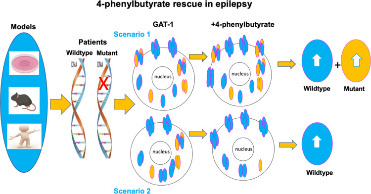
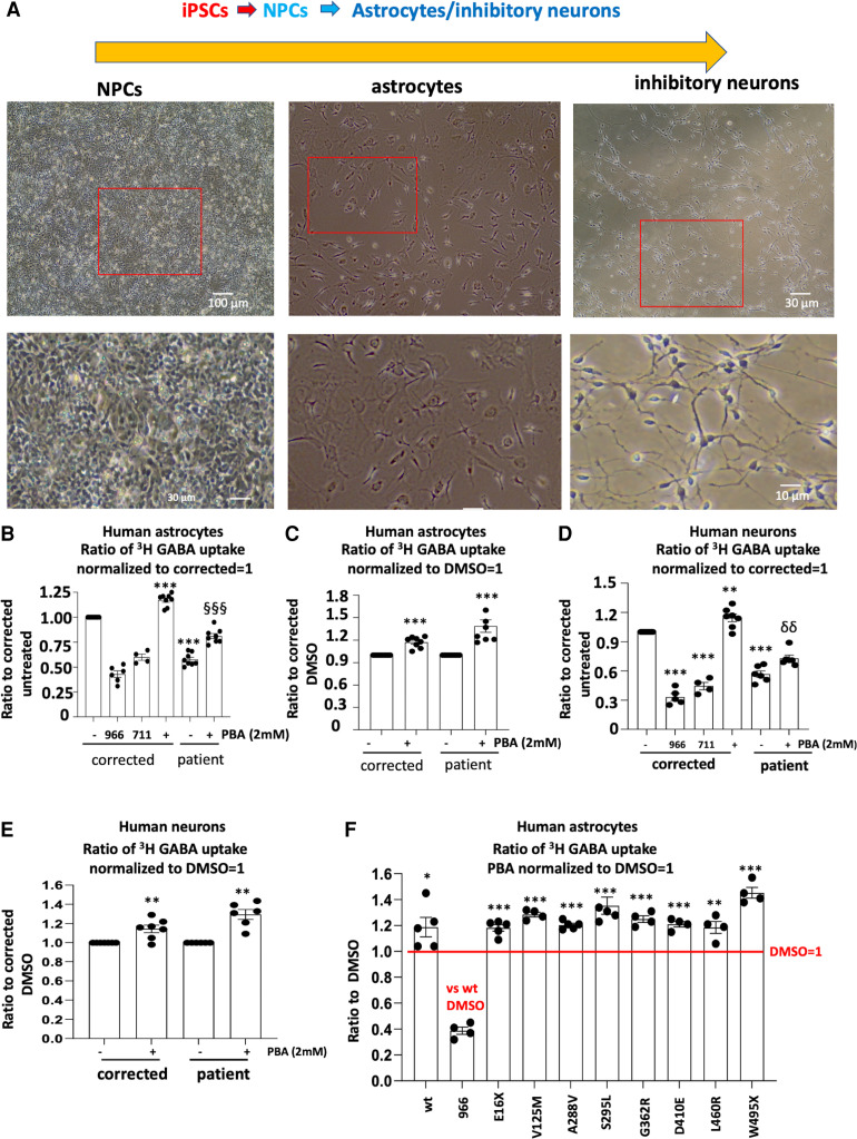
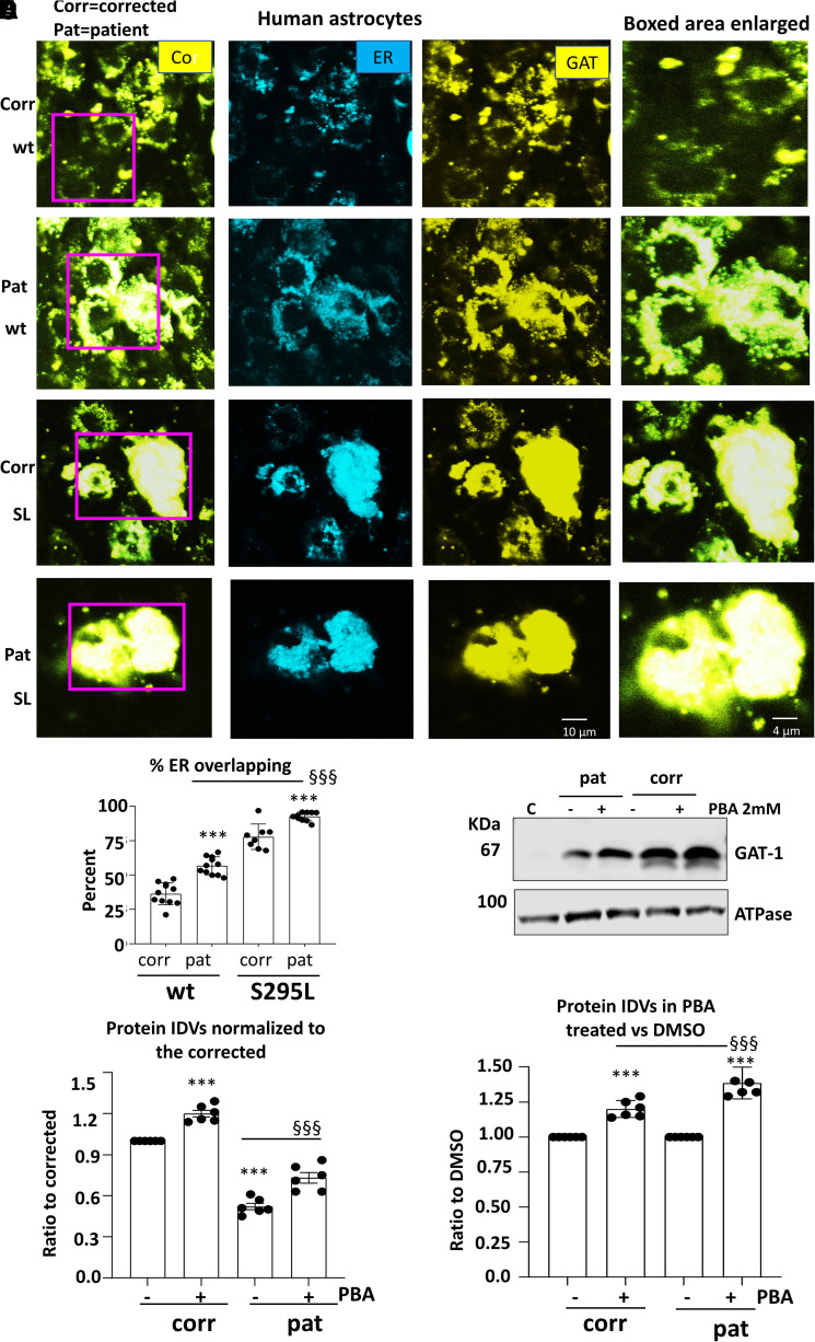
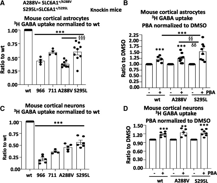
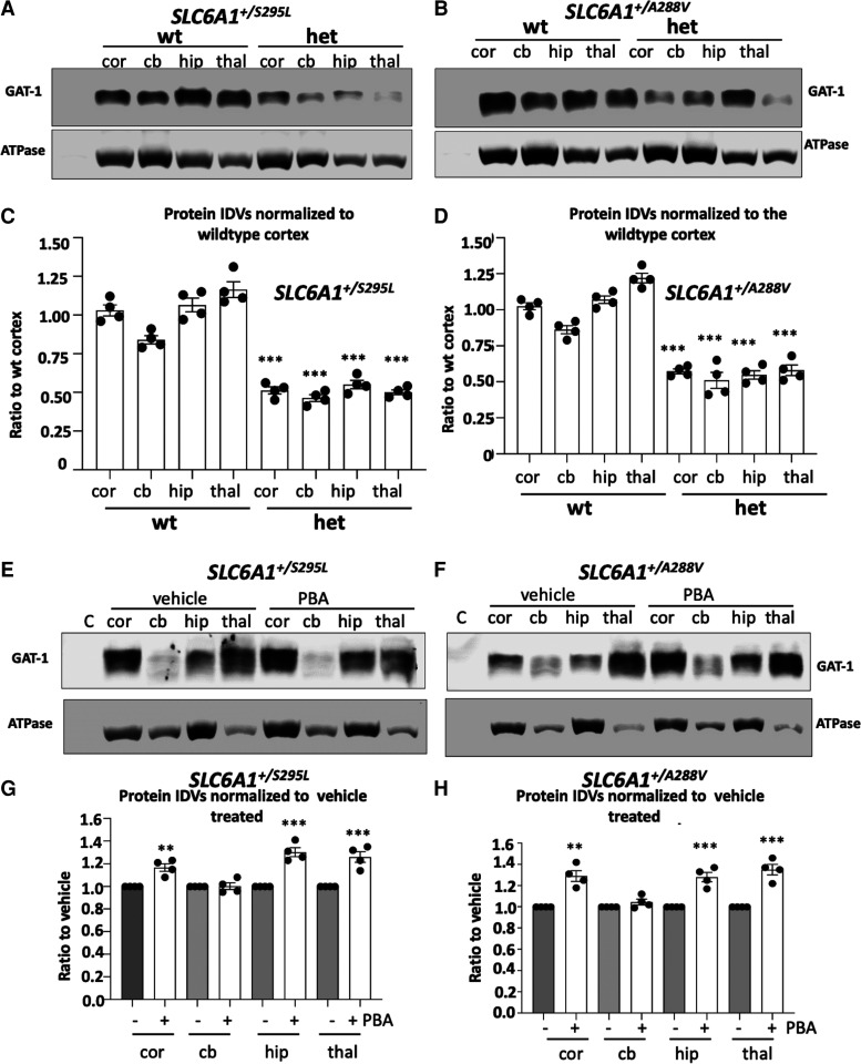
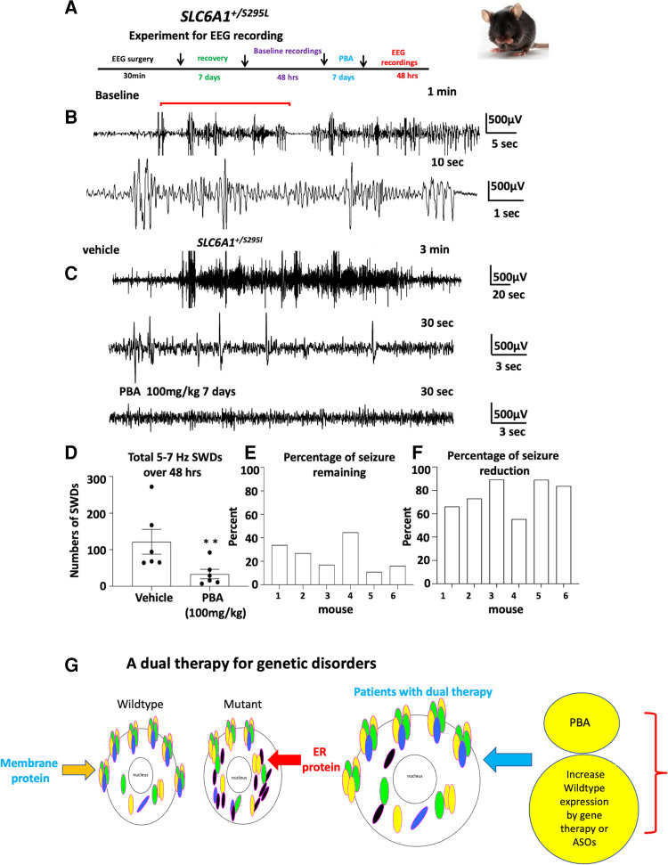
We have studied the molecular mechanisms of variants in solute carrier Family 6 Member 1 associated with neurodevelopmental disorders, including various epilepsy syndromes, autism and intellectual disability. Based on functional assays of solute carrier Family 6 Member 1 variants, we conclude that partial or complete loss of γ-amino butyric acid uptake due to reduced membrane γ-amino butyric acid transporter 1 trafficking is the primary aetiology. Importantly, we identified common patterns of the mutant γ-amino butyric acid transporter 1 protein trafficking from biogenesis, oligomerization, glycosylation and translocation to the cell membrane across variants in different cell types such as astrocytes and neurons. We hypothesize that therapeutic approaches to facilitate membrane trafficking would increase γ-amino butyric acid transporter 1 protein membrane expression and function. 4-Phenylbutyrate is a Food and Drug Administration-approved drug for paediatric use and is orally bioavailable. 4-Phenylbutyrate shows promise in the treatment of cystic fibrosis. The common cellular mechanisms shared by the mutant γ-amino butyric acid transporter 1 and cystic fibrosis transmembrane conductance regulator led us to hypothesize that 4-phenylbutyrate could be a potential treatment option for solute carrier Family 6 Member 1 mutations. We examined the impact of 4-phenylbutyrate across a library of variants in cell and knockin mouse models. Because γ-amino butyric acid transporter 1 is expressed in both neurons and astrocytes, and γ-amino butyric acid transporter 1 deficiency in astrocytes has been hypothesized to underlie seizure generation, we tested the effect of 4-phenylbutyrate in both neurons and astrocytes with a focus on astrocytes. We demonstrated existence of the mutant γ-amino butyric acid transporter 1 retaining wildtype γ-amino butyric acid transporter 1, suggesting the mutant protein causes aberrant protein oligomerization and trafficking. 4-Phenylbutyrate increased γ-amino butyric acid uptake in both mouse and human astrocytes and neurons bearing the variants. Importantly, 4-phenylbutyrate alone increased γ-amino butyric acid transporter 1 expression and suppressed spike wave discharges in heterozygous knockin mice. Although the mechanisms of action for 4-phenylbutyrate are still unclear, with multiple possibly being involved, it is likely that 4-phenylbutyrate can facilitate the forward trafficking of the wildtype γ-amino butyric acid transporter 1 regardless of rescuing the mutant γ-amino butyric acid transporter 1, thus increasing γ-amino butyric acid uptake. All patients with solute carrier Family 6 Member 1 variants are heterozygous and carry one wildtype allele, suggesting a great opportunity for treatment development leveraging wildtype protein trafficking. The study opens a novel avenue of treatment development for genetic epilepsy via drug repurposing.

Keywords: 4-phenylbutyrate acid; GABA transporter 1 (GAT-1); autism; chaperone; epilepsy.


Figures

Graphical abstract
Graphical abstract
Figure 1
Figure 1
Reduced function and trafficking of the mutant GABA transporter 1 encoded by SLC6A1 variants associated with epilepsy, autism, ADHD and intellectual delay. (A) Schematic presentation of mutant GABA transporter 1 (GAT-1) protein topology and locations of representative variants in human SLC6A1 associated with various epilepsy syndromes and neurodevelopmental disorders as described in our previous work. These variants are distributed in various locations and domains of the encoded GAT-1 protein peptide. The large dots represent the eight variants evaluated in the study. (B–D) HEK293T cells were transfected with the wildtype or the mutant GAT-1YFP for 48 h. (B) The graph represents the altered GABA reuptake function of the mutant GAT-1 encoded by eight different SLC6A1 variants in HEK293T cells measured by the high-throughput 3H radio-labelling GABA uptake on a liquid scintillator with QuantaSmart. Around 966 stands for the wildtype treated with GAT-1 inhibitor Cl-966 (50 µM) and NNC-711 for the wildtype treated with NNC-711 (35 µM) for 30 min before preincubation. (C, D) The flow cytometry histograms depict the relative surface (C) or total (D) expression of the wildtype and the mutant GAT-1. The relative total expression level of GAT-1 in each mutant transporter was normalized to that obtained from cells with transfection of the wildtypes. N = 4–5 different transfections, δδδ P < 0.001 overall mutations versus wt, *P < 0.05, **P < 0.01, ***P < 0.001 versus wt, one-way analysis of variance and Newman–Keuls test. Values were expressed as mean ± SEM.
Figure 2
Figure 2
All surveyed mutant GAT-1 transporters had less mature but more immature form of the GAT-1 protein. (A, B) The total lysates of HEK293T cells expressing the wildtype or variant GAT-1 were undigested (U) or digested with Endo-H (H) and then analysed by SDS-PAGE. The membrane was immunoblotted with a rabbit anti-GAT-1. The boxed region represents the mature form of GAT-1 in cells. CHO stands for Chinese hamster ovary cells. CHO cells were used for control because of the low level of the endogenous GAT-1 expression. (C) The graph represents the normalized integrated protein density values (IDVs) of the mature form of GAT-1 defined by being Endo-H resistant normalized to the wildtype mature form of GAT-1 (bands 1 + 2). (D) The graph represents the normalized IDVs of the immature form of GAT-1 defined by being Endo-H unresistant (shifted to a lower level after H digestion) normalized to the wildtype immature form of GAT-1 (Band 3 shifted to Band 4). N = 4–5 different transfections, δδδ P < 0.001 overall mutations versus wt, *P < 0.05, **P < 0.01, ***P < 0.001 versus wt, one-way analysis of variance and Newman–Keuls test. Values were expressed as mean ± SEM.
Figure 3
Figure 3
4-Phenylbutyric acid (PBA) concentration and time dependently increased the GABA uptake in cells expressing the wildtype or the mutant GAT-1. (A, B) HEK293T cells were transfected with wildtype GAT-1YFP (wt) for 48 h, PBA (2 mM) was applied dropwise to each dish at different concentrations (A) and incubated for time durations (B). PBA from stocking solution (2 M) was diluted with DMEM 100 µL to desired concentration. (A) The graphs represent the altered GABA reuptake function of the wildtype GAT-1YFP in HEK293T cells treated with PBA for a series of different concentrations. The GABA uptake activity of cells treated with PBA of different concentrations was normalized to the sister cultures treated with DMSO alone for 24 h. (B) The graphs represent the altered GABA reuptake function of the wildtype GAT-1 in HEK293T cells treated with PBA for a series of different time duration over treated with DMSO alone for 24 h. GABA uptake activity was measured by the high-throughput 3H radio-labelling GABA uptake on a liquid scintillator with QuantaSmart. (C) Cartoon showing the mutant allele only was expressed. HEK293T cells were transfected with the wildtype or the mutant GAT-1YFP cDNAs alone for 48 h. (D) Cartoon showing the coexistence condition of the wildtype and the mutant allele in patients and both the wildtype and the mutant alleles were expressed. HEK293T cells were transfected with the wildtype GAT-1YFP alone or in mixture of the wildtype or the mutant cDNAs for 48 h. In the mixed condition, the ratio of the wildtype GAT-1 with pcDNA or the mutant cDNAs are 1:1 with the total cDNA amount of 0.5 µg. PBA (2 mM) was applied for 24 h, while DMSO was applied as control. Both wt and the mutant were normalized to its own DMSO-treated conditions. Around 966 stands for the wildtype treated with Cl-966 (50 µM), while 711 stands for NNC-711 (35 µM). N = 4–7 different transfections. In A and B, *P < 0.05, **P < 0.01, ***P < 0.001 versus wt 0. In C and D, *P < 0.05, **P < 0.01, ***P < 0.001 versus its own DMSO treated. In C, ns stands for no significance. One-way analysis of variance and Newman–Keuls test. Values were expressed as mean ± SEM.
Figure 4
Figure 4
4-Phenylbutyrate acid (PBA) rescued the GABA uptake function in the patient astrocytes and neurons. (A) Human astrocytes and inhibitory neurons were differentiated from the neural progenitor cells (NPCs) from the patient and the CRISPR corrected isogenic control line. Live images of human NPCs, astrocytes and GABAergic inhibitory neurons differentiated from the human-induced pluripotent stem cells (iPSCs) on the day of experiment. (B–E) Astrocytes at Days 30–35 (B, C) or neurons at Days 60–65 (D, E) after differentiation were treated with PBA (2 mM) for 24 h before 3H radioactive GABA uptake assay. The DMSO-treated corrected or patient cells were taken as 1. (F) Corrected astrocytes at Days 30–35 after differentiation were transfected with the wildtype or the mutant GAT-1YFP cDNAs (1 µg per a 35 mm2 dish) for 48 h before 3H radioactive GABA uptake assay. PBA (2 mM) was applied for 24 h before GABA uptake assay experiment. GABA flux was measured after 30 min transport at room temperature. The influx of GABA, expressed in pmol/µg protein/min, was averaged from duplicates for each condition and for each transfection. The average counting was DMSO-treated condition taken as 1. In B, D and F, 966 stands for Cl-966 (50 µM) and 711 stands for NNC-711 (35 µM) that was applied 30 min to the astrocytes transfected with the wildtype GAT-1YFP before preincubation and removed during preincubation. In B and D, ***P < 0.001 versus corrected DMSO treated. §§<0.01, §§§P < 0.001 versus patient DMSO treated. In C and E, the PBA-treated corrected or patient GABA uptake was normalized to its DMSO treated. In C, E and F, the PBA-treated corrected/wildtype or patient/mutant GABA uptake was normalized to its DMSO treated. *P < 0.05; **P < 0.01; ***P < 0.001 versus DMSO treated in its own group. In B, C, D and E, n = 4–8 batches of cells. In F, n = 4–5 different transfections. Unpaired t test was used for C and E. One-way analysis of variance followed by a Dunnett post hoc multiple comparison test was used in B, D and F. Values were expressed as mean ± SEM.
Figure 5
Figure 5
The patient astrocytes caused ER retention of the wildtype and exacerbated the mutant GAT-1 ER retention while 4-phenylbutyrate acid (PBA) increased GAT-1 protein expression. (A, B) Human patient corrected (isogenic control, Corr) or uncorrected patient (Pat) astrocytes at Days 30–35 after differentiation from iPSCs were co-transfected with the endoplasmic reticulum (ER) marker ERCFP in combination with the wildtype or the mutant GAT-1YFP cDNAs (0.5 µg:0.5 µg per a 35 mm2 dish) for 48 h before confocal microscopy analysis. Confocal images were acquired in live astrocytes under 63X objective with zoom under 2.5. (A) Boxed regions were enlarged. (B) The graph represents the ER overlapping signal of GAT-1YFP analysed by Metamorph. (C, D) Total cell lysates from astrocytes cultured in 100 mm2 dishes treated with DMSO or PBA (2 mM) for 24 h and were analysed with SDS-PAGE. Membranes were blotted with a rabbit polyclonal atnti-GAT-1 antibody (C). The lysates of Chinese hamster ovary (CHO) cells were used as control. (D, E) The protein IDVs of the corrected or patient GAT-1 in human astrocytes were normalized to the corrected astrocytes treated with DMSO, the GAT-1 protein was normalized to its own internal control ATPase or GAPDH and then to the DMSO-treated corrected levels, which is arbitrarily taken as 1 (D) or its own genotype but DMSO treated, which is taken as 1 (E). Values were expressed as mean ± SEM. In B, N = 8–11 culture replicates. In C, D and E, N = 6 batch of cells. In B, ***P < 0.001 versus corrected. §§§P < 0.001 versus wt in patient cells. In D, ***P < 0.001 corrected untreated. §§§P < 0.001 versus patient untreated. In E, ***P < 0.001 versus its own untreated; §§§P < 0.001 versus corrected PBA treated, two-way analysis of variance followed by Bonferroni multiple comparison test.
Figure 6
Figure 6
4-Phenylbutyrate acid (PBA) rescued the GABA uptake in cortical astrocytes and neurons in SLC6A1+/A288V and SLC6A1+/S295L mice. Mouse cortical astrocytes or neurons were cultured from postnatal pus at Days 0–3 for astrocytes and Day 0 for neurons from the SLC6A1+/A288V or SLC6A1+/S295L mouse line. (A, B) Astrocytes under Passage 2 were grown in 100 mm2 dishes and passaged into 35 mm2 dishes before GABA uptake assay. (C, D) Neurons were directly cultured in the 35 mm2 dishes and GABA uptake was evaluated between Days 15 and 17 after culture. Cl-966 (50 µM) and NNC711 (35 µM) were applied 30 min before preincubation and removed during preincubation. GAT-3 inhibitor SNAP5114 (30 µM) was applied during GABA uptake to make sure only GAT-1 activity was measured. The wildtype astrocytes (A, B) or cortical neurons (C, D) of either SLC6A1+/A288V or SLC6A1+/S295L were taken as 1. The cultures derived from each mutant mouse were compared with the culture from its own wildtype littermates. In B and D, sister cultures of astrocytes or neurons from different mouse lines were incubated with DMSO or PBA 2 mM for 24 h before GABA uptake. The wildtype data were pooled from two mouse lines. The GABA uptake activity of PBA treated was normalized to its own DMSO conditions. The graph represents the relative GABA uptake level normalized to cells of its own genotype treated with DMSO. Cl-966 (50 µM) or NNC-711 (35 µM) was applied 30 min before preincubation and removed during preincubation. Two-way analysis of variance followed by Bonferroni multiple comparison test was used. Values were expressed as mean ± SEM. In A and C, ***P < 0.001 versus wt; §§§P < 0.001 S295L versus A288V. In B and D, ***P < 0.001 versus untreated; §§P < 0.01 S295L versus wt treated; δδ P < 0.01 versus A288V treated. N = 4–9 batches of astrocytes from four pairs of littermates for A. N = 5–8 batches of astrocytes from four pairs of littermates for B; N = 4–7 batches of neuron cultures from 5 litters of A288V and 6 litters of S295L in C and D.
Figure 7
Figure 7
Both SLC6A1+/A288V and SLC6A1+/S295L mice had reduced GAT-1 protein that was partially restored by 4-phenylbutyrate acid (PBA). (A–H) Lysates from different brain regions [cortex (cor), cerebellum (cb), hippocampus (hip) and thalamus (thal)] from the wildtype (wt) and heterozygous (het) mice at 4–6 months old, untreated (A, B) or treated with vehicle or PBA (100 mg/kg) for 7 days (E, F) were subjected to SDS-PAGE and immunoblotted with anti-GAT-1 antibody. (C, D) Integrated density values (IDVs) for total GAT-1 from wildtype and het KI were normalized to the Na+/K+ ATPase or anti-glyceraldehyde-3-phosphate dehydrogenase (GAPDH) loading control (LC) in each specific brain region and plotted. N = 4 from four pairs of mice. (G, H) Integrated density values (IDVs) for total GAT-1 from het KI treated with vehicle or treated with PBA were normalized to the Na+/K+ ATPase or anti-glyceraldehyde-3-phosphate dehydrogenase (GAPDH) loading control (LC). The IDVs of the heterozygous treated with PBA were then normalized to vehicle treated. The vehicle treated in each brain region was taken as 1. N = 4 from four pairs of mice for C, D, G and H. Values were expressed as mean ± SEM. One-way analysis of variance or unpaired t test. In C and D, ***P < 0.001 versus wt, in G and H, **P < 0.01; ***P < 0.001 versus vehicle treated.
Figure 8
Figure 8
4-Phenylbutyrate acid (PBA) alone reduced seizures in mutation knockin mice. (A) Schematic depiction of experimental paradigm for EEG recordings and PBA treatment. (B) Representative EEG recordings show that the heterozygous SLC6A1+/S295L (het) KI mice had frequent absence like spike wave discharges (SWDs) and some myoclonic jerks during baseline recordings. (C) Comparison of EEG traces recorded after the vehicle (normal saline 100 µL) treated or after treatment with PBA (100 mg/kg, i.p., single dose, daily) for 7 days. (D) Graph showing the total number of 5–7 Hz SWDs calculated by Seizure Pro during 48 h recordings after vehicle or PBA treatment. PBA treatment (100 mg/kg, i.p., single dose, daily) for 7 days reduced seizure activity. **P < 0.01; versus vehicle treated, paired t test, N = 6 animals. Values were expressed as mean ± SEM. (E, F) Graph showing the percentage of seizure remaining (E) and seizure reduction (F) in each mouse after PBA treatment (N = 6 animals, paired t test). (G) We propose a dual therapy as a feasible approach for treating SLC6A1 variants and other genetic disorders by boosting the wildtype allele, removing the mutant allele and ER stress with PBA.

Similar articles

Cited by

References

    1. Kang JQ, Shen W, Zhou C, Xu D, Macdonald RL. The human epilepsy mutation GABRG2(Q390X) causes chronic subunit accumulation and neurodegeneration. Nat Neurosci. 2015;18(7):988–996. - PMC - PubMed
    1. Soto C, Pritzkow S. Protein misfolding, aggregation, and conformational strains in neurodegenerative diseases. Nat Neurosci. 2018;21(10):1332–1340. - PMC - PubMed
    1. Ross CA, Poirier MA. Protein aggregation and neurodegenerative disease. Nat Med. 2004;10:S10–S17. - PubMed
    1. Goodspeed K, Pérez-Palma E, Iqbal S, et al. . Current knowledge of SLC6A1-related neurodevelopmental disorders. Brain Commun. 2020;2(2):fcaa170. - PMC - PubMed
    1. Carvill GL, McMahon JM, Schneider A, et al. . Mutations in the GABA transporter SLC6A1 cause epilepsy with myoclonic-atonic seizures. Am J Hum Genet. 2015;96(5):808–815. - PMC - PubMed

LinkOut - more resources