. 2022 Jul 15:9:901286.
doi: 10.3389/fcvm.2022.901286. eCollection 2022.

Low α2-Plasmin Inhibitor Antigen Levels on Admission Are Associated With More Severe Stroke and Unfavorable Outcomes in Acute Ischemic Stroke Patients Treated With Intravenous Thrombolysis

Affiliations

Low α2-Plasmin Inhibitor Antigen Levels on Admission Are Associated With More Severe Stroke and Unfavorable Outcomes in Acute Ischemic Stroke Patients Treated With Intravenous Thrombolysis

Edina Gabriella Székely et al. Front Cardiovasc Med. .

Abstract

Background: Intravenous administration of recombinant tissue plasminogen activator (rt-PA) fails to succeed in a subset of acute ischemic stroke (AIS) patients, while in approximately 6-8% of cases intracerebral hemorrhage (ICH) occurs as side effect.

Objective: Here, we aimed to investigate α2-plasmin inhibitor (α2-PI) levels during thrombolysis and to find out whether they predict therapy outcomes in AIS patients.

Patients/methods: In this prospective, observational study, blood samples of 421 AIS patients, all undergoing i.v. thrombolysis by rt-PA within 4.5 h of their symptom onset, were taken before and 24 h after thrombolysis. In a subset of patients (n = 131), blood was also obtained immediately post-lysis. α2-PI activity and antigen levels were measured by chromogenic assay and an in-house ELISA detecting all forms of α2-PI. α2-PI Arg6Trp polymorphism was identified in all patients. Stroke severity was determined by NIHSS on admission and day 7. Therapy-associated ICH was classified according to ECASSII. Long-term outcomes were defined at 3 months post-event by the modified Rankin Scale (mRS).

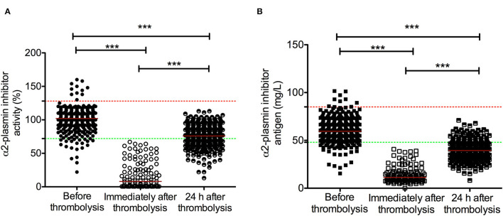
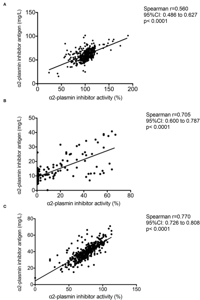
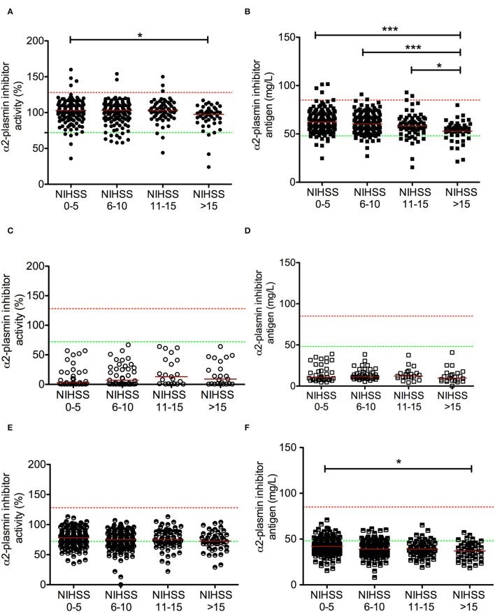
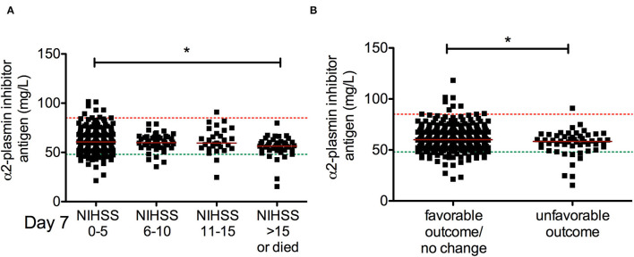
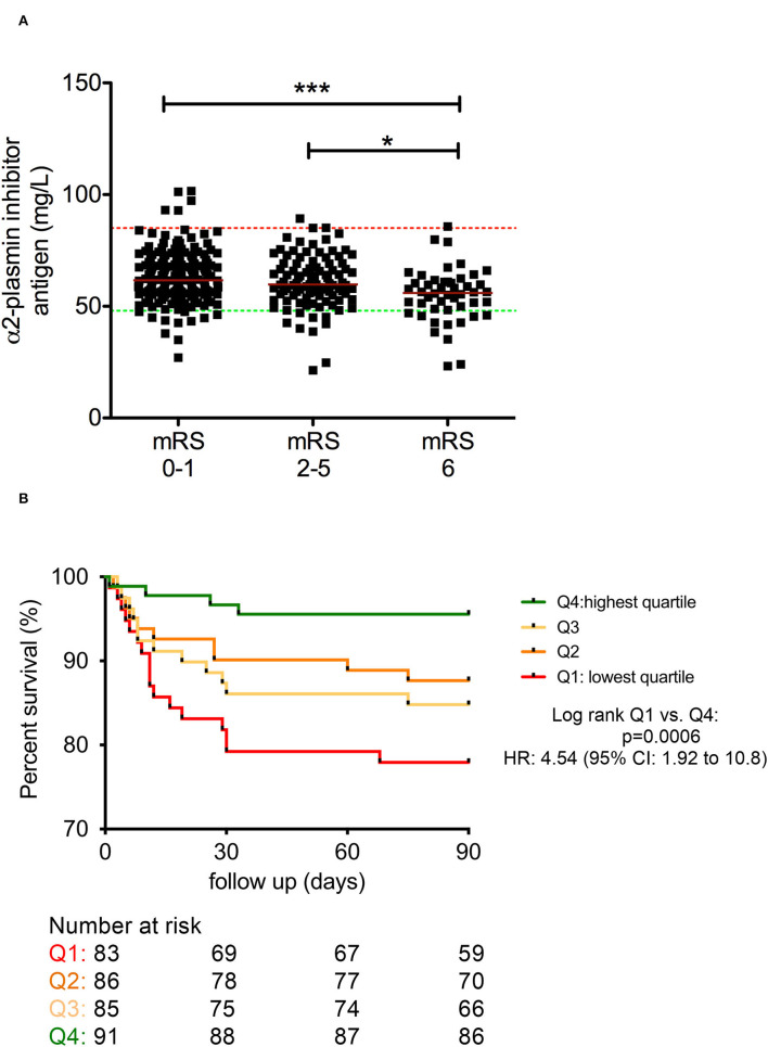
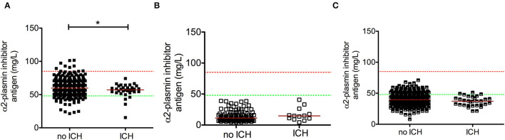
Results: Median α2-PI activity and antigen levels showed a significant drop immediately post-lysis and increased to subnormal levels at 24 h post-event. Admission α2-PI levels showed a significant negative stepwise association with stroke severity. Patients with favorable long-term outcomes (mRS 0-1) had significantly higher admission α2-PI antigen levels (median:61.6 [IQR:55.9-70.5] mg/L) as compared to patients with poor outcomes (mRS 2-5: median:59.7 [IQR:54.5-69.1] and mRS 6: median:56.0 [IQR:48.5-61.0] mg/L, p < 0.001). In a Kaplan-Meier survival analysis, patients with an α2-PI antigen in the highest quartile on admission showed significantly better long-term survival as compared to those with α2-PI antigen in the lowest quartile (HR: 4.54; 95%CI:1.92-10.8, p < 0.001); however, in a multivariate analysis, a low admission α2-PI antigen did not prove to be an independent risk factor of poor long-term outcomes. In patients with therapy-related ICH (n = 34), admission α2-PI antigen levels were significantly, but only marginally, lower as compared to those without hemorrhage.

Conclusions: Low α2-PI antigen levels on admission were associated with more severe strokes and poor long-term outcomes in this cohort. Our results suggest that in case of more severe strokes, α2-PI may be involved in the limited efficacy of rt-PA thrombolysis.

Keywords: acute ischemic stroke; fibrinolysis; intracerebral hemorrhage (ICH); outcome; recombinant tissue plasminogen activator (rt-PA); thrombolysis; α2-plasmin inhibitor.


Conflict of interest statement

The authors declare that the research was conducted in the absence of any commercial or financial relationships that could be construed as a potential conflict of interest.

Figures

Figure 1
Figure 1
α2-plasmin inhibitor activity (A) and antigen levels (B) in acute ischemic stroke patients during the course of rt-PA thrombolysis. α2-plasmin inhibitor activity (circles) and antigen levels (squares) were assessed on admission (solid symbols), immediately after thrombolysis (immediately after administering full dose of rt-PA, empty symbols) and 24 h after thrombolysis (half-colored symbols). Red horizontal lines indicate median in each group. Upper and lower limits of reference are indicated with red and green dashed lines, respectively. rt-PA, recombinant tissue plasminogen activator. ***p < 0.001.
Figure 2
Figure 2
Correlation between α2-plasmin inhibitor activity and antigen levels as assessed on admission (A) immediately after thrombolysis (B) and 24 h after rt-PA thrombolysis (C) in acute ischemic stroke patients.
Figure 3
Figure 3
Association between α2-plasmin inhibitor activity and antigen levels at different time points during rt-PA thrombolysis with stroke severity on admission. α2-plasmin inhibitor activity was assessed on admission [(A) solid circles], immediately after thrombolysis [immediately after administering full dose of rt-PA, (C) empty circles], and 24 h after thrombolysis [(E) half-colored circles]. α2-plasmin inhibitor antigen levels were assessed on admission [(B) solid squares], immediately after thrombolysis [immediately after administering full dose of rt-PA, (D) empty squares], and 24 h after thrombolysis [(F) half-colored squares]. Red horizontal lines indicate median in each group. Stroke severity on admission was grouped according to NIHSS. Upper and lower limits of reference are indicated with red and green dashed lines, respectively. NIHSS, National Institutes of Health Stroke Scale, rt-PA, recombinant tissue plasminogen activator. *p < 0.05, ***p < 0.001.
Figure 4
Figure 4
Association between α2-plasmin inhibitor antigen levels on admission and short-term outcome of thrombolysis. α2-plasmin inhibitor antigen levels were assessed on admission and compared to stroke severity on day 7 according to NIHSS (A) or to the change in the NIHSS by day 7 (B). Favorable outcome (neurologic improvement) is defined as a decrease in NIHSS score by at least 4 points or to 0 by day 7, while an increase in NIHSS score by at least 4 points is defined as unfavorable outcome. Upper and lower limits of reference are indicated with red and green dashed lines, respectively. Red horizontal lines indicate median in each group. NIHSS, National Institutes of Health Stroke Scale, rt-PA, recombinant tissue plasminogen activator. *p < 0.05.
Figure 5
Figure 5
Association between α2-plasmin inhibitor antigen levels on admission and long-term outcome of thrombolysis (A). Kaplan–Meier survival curves of patients based on quartiles of α2-plasmin inhibitor antigen levels as measured on admission (B). α2-plasmin inhibitor antigen levels were assessed on admission (solid squares) and compared to long-term functional outcomes of thrombolysis based on the modified Rankin scale (mRS) (A). Red horizontal lines indicate median in each group. Upper and lower limits of reference are indicated with red and green dashed lines, respectively. mRS 0–1 was considered as favorable outcome, and mRS 6 indicates mortality. Kaplan–Meier survival curves of patients are indicated according to quartiles of α2-plasmin inhibitor antigen levels on admission (B). The number at risk is indicated below the graph. Upper quartile (Q4, green line), middle quartiles (Q3 and Q2, light orange and orange lines, respectively), and lowest quartile (Q1, red line) results were grouped based on the following cutoff values: 67.4 mg/L, 60 mg/L, and 54.1 mg/L α2-plasmin inhibitor antigen level, respectively. HR, hazard ratio; mRS, modified Rankin Scale. *p < 0.05, ***p < 0.001.
Figure 6
Figure 6
Association between α2-plasmin inhibitor antigen levels and post-lysis intracerebral bleeding (ICH). α2-plasmin inhibitor antigen levels were assessed on admission (A), immediately after thrombolysis (B), and 24 h after rt-PA thrombolysis (C), and levels were compared in patients without or with intracerebral hemorrhage on day 1 post-lysis. Upper and lower limits of reference are indicated with red and green dashed lines, respectively. Red horizontal lines indicate median in each group. *p < 0.05.

Similar articles

Cited by

  • Coagulation biomarkers for ischemic stroke.
    Barakzie A, Jansen AJG, Ten Cate H, de Maat MPM. Barakzie A, et al. Res Pract Thromb Haemost. 2023 Apr 23;7(4):100160. doi: 10.1016/j.rpth.2023.100160. eCollection 2023 May. Res Pract Thromb Haemost. 2023. PMID: 37274178 Free PMC article.

References

    1. Katan M, Luft A. Global burden of stroke. Semin Neurol. (2018) 38:208–11. 10.1055/s-0038-1649503 - DOI - PubMed
    1. Powers WJ, Rabinstein AA, Ackerson T, Adeoye OM, Bambakidis NC, Becker K, et al. . 2018 guidelines for the early management of patients with acute ischemic stroke: a guideline for healthcare professionals from the American Heart Association/American Stroke Association. Stroke. (2018) 49:e46–e110. 10.1161/STR.0000000000000158 - DOI - PubMed
    1. National National Institute of Neurological Disorders. Stroke rt-PA SSG. Tissue plasminogen activator for acute ischemic stroke. N Engl J Med. (1995) 333:1581–7. 10.1056/NEJM199512143332401 - DOI - PubMed
    1. Hacke W, Kaste M, Bluhmki E, Brozman M, Davalos A, Guidetti D, et al. . Thrombolysis with alteplase 3 to 4.5 hours after acute ischemic stroke. N Engl J Med. (2008) 359:1317–29. 10.1056/NEJMoa0804656 - DOI - PubMed
    1. Henderson SJ, Weitz JI, Kim PY. Fibrinolysis: strategies to enhance the treatment of acute ischemic stroke. J Thromb Haemost. (2018) 16:1932–40. 10.1111/jth.14215 - DOI - PubMed