

Alpha rhythm of electroencephalography was modulated differently by three transcranial direct current stimulation protocols in patients with ischemic stroke
- PMID: 35911595
- PMCID: PMC9334563
- DOI: 10.3389/fnhum.2022.887849
Alpha rhythm of electroencephalography was modulated differently by three transcranial direct current stimulation protocols in patients with ischemic stroke
Abstract
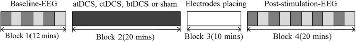
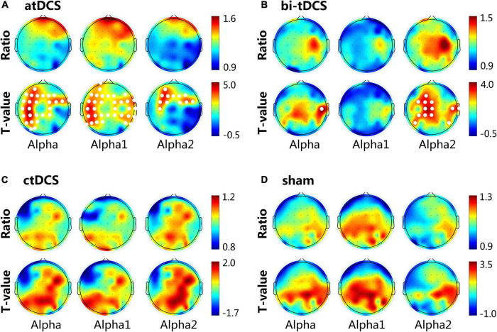
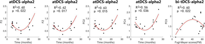
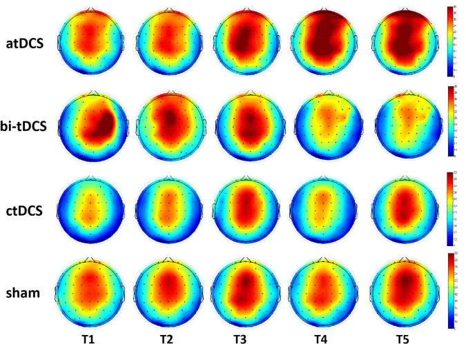
The heterogeneity of transcranial direct current stimulation (tDCS) protocols and clinical profiles may explain variable results in modulating excitability in the motor cortex after stroke. However, the cortical electrical effects induced by different tDCS protocols remain unclear. Here, we aimed to compare rhythm changes in electroencephalography (EEG) induced by three tDCS position protocols and the association between tDCS effects and clinical factors in stroke. Nineteen patients with chronic ischemic stroke underwent four experimental sessions with three tDCS protocols [anodal (atDCS), cathodal (ctDCS), and bilateral (bi-tDCS)] and a sham protocol, according to a single-blind randomized crossover design. Resting-state EEG was acquired before and after each protocol. First, a paired-sample t-test was used to examine the difference in spectral power between pre- and post-stimulation. Then, linear and quadratic regression models were used separately to describe the association between the clinical factors of stroke and changes in spectral power which was significantly different between pre- and post-tDCS. Finally, repeated measures analysis of variance with lesion hemisphere, stimulation protocol, and the location was performed to investigate the effects of tDCS over time. The induced effect of tDCS was mainly reflected in the alpha rhythms. The alpha power was increased by atDCS, especially low-alpha (8-10 Hz), in localized areas of the central and distant areas of the frontal and parietal lobes. Bi-tDCS also affected alpha power but in a smaller area that mainly focused on high-alpha rhythms (10-13 Hz). However, ctDCS and sham had no significant effects on any EEG rhythm. The clinical factors of time since stroke and motor impairment level were related to the change in high-alpha induced by atDCS and bi-tDCS following quadratic regression models. The above-mentioned modulation effect lasted for 20 min without attenuation. In conclusion, our findings provide evidence that the alpha rhythm of EEG is modulated differently by different tDCS protocols and that high alpha is affected by clinical characteristics such as post-stroke time and motor deficits, which is of great significance for understanding the modulation effect of different tDCS protocols on stroke and the guidance of protocols to promote motor recovery following stroke.
Keywords: alpha rhythm; chronic stroke; quantitative EEG; spectral power; transcranial direct current stimulation.
Copyright © 2022 Chen, Wang, Song, Sun, Zhang, Zhao and Du.
Conflict of interest statement
The authors declare that the research was conducted in the absence of any commercial or financial relationships that could be construed as a potential conflict of interest.
Figures
Similar articles
-
Varied Response of EEG Rhythm to Different tDCS Protocols and Lesion Hemispheres in Stroke Subjects with Upper Limb Dysfunction.Neural Plast. 2022 Jul 30;2022:7790730. doi: 10.1155/2022/7790730. eCollection 2022. Neural Plast. 2022. PMID: 35941932 Free PMC article. Clinical Trial.
-
Effects of anodal transcranial direct current stimulation over motor cortex on resting-state brain activity in the early subacute stroke phase: A power spectral density analysis.Clin Neurol Neurosurg. 2024 Feb;237:108134. doi: 10.1016/j.clineuro.2024.108134. Epub 2024 Jan 26. Clin Neurol Neurosurg. 2024. PMID: 38335706 Clinical Trial.
-
Local and remote effects of transcranial direct current stimulation on the electrical activity of the motor cortical network.Hum Brain Mapp. 2014 May;35(5):2220-32. doi: 10.1002/hbm.22322. Epub 2013 Aug 2. Hum Brain Mapp. 2014. PMID: 23913800 Free PMC article.
-
Does Cathodal vs. Sham Transcranial Direct Current Stimulation Over Contralesional Motor Cortex Enhance Upper Limb Motor Recovery Post-stroke? A Systematic Review and Meta-analysis.Front Neurol. 2021 Apr 15;12:626021. doi: 10.3389/fneur.2021.626021. eCollection 2021. Front Neurol. 2021. PMID: 33935936 Free PMC article.
-
A Literature Review on Optimal Stimulation Parameters of Transcranial Direct Current Stimulation for Motor Recovery After Stroke.Brain Neurorehabil. 2024 Nov 28;17(3):e24. doi: 10.12786/bn.2024.17.e24. eCollection 2024 Nov. Brain Neurorehabil. 2024. PMID: 39649716 Free PMC article. Review.
Cited by
-
Effect of transcranial direct current stimulation and narrow-band auditory stimulation on the intraoperative electroencephalogram: an exploratoratory feasibility study.Front Psychiatry. 2024 Jul 16;15:1362749. doi: 10.3389/fpsyt.2024.1362749. eCollection 2024. Front Psychiatry. 2024. PMID: 39081532 Free PMC article.
-
Deciphering Post-Stroke Sleep Disorders: Unveiling Neurological Mechanisms in the Realm of Brain Science.Brain Sci. 2024 Mar 25;14(4):307. doi: 10.3390/brainsci14040307. Brain Sci. 2024. PMID: 38671959 Free PMC article. Review.
-
Editorial: Perspectives in non-invasive brain stimulation and neuromodulation.Front Hum Neurosci. 2023 Nov 3;17:1324517. doi: 10.3389/fnhum.2023.1324517. eCollection 2023. Front Hum Neurosci. 2023. PMID: 38021237 Free PMC article. No abstract available.
-
Transcranial direct current stimulation-induced changes in motor cortical connectivity are associated with motor gains following ischemic stroke.Sci Rep. 2024 Jul 8;14(1):15645. doi: 10.1038/s41598-024-66464-5. Sci Rep. 2024. PMID: 38977806 Free PMC article. Clinical Trial.
References
LinkOut - more resources
Full Text Sources