

Helicobacter suis-Associated Gastritis Mimicking Conventional H. pylori-Associated Atrophic Gastritis
- PMID: 35911659
- PMCID: PMC9329004
- DOI: 10.1155/2022/4254605
Helicobacter suis-Associated Gastritis Mimicking Conventional H. pylori-Associated Atrophic Gastritis
Abstract
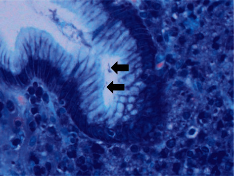
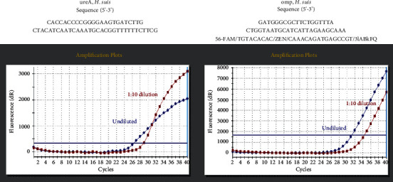
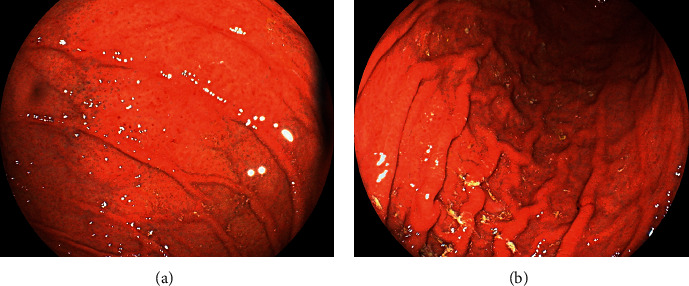
A 45-year-old Japanese man underwent esophagogastroduodenoscopy, which revealed spotty redness at the gastric fornix, mucosal swelling, diffuse redness in the corpus, and mucosal atrophy in the gastric angle and antrum. Histological examination showed rod-shaped bacteria that appeared larger than Helicobacter pylori. The patient tested positive for rapid urease test, and serum anti-H. pylori IgG antibody test results were negative. Further examination of the bacteria revealed that H. suis antibody test was positive, and the presence of H. suis was confirmed using H. suis-specific real-time PCR. H. suis was successfully eradicated after triple therapy with vonoprazan, amoxicillin, and clarithromycin. This case reinforces the notion that non-H. pylori Helicobacter species such as H. suis and H. heilmannii may be involved in the pathogenesis of active gastritis in patients who test negative for H. pylori antibodies.
Copyright © 2022 Masaya Iwamuro et al.
Conflict of interest statement
The authors declare that they have no conflicts of interest.
Figures

Similar articles
-
Investigation of endoscopic findings in nine cases of Helicobacter suis-infected gastritis complicated by gastric mucosa-associated lymphoid tissue lymphoma.Helicobacter. 2022 Jun;27(3):e12887. doi: 10.1111/hel.12887. Epub 2022 Apr 1. Helicobacter. 2022. PMID: 35363918
-
Gene expression of ornithine decarboxylase, cyclooxygenase-2, and gastrin in atrophic gastric mucosa infected with Helicobacter pylori before and after eradication therapy.Dig Dis Sci. 2003 Jan;48(1):36-46. doi: 10.1023/a:1021774029089. Dig Dis Sci. 2003. PMID: 12645788
-
Factors for Negative Result in Serum Anti-Helicobacter pylori IgG Antibody Test in Adult Subjects With Nodular Gastritis: A Single-center Study.Cureus. 2021 Jun 14;13(6):e15651. doi: 10.7759/cureus.15651. eCollection 2021 Jun. Cureus. 2021. PMID: 34306861 Free PMC article.
-
Elevated carbohydrate antigen 19-9 following Helicobacter suis gastritis and normalisation after eradication: first case report and review of the literature.Acta Gastroenterol Belg. 2022 Apr-Jun;85(2):403-405. doi: 10.51821/85.2.8826. Acta Gastroenterol Belg. 2022. PMID: 35709787 Review.
-
Gastric Helicobacter species associated with dogs, cats and pigs: significance for public and animal health.Vet Res. 2022 Jun 13;53(1):42. doi: 10.1186/s13567-022-01059-4. Vet Res. 2022. PMID: 35692057 Free PMC article. Review.
Cited by
-
The central role of gastrin in gastric cancer.Front Oncol. 2023 Oct 24;13:1176673. doi: 10.3389/fonc.2023.1176673. eCollection 2023. Front Oncol. 2023. PMID: 37941554 Free PMC article. Review.
-
Development of serological assays to identify Helicobacter suis and H. pylori infections.iScience. 2023 Mar 29;26(4):106522. doi: 10.1016/j.isci.2023.106522. eCollection 2023 Apr 21. iScience. 2023. PMID: 37123222 Free PMC article.
References
-
- Padra M., Adamczyk B., Flahou B., et al. Helicobacter suis infection alters glycosylation and decreases the pathogen growth inhibiting effect and binding avidity of gastric mucins. Mucosal Immunology . 2019;12(3):784–794. - PubMed
Publication types
LinkOut - more resources
Full Text Sources
