. 2022 Jul 12:13:909831.
doi: 10.3389/fimmu.2022.909831. eCollection 2022.

Placental Malaria is Associated with Higher LILRB2 Expression in Monocyte Subsets and Lower Anti-Malarial IgG Antibodies During Infancy

Affiliations

Placental Malaria is Associated with Higher LILRB2 Expression in Monocyte Subsets and Lower Anti-Malarial IgG Antibodies During Infancy

Celia Dechavanne et al. Front Immunol. .

Abstract

Background: Placental malaria (PM) is associated with a higher susceptibility of infants to Plasmodium falciparum (Pf) malaria. A hypothesis of immune tolerance has been suggested but no clear explanation has been provided so far. Our goal was to investigate the involvement of inhibitory receptors LILRB1 and LILRB2, known to drive immune evasion upon ligation with pathogen and/or host ligands, in PM-induced immune tolerance.

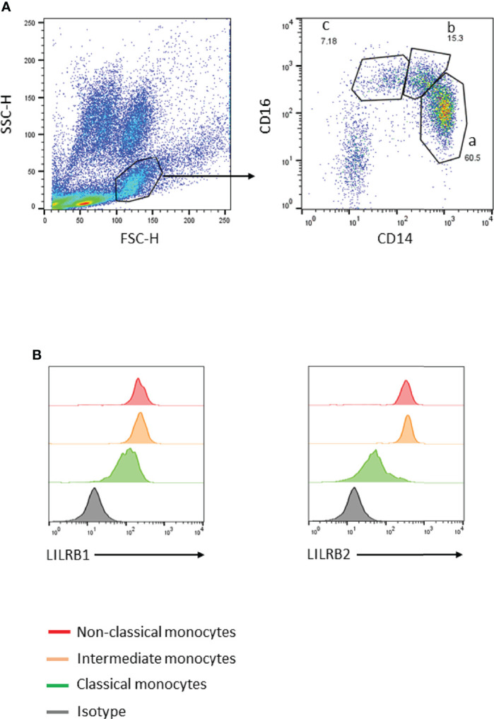
Method: Infants of women with or without PM were enrolled in Allada, southern Benin, and followed-up for 24 months. Antibodies with specificity for five blood stage parasite antigens were quantified by ELISA, and the frequency of immune cell subsets was quantified by flow cytometry. LILRB1 or LILRB2 expression was assessed on cells collected at 18 and 24 months of age.

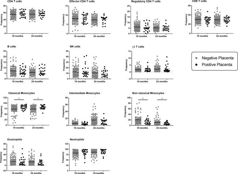
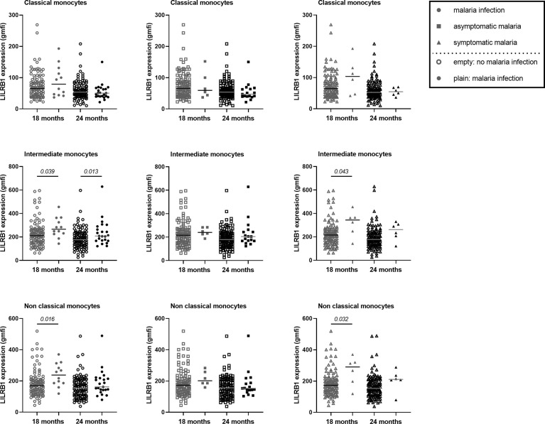
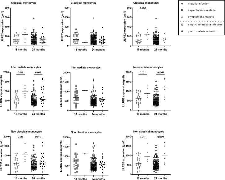
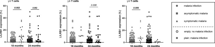
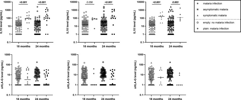
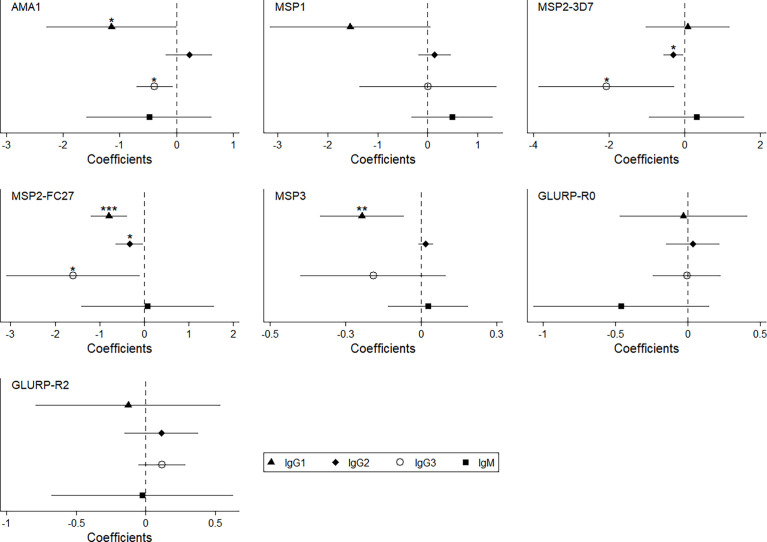
Findings: Infants born to women with PM had a higher risk of developing symptomatic malaria than those born to women without PM (IRR=1.53, p=0.040), and such infants displayed a lower frequency of non-classical monocytes (OR=0.74, p=0.01) that overexpressed LILRB2 (OR=1.36, p=0.002). Moreover, infants born to women with PM had lower levels of cytophilic IgG and higher levels of IL-10 during active infection.

Interpretation: Modulation of IgG and IL-10 levels could impair monocyte functions (opsonisation/phagocytosis) in infants born to women with PM, possibly contributing to their higher susceptibility to malaria. The long-lasting effect of PM on infants' monocytes was notable, raising questions about the capacity of ligands such as Rifins or HLA-I molecules to bind to LILRB1 and LILRB2 and to modulate immune responses, and about the reprogramming of neonatal monocytes/macrophages.

Keywords: Gd T cell; HLA-G; LILRB1; LILRB2; Plasmodium falciparum; immune tolerance; malaria candidate vaccine; monocytes.


Conflict of interest statement

The authors declare that the research was conducted in the absence of any commercial or financial relationships that could be construed as a potential conflict of interest.

Figures

Figure 1
Figure 1
Gating strategy to identify peripheral blood monocyte subsets. (A) Monocytes were gated using forward- and side-scatter properties. The 3 subsets were characterized through expression of CD14 and CD16 markers (a: classical CD14++CD16, b: intermediate CD14++CD16+, and c: non-classical CD14+CD16++). (B) LILRB1 or LILRB2 expression levels on the 3 monocyte subsets were defined using histogram geometric mean.
Figure 2
Figure 2
Immune cell populations at 18 and 24 months of age according to placenta malaria. The frequency of each immune cell population at either 18 or 24 months of age was represented in one graphic. Cell subset frequencies were determined as followed: CD4, CD8 and γδ T cells among total CD3+ T cells; regulatory and effector CD4+ T cells among CD4+ CD3+ T cells; Monocyte subsets among total monocytes, B or NK cells among lymphoid cells and neutrophils or eosinophils among granulocytes gated using forward- and side-scatter properties. The frequency of immune cells in infants born form mother with or without placental malaria are represented in gray and dark plain round respectively. The horizontal dark line in the beeswarm plot represents the median value. A linear regression was performed to compare the frequencies between the two groups of infants (infants born from a mother with an active placental malaria infection (n=27) vs infants born from a mother without placental malaria infection (n=127)). *: p value lower than 0.05 (and higher than 0.01).
Figure 3
Figure 3
Higher expression of LILRB1 on intermediate and non-classical monocytes at 18 and 24 months of age during active malaria. The expression of LILRB1 on immune cells was measured with the geometric mean of fluorescence intensity (gmfi) between Pf-infected and not-infected infants at 18 or 24 months of age. Total, asymptomatic or symptomatic malaria infections were represented with plain circle, square and triangle respectively. No infection at the visit was represented with empty symbols. The horizontal dark line in the beeswarm plot represents the median value. The number of infants with total, asymptomatic or symptomatic malaria infections varies between 6 and 23 for all monocyte subtypes. A linear regression (univariate analysis) was performed to compare the expression between the two groups of infants. P values are indicated in italic.
Figure 4
Figure 4
Higher expression of LILRB2 on intermediate and non-classical monocytes at 18 and 24 months during active malaria. The expression of LILRB2 on immune cells was measured with the geometric mean of fluorescence intensity (gmfi) between infected and not-infected infants at 18 or 24 months of age. Respectively 16 and 25 Pf infections were reported at 18 and 24 months among which 10 and 6 were symptomatic Pf infections. Total, asymptomatic or symptomatic malaria infections were represented with plain circle, square and triangle respectively. No infection at the visit was represented with empty symbols. The horizontal dark line in the beeswarm plot represents the median value. The number of infants with total, asymptomatic or symptomatic malaria infections varies between 1 and 23 for all monocyte subtypes. A linear regression (univariate analysis) was performed to compare the expression between the two groups of infants. P values are indicated in italic. In bold are the associations still significant after Bonferroni correction.
Figure 5
Figure 5
Modulation of LILRB1 expression on γδT cell surface in infant with malaria infections. The expression of LILRB1 on immune cells was measured with the geometric mean of fluorescence (gmfi) in infants at 18 and 24 months of age. Empty circle, square and triangle (gray or black) represented infant without malaria infection at the sample collect. Plain circle, square and triangle (gray or black) represented infant with malaria infection, symptomatic malaria and asymptomatic infection, respectively. The horizontal dark line in the beeswarm plot represents the median value. Respectively 16 and 25 Pf infections were reported at 18 and 24 months among which 10 and 6 were symptomatic Pf infections. P-values in bold were the one that were still considered significant after Bonferroni correction.
Figure 6
Figure 6
Higher IL10 level in infants with either symptomatic or asymptomatic malaria infection. The level of IL10 in pg/mL from the plasma of infant at 18 and 24 months of age was represented on the first row of graphics. HLA-G level in ng/mL from the plasma of infant at 18 and 24 months of age was represented on the second row of the graphics. Empty circle, square and triangle (gray or black) represented infants without malaria infection at the time of sample collection. Plain circle, square and triangle (gray or black) represented infant with malaria infection, symptomatic malaria and asymptomatic infection, respectively. The horizontal dark line in the beeswarm plot represents the median value. P-values in bold were the one that were still considered significant after Bonferroni correction.
Figure 7
Figure 7
Lower level of specific anti-malarial IgG1 and IgG3 in infant born to women with PM and with malaria infection. This coefplot showed the synergic effect of PM and Pf active infection (interaction term) on the levels of anti-malarial antibodies in infant between 18 and 24 months of age. The models were adjusted on age, gender, birth weight, maternity, ethnicity, maternal anemia, maternal IPTp and spatiotemporal malaria exposure risk.

References

    1. Rogerson SJ, Desai M, Mayor A, Sicuri E, Taylor SM, van Eijk AM. Burden, Pathology, and Costs of Malaria in Pregnancy: New Developments for an Old Problem. Lancet Infect Dis (2018) 18:e107–e18. doi: 10.1016/S1473-3099(18)30066-5 - DOI - PubMed
    1. White NJ, Pukrittayakamee S, Hien TT, Faiz MA, Mokuolu OA, Dondorp AM. Malaria. Lancet (2014)383(9918):723–35. doi: 10.1016/B978-0-7020-5101-2.00044-3 - DOI - PubMed
    1. Fried M, Duffy PE. Malaria During Pregnancy. Cold Spring Harb Perspect Med (2017) 7(6). doi: 10.1101/cshperspect.a025551 - DOI - PMC - PubMed
    1. Desai M, ter Kuile FO, Nosten F, McGready R, Asamoa K, Brabin B, et al. . Epidemiology and Burden of Malaria in Pregnancy. Lancet Infect Dis (2007) 7(2):93–104. doi: 10.1016/S1473-3099(07)70021-X - DOI - PubMed
    1. Le Hesran JY, Cot M, Personne P, Fievet N, Dubois B, Beyeme M, et al. . Maternal Placental Infection With Plasmodium Falciparum and Malaria Morbidity During the First 2 Years of Life. Am J Epidemiol (1997) 146(10):826–31. doi: 10.1093/oxfordjournals.aje.a009200 - DOI - PubMed

Publication types

MeSH terms