

Restorative therapy using microglial depletion and repopulation for central nervous system injuries and diseases
- PMID: 35911768
- PMCID: PMC9329909
- DOI: 10.3389/fimmu.2022.969127
Restorative therapy using microglial depletion and repopulation for central nervous system injuries and diseases
Abstract
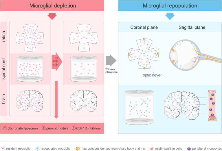
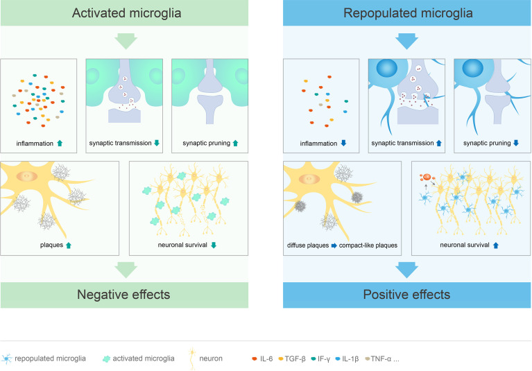
Microglia are important resident immune cells in the central nervous system (CNS) and play an important role in its development, homeostasis, and disease treatments. Activated microglia perform diverse functions in mouse models of CNS neurodegenerative diseases or deficits. In humans, microglia have been linked to various neurodegenerative diseases. Following brain or spinal cord injury, microglia express pro- and anti-inflammatory phenotypes at different stages of recovery. With the development of pharmacological and genetic tools for microglial depletion, studies have demonstrated that microglial depletion exerts both positive and negative effects in the treatment of CNS diseases. Notably, microglial depletion provides an empty niche that stimulates production of new microglia. Microglial depletion and repopulation can not only treat diseases by eliminating dysfunctional microglia but can also provide an indication of the molecular mechanisms of diseases. Although this approach has shown impressive results, its use is still in its infancy. In this review, we summarize the current pharmacological and genetic tools for microglial depletion and highlight recent advances in microglial repopulation therapy for the treatment and functional recovery of neurological diseases and deficits. Finally, we briefly discuss the therapeutic challenges and prospective uses of microglial repopulation therapy.
Keywords: central nervous system; depletion; diseases; microglia; repopulation.
Copyright © 2022 Shi, Zhang, Shang, Zhang, Xia, Fu and Yu.
Conflict of interest statement
The authors declare that the research was conducted in the absence of any commercial or financial relationships that could be construed as a potential conflict of interest.
Figures
Similar articles
-
The Role of Microglial Depletion Approaches in Pathological Condition of CNS.Cell Mol Neurobiol. 2023 Aug;43(6):2459-2471. doi: 10.1007/s10571-023-01326-8. Epub 2023 Feb 4. Cell Mol Neurobiol. 2023. PMID: 36738403 Free PMC article. Review.
-
Enforced microglial depletion and repopulation as a promising strategy for the treatment of neurological disorders.Glia. 2019 Feb;67(2):217-231. doi: 10.1002/glia.23529. Epub 2018 Oct 30. Glia. 2019. PMID: 30378163 Free PMC article. Review.
-
Novel Microglia-based Therapeutic Approaches to Neurodegenerative Disorders.Neurosci Bull. 2023 Mar;39(3):491-502. doi: 10.1007/s12264-022-01013-6. Epub 2023 Jan 3. Neurosci Bull. 2023. PMID: 36593381 Free PMC article. Review.
-
Microglial dynamics and emerging therapeutic strategies in CNS homeostasis and pathology.Front Pharmacol. 2025 May 13;16:1577809. doi: 10.3389/fphar.2025.1577809. eCollection 2025. Front Pharmacol. 2025. PMID: 40432891 Free PMC article. Review.
-
A limited capacity for microglial repopulation in the adult brain.Glia. 2018 Nov;66(11):2385-2396. doi: 10.1002/glia.23477. Epub 2018 Oct 28. Glia. 2018. PMID: 30370589 Free PMC article.
Cited by
-
Targeting Persistent Changes in Neuroimmune and Epigenetic Signaling in Adolescent Drinking to Treat Alcohol Use Disorder in Adulthood.Pharmacol Rev. 2023 Mar;75(2):380-396. doi: 10.1124/pharmrev.122.000710. Epub 2022 Dec 12. Pharmacol Rev. 2023. PMID: 36781218 Free PMC article. Review.
-
Microglia-mediated neuroinflammation in traumatic brain injury: a review.Mol Biol Rep. 2024 Oct 19;51(1):1073. doi: 10.1007/s11033-024-09995-4. Mol Biol Rep. 2024. PMID: 39425760 Review.
-
New insight on microglia activation in neurodegenerative diseases and therapeutics.Front Neurosci. 2023 Dec 22;17:1308345. doi: 10.3389/fnins.2023.1308345. eCollection 2023. Front Neurosci. 2023. PMID: 38188026 Free PMC article. Review.
-
Microglial repopulation restricts ocular inflammation and choroidal neovascularization in mice.Front Immunol. 2024 Apr 22;15:1366841. doi: 10.3389/fimmu.2024.1366841. eCollection 2024. Front Immunol. 2024. PMID: 38711521 Free PMC article.
-
Prolonged exposure of neonatal mice to sevoflurane leads to hyper-ramification in microglia, reduced contacts between microglia and synapses, and defects in adult behavior.Front Neurol. 2023 Mar 21;14:1142739. doi: 10.3389/fneur.2023.1142739. eCollection 2023. Front Neurol. 2023. PMID: 37025197 Free PMC article.
References
Publication types
MeSH terms
LinkOut - more resources
Full Text Sources
Medical