. 2022 Jul 11:13:849701.
doi: 10.3389/fimmu.2022.849701. eCollection 2022.

ICAM-1 on Breast Cancer Cells Suppresses Lung Metastasis but Is Dispensable for Tumor Growth and Killing by Cytotoxic T Cells

Affiliations

ICAM-1 on Breast Cancer Cells Suppresses Lung Metastasis but Is Dispensable for Tumor Growth and Killing by Cytotoxic T Cells

Ofer Regev et al. Front Immunol. .

Abstract

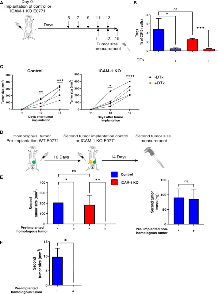
Breast tumors and their derived circulating cancer cells express the leukocyte β2 integrin ligand Intercellular adhesion molecule 1 (ICAM-1). We found that elevated ICAM-1 expression in breast cancer cells results in a favorable outcome and prolonged survival of breast cancer patients. We therefore assessed the direct in vivo contribution of ICAM-1 expressed by breast cancer cells to breast tumorigenesis and lung metastasis in syngeneic immunocompetent mice hosts using spontaneous and experimental models of the lung metastasis of the C57BL/6-derived E0771 cell line, a luminal B breast cancer subtype. Notably, the presence of ICAM-1 on E0771 did not alter tumor growth or the leukocyte composition in the tumor microenvironment. Interestingly, the elimination of Tregs led to the rapid killing of primary tumor cells independently of tumor ICAM-1 expression. The in vivo elimination of a primary E0771 tumor expressing the ovalbumin (OVA) model neoantigen by the OVA-specific OVA-tcr-I mice (OT-I) transgenic cytotoxic T lymphocytes (CTLs) also took place normally in the absence of ICAM-1 expression by E0771 breast cancer target cells. The whole lung imaging of these cells by light sheet microscopy (LSM) revealed that both Wild type (WT)- and ICAM-1-deficient E0771 cells were equally disseminated from resected tumors and accumulated inside the lung vasculature at similar magnitudes. ICAM-1-deficient breast cancer cells developed, however, much larger metastatic lesions than their control counterparts. Strikingly, the vast majority of these cells gave rise to intravascular tumor colonies both in spontaneous and experimental metastasis models. In the latter model, ICAM-1 expressing E0771- but not their ICAM-1-deficient counterparts were highly susceptible to elimination by neutrophils adoptively transferred from E0771 tumor-bearing donor mice. Ex vivo, neutrophils derived from tumor-bearing mice also killed cultured E0771 cells via ICAM-1-dependent interactions. Collectively, our results are a first indication that ICAM-1 expressed by metastatic breast cancer cells that expand inside the lung vasculature is involved in innate rather than in adaptive cancer cell killing. This is also a first indication that the breast tumor expression of ICAM-1 is not required for CTL-mediated killing but can function as a suppressor of intravascular breast cancer metastasis to lungs.

Keywords: adhesion; integrins; killing; metastasis; neutrophils; vasculature.


Conflict of interest statement

NA is a paid consultant for companies with an interest in liquid biopsy. The remaining authors declare that the research was conducted in the absence of any commercial or financial relationships that could be construed as a potential conflict of interest.

Figures

Figure 1
Figure 1
Tumor growth in vitro and in vivo is not affected by ICAM-1 expression on E0771 breast cancer cells. (A) Effect of E0771 ICAM-1 deletion on cell growth in vitro. 2 × 10⁴ control or ICAM-1 KO E0771 cells were seeded in 6-well plates in duplicates, and the growth curves of each experimental group were determined. The experiment was repeated twice. The results are an average of both experiments. **P< 0.005. (B) Effect of tumor ICAM-1 deletion on E0771 cell growth inhibition by the DNA damage agent etoposide. 1 × 10⁴ control or ICAM-1 KO E0771 cells were seeded in 96-well plates and exposed 1 day later to the indicated concentrations of etoposide. Approximately 48 h later, the cells were counted, and growth inhibition was determined and expressed as the percentage of triplicate values. The experiment was repeated twice. The results are an average of both experiments. The values are the mean ± SEM. (C) Effect of ICAM-1 deletion on E0771 growth in vivo. 1 × 10³ control or ICAM-1 KO E0771 cells suspended in Matrigel were implanted into the mammary fat pad of C57BL\6 female mice. The tumor volume was measured on the indicated days. The values are the mean ± SEM. n=5. (D) Primary tumors were implanted as in C and, 2 weeks later, were harvested and cut into 4-μm paraffin sections. Sections were immunostained for ICAM-1 and DAPI. A representative of 3–4 sections. (E) Myeloid and lymphoid compositions of the tumor microenvironment of primary control and ICAM-1 KO E0771 tumors. Tumors were implanted as in (C). Approximately 14 days later, the tumors were excised for single-cell suspension preparation. The numbers of DCs (CD45+/MHC-IIhi/CD11Chi), B cells (CD45+/CD19hi), CD8 T cells (CD45+/CD3+/CD8+), tumor-associated macrophages (CD45+/F4/80hi), neutrophils (CD45+/Ly6G+), and Tregs (CD45+/CD4+/CD25+/FOXP3+) were determined by FACS and were normalized to total CD45+ cells recovered from the tumors. N=7. (F, G) The numbers of DCs, B cells, and CD8+ T cells normalized to total CD45+ cells in the inguinal breast tumor-draining LNs (F) or the spleen (G). Primary control and ICAM-1 KO E0771 tumors were implanted as in C, and 14 days later, the inguinal TdLNs and the spleen were harvested, and their content of DCs, B cells and CD8+ T cells was determined by FACs. The bars in E–G depict the average values ± SEM. n=3. *p< 0.05.
Figure 2
Figure 2
Treg depletion and homologous tumor preimplantation eliminate primary E0771 tumors independently of ICAM-1 expression on the breast cancer cells. (A) A scheme depicting the experiment. 1 × 10³ control or ICAM-1 KO E0771 cells were implanted in the mammary fat pad of C57BL/6 mice. DTx was injected every 2 days at the indicated time points, and the tumor size was measured on the indicated days. (B) Treg depletion by DTx injections was validated on day 17 in the TdLNs of mice implanted with control or the ICAM-1 KO E0771 cells. n=3. *p<0.05; ***p<0.0005; (C) The graphs depict the tumor size in the differently treated mice at the indicated time points. Black: untreated mice. Red: DTx-treated mice. n=5. * p<0.05; **p<0.005; ***p<0.0005; ****p<0.0001. At all time points, no significant differences were found between control and ICAM-1 KO E0771 tumor sizes. (D) The experimental scheme: a WT E0771 tumor/Matrigel or a Matrigel suspension was pre-implanted in the left mammary fat pad (green-filled circle). Approximately 10 days later, a second primary tumor composed of either control or ICAM-1 KO E0771 cells (in Matrigel) was implanted in the right mammary fat pad (yellow-filled circle), and 14 days later, the size of the right tumor was measured. (E) The mean volume of the second primary tumors is depicted for the four experimental groups. n=5. The bars depict the average values ± range of either the tumor volume (left panel) or mass (right panel). *p<0.05; ***p<0.0005. (F) A primary B16-G-CSF tumor (1 × 10³ cells) or control Matrigel was implanted in the left mammary fat pad. Approximately 10 days later, a primary tumor of control E0771 cells was implanted in the right mammary fat pad as in (E) Approximately 12 days later, the E0771 tumor weight was measured. The bars depict the mean values ± SEM. n=3. *p<0.05.
Figure 3
Figure 3
Killing of OVA-expressing breast cancer E0771 cells in vivo by ex vivo expanded OT-1 CTLs is ICAM-1 independent. (A) Expression of the OVA-derived SIINFEKL peptide complexed with H-2Kb on control and ICAM-1 KO E0771 cells infected with an OVA-mCherry encoding vector. The SIINFEKL/H-2Kb expression was detected with the 25-D1.16 mAb (32) and a secondary Ab (Blue). Staining with the control secondary Ab is shown in red. (B) ICAM-1 on E0771 cells is functionally adhesive. OT-1 effector CTLs were settled on the monolayers of control or ICAM-1 KO E0771 cells for 4 min and subsequently subjected to progressively increasing shear stresses as described in the Materials and Methods section. The fractions of CTLs that remained adherent to the monolayers at the highest shear stress were determined in three fields of view. *p<0.05. (C) The killing of control or ICAM-1 KO OVA- expressing E0771 cells or non-OVA expressing control E0771 cells by OT-I effector CTLs was tested in vitro. Tumor cells were seeded in triplicate in 96- well plates, and 24 h later, CTLs were added to the wells in different effectors to E0771 target ratios. The number of viable OVA-expressing tumor cells in the wells was counted 24 h later, and the percentage of killing was calculated as explained in the Materials and Methods section. n=4. **p<0.005; ***p<0.0005. ****p<0.0001 for control and ICAM-1 KO OVA E0771 groups. (D) A scheme depicting the experiment. Mice were implanted with control or ICAM-1 KO OVA expressing E0771 cells as in Figure 1C. Approximately 3 days later, 107 of the OT-I effector CTLs were injected i.v. to each of the indicated experimental groups. (E) The tumor size was measured on days 12, 14, and 16 postimplantation. n=5. * p<0.05; **p<0.005; ***p<0.0005; ****p<0.0001.
Figure 4
Figure 4
Dissemination and early accumulation inside the blood vessels of E0771 breast cancer cells are not affected by their ICAM-1 expression in a spontaneous model of metastasis to lungs. (A) A scheme depicting the experiment. 3 × 10⁵ control or ICAM-1 KO RFP-expressing E0771 cells were implanted in the mammary fat pads of recipient mice. Two weeks later, lungs were harvested and processed for either the FACS analysis of total cell suspensions or for the fixation and clearance of lungs for imaging by three-dimensional (3D) light sheet fluorescence microscopy (LSM). For imaging, mice were intravenously stained with Alexa fluor 647 labeled CD31 mAb injected i.v. 15 min before euthanasia and lung harvesting. (B) The number of RFP-positive E0771 cells recovered in the lungs determined by FACS. n=4. (C) A representative LSM image of a whole lung lobe taken from Movie 8. The white cube represents the lung volume (2,160 µm × 2,560 µm × 2,000 µm) imaged in D. (D) A representative LSM image of RFP cells disseminated from the primary breast tumor and accumulated in the lung lobes. Green: lung auto-fluorescence. Red: E0771 cells in the imaged cube depicted in C, representative of four lung lobes isolated from two mice. The right images are the magnifications of the yellow rectangles marked in the middle images. Top images: control E0771 cells. Bottom images: ICAM-1 KO E0771 cells. The spots representing the RFP signal were <3,000 µm3 in volume. The bars in the right panel depict the average number ± SEM of control and ICAM-1 KO E0771 cells (spots) detected in 4 lung cubes (2,160 µm × 2,560 µm × 2,000 µm in dimension). (E) The left and right images depict, respectively, individual control and ICAM-1 KO E0771 cells (red) and CD31-labeled lung vessels (cyan). Images were taken from the LSM-analyzed lung samples represented by the examples in Movie 3. The bars in the right panel depict the percentage of control or ICAM-1 KO E0771 cells detected inside the CD31-labeled lung vessels. The bars in (B, D, E) depict the mean values ± SEM of the same 4 lung cubes analyzed in D.
Figure 5
Figure 5
ICAM-1 expression by E0771 breast cancer cells suppresses the late growth of disseminated cancer cells in a spontaneous model of breast cancer metastasis to lung. (A) A scheme depicting the experiment. 3×10⁵ control or ICAM-1 KO E0771 cells were implanted in the mammary fat pads of recipient mice. Tumors were resected 2 weeks later, and 2 weeks after resection, the lungs were harvested and processed for either the FACS analysis of total cell suspensions or of lungs subjected to fixation and clearance for imaging by LSM. The mice were intravenously stained with Alexa fluor 647-labeled CD31 mAb 15 min before lung harvesting as in Figure 4. (B) The number of RFP-positive E0771 cells recovered in the lungs determined by FACS. The bars depict the mean values ± SEM n=6. (C) Two weeks after tumor resection, lungs were harvested and the number of tumor foci detected on the lung surface was determined. The left panel depicts representative lung images. The right panel depicts the number of tumor foci on the lung surface ± SEM. n=4. ​**p<0.005. (D) Representative LSM images of RFP control and ICAM-1 KO cells disseminated from their respective primary breast tumors and accumulated in the lung lobes. The rectangles in the ICAM-1 KO lung image contain representative single tumor cells (i.e., spots of <3,000 µm3, top rectangle) or either oligocellular lesions or macrolesions (>40,000 µm3, bottom rectangle). The rectangles are each magnified in the two right images. Green: lung auto-fluorescence. Red: E0771 cells. (E) The left graph depicts the mean ± SEM number of RFP E0771 spots detected in 8 imaged lung cubes (each 2,160 µm × 2,560 µm × 2,000 µm in dimension). The middle graph depicts the mean ± SEM number of macrolesions detected in 8 lung cubes. The right graph depicts the mean ± SEM percentages of macrolesions out of all E0771 spots detected. *p<0.05; **p<0.005. (F) The left and right images depict, respectively, control and ICAM-1 KO E0771 cells (red) entrapped inside CD31-labeled lung blood vessels (cyan). The bars in the right panel depict the percentage of control or ICAM-1 KO E0771 cell spots detected inside the CD31-labeled lung vessels. Results are mean ± SEM of the same 8 lung cubes analyzed in E.
Figure 6
Figure 6
E0771 breast cancer ICAM-1 suppresses experimental metastasis to lung. (A) Visualization of singular and clustered E0771 cells accumulated inside lungs imaged by LSM. 1 × 10⁴ WT E0771 cells were injected i.v. Nuclei were labeled with REEDOT1. Nuclei (purple) and CMTMR-labeled E0771 breast cancer cells (red) were imaged by the LSM of cleared lungs lobes as outlined in Figures 4, 5. Bars = 20 µm. The right panel depicts the number of single cells and clusters detected in each imaged lung cube as in Figures 4, 5. The left lobes of two mice were imaged. The bars depict the mean values ± SEM. (B) LSM images of cleared lungs labeled with anti-CD31 mAb as in Figures 4, 5, 1 h postinjection of 1 × 10⁴ CMTMR-labeled E0771 cells (left panel) or 2 weeks postinjection of 104 RFP E0771 (right panel). (C) A scheme of the experimental metastasis model. Approximately 104 control or ICAM-1 KO E0771 cells were injected i.v., and 14 days later, lungs were harvested and the number of E0771 lesions was determined by the H&E staining of 5-µm-thin lung sections. (D) Left panel: The number of metastatic lesions detected in 100 lung sections of 20 mice. A total of 5 lung sections 100 μm apart were isolated from the left lung lobe of each recipient mouse. The bars depict the mean values ± SEM. **p<0.005. The right panel depicts the representative histological sections of the two different experimental groups. The arrowheads depict either small or large lesions. (E) H&E staining of the representative lung sections of lung lobes containing ICAM-1 KO lesions. The arrows depict intravascular lesions in small (left) and large (right) vessels.
Figure 7
Figure 7
Adoptively transferred neutrophils eliminate E0771 cells entering the lungs via breast cancer expressed ICAM-1. (A) A scheme of the experimental outline. Metastatic lesions developed by either control or ICAM-1 KO E0771 cells injected into the lungs of recipient mice was determined in the mice pretreated with either naïve or tumor entrained splenocytes isolated from donor spleens and introduced i.v. 24 h after E0771 injection. At the indicated groups, CD8+ T cells or neutrophils were depleted in donor mice with an i.p. injection of anti CD8 or Ly6G, respectively, 2 days before the adoptive transfer. (B) The number of metastatic lesions developed in recipient lungs subjected to the indicated treatments determined by H&E staining as in previous figures. The first experimental group was transferred with naïve splenocytes. Each group consisted of 3 mice. *p<0.05. **p<0.005 NS = non-significant. (C) In vitro killing of control or ICAM-1 KO E0771 cells by the blood-derived neutrophils of tumor-bearing mice determined in triplicates. n=12. **p<0.005. The bars in B and C depict the mean values ± SEM.

Similar articles

Cited by

References

    1. Allingham MJ, van Buul JD, Burridge K. ICAM-1-Mediated, Src- and Pyk2-Dependent Vascular Endothelial Cadherin Tyrosine Phosphorylation is Required for Leukocyte Transendothelial Migration. J Immunol (2007) 179:4053–64. doi: 10.4049/jimmunol.179.6.4053 - DOI - PubMed
    1. Hu G, Vogel SM, Schwartz DE, Malik AB, Minshall RD. Intercellular Adhesion Molecule-1-Dependent Neutrophil Adhesion to Endothelial Cells Induces Caveolae-Mediated Pulmonary Vascular Hyperpermeability. Circ Res (2008) 102:e120–31. doi: 10.1161/CIRCRESAHA.107.167486 - DOI - PMC - PubMed
    1. Juliano RL. Signal Transduction by Cell Adhesion Receptors and the Cytoskeleton: Functions of Integrins, Cadherins, Selectins, and Immunoglobulin-Superfamily Members. Annu Rev Pharmacol Toxicol (2002) 42:283–323. doi: 10.1146/annurev.pharmtox.42.090401.151133 - DOI - PubMed
    1. Yang L, Froio RM, Sciuto TE, Dvorak AM, Alon R, Luscinskas FW. ICAM-1 Regulates Neutrophil Adhesion and Transcellular Migration of TNF-Alpha-Activated Vascular Endothelium Under Flow. Blood (2005) 106:584–92. doi: ICAM-1 Regulates Neutrophil Adhesion and Transcellular Migration of TNF-Alpha-Activated Vascular Endothelium Under Flow - PMC - PubMed
    1. Abrahamt G, Colonno RJ. Many Rhinovirus Serotypes Share the Same Cellular Receptor. (1984) 51:340–5. doi: 10.1128/jvi.51.2.340-345.1984 - DOI - PMC - PubMed

Publication types

Substances