

Comparative assessment of cephalometric with its analogous photographic variables
- PMID: 35911811
- PMCID: PMC9326206
- DOI: 10.4103/njms.NJMS_267_20
Comparative assessment of cephalometric with its analogous photographic variables
Abstract
Objective: The purpose of this study was to evaluate the accuracy of photographic measurements and compare it with its analogous cephalometric variables.
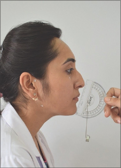
Materials and methods: Lateral cephalograms and standardized facial profile photographs were obtained from a sample of 120 subjects (92 females, 28 males; age 12-22 years with mean age of 17.5 years). A total of 4 linear and 7 angular measurements along with 3 ratios analogous to one another were measured on both. Descriptive statistics for all measurements were computed. Pearson's correlation coefficients were computed between analogous measurements, and regression analysis was done for each variable measured on the photograph to accurately predict the cephalometric variable.
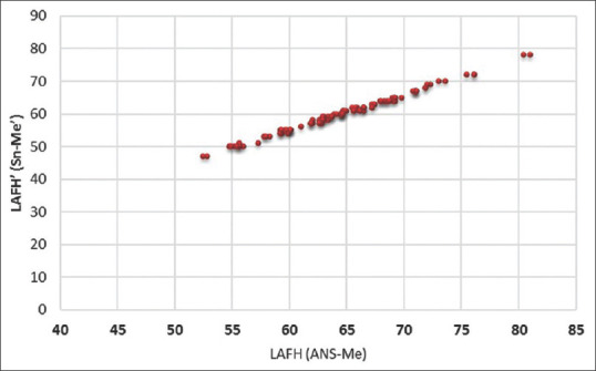
Results: The reliability of the standardized photographic technique was satisfactory. Most photographic measurements showed highly significant correlations (P < 0.001) with cephalometric variables. Among all measurements used, the A'N'B' angle was the most effective in explaining the variability of its analogous cephalometric (r2= 0.35). The Frankfort-mandibular plane angle' angle showed best results for vertical assessment (r2= 0.81) along with anterior face height (AFH) and lower anterior facial height (r2= 0.859) and ratio lower posterior facial height/AFH (r2= 0.702).
Conclusions: Although we cannot rule out lateral cephalogram as the primary record in orthodontics, photographic assessment can always be used through proper standardization, as an alternative diagnostic aid, and also for large-scale epidemiological purposes and places with unavailability of cephalostat.
Keywords: Diagnostic aid; lateral cephalograms; standardized facial profile photographs.
Copyright: © 2022 National Journal of Maxillofacial Surgery.
Figures


Similar articles
-
Photographic Assessment of Cephalometric Measurements in Skeletal Class II Cases: A Comparative Study.J Clin Diagn Res. 2017 Jun;11(6):ZC60-ZC64. doi: 10.7860/JCDR/2017/25042.10075. Epub 2017 Jun 1. J Clin Diagn Res. 2017. PMID: 28764295 Free PMC article.
-
Photographic assessment of cephalometric measurements.Angle Orthod. 2013 Nov;83(6):1049-58. doi: 10.2319/120712-925.1. Epub 2013 Apr 18. Angle Orthod. 2013. PMID: 23597034 Free PMC article.
-
Correlations between cephalometric and facial photographic measurements of craniofacial form.Am J Orthod Dentofacial Orthop. 2007 Jan;131(1):67-71. doi: 10.1016/j.ajodo.2005.02.033. Am J Orthod Dentofacial Orthop. 2007. PMID: 17208108
-
Soft-tissue and dentoskeletal profile changes associated with mandibular setback osteotomy.Am J Orthod Dentofacial Orthop. 1991 Oct;100(4):312-23. doi: 10.1016/0889-5406(91)70068-8. Am J Orthod Dentofacial Orthop. 1991. PMID: 1927981 Review.
-
The extraction of permanent second molars and its effect on the dentofacial complex of patients treated with the Tip-Edge appliance.Eur J Orthod. 2002 Oct;24(5):501-18. doi: 10.1093/ejo/24.5.501. Eur J Orthod. 2002. PMID: 12407946 Review.
Cited by
-
Comparative Evaluation of Photogrammetric, Radiographic, and Direct Measurements in Facial Analysis: A Cross-Sectional Study.Cureus. 2025 Mar 30;17(3):e81445. doi: 10.7759/cureus.81445. eCollection 2025 Mar. Cureus. 2025. PMID: 40303517 Free PMC article.
-
Differences in linear and angular measurement of the lower facial third in lateral cephalometry and lateral photometry: A comparative study.Saudi Dent J. 2024 Nov;36(11):1438-1441. doi: 10.1016/j.sdentj.2024.08.010. Epub 2024 Aug 22. Saudi Dent J. 2024. PMID: 39619709 Free PMC article.
References
-
- Broadbent BH. A newxray technique and its application to orthodontics. Angle Orthod. 1931;1:45–66.
-
- Zhang X, Hans MG, Graham G, Kirchner HL, Redline S. Correlations between cephalometric and facial photographic measurements of craniofacial form. Am J Orthod Dentofacial Orthop. 2007;131:67–71. - PubMed
-
- Smith NJ. Risk assessment: The philosophy underlying radiation protection. Int Dent J. 1987;37:43–51. - PubMed
-
- Staudt CB, Kiliaridis S. A nonradiographic approach to detect Class III skeletal discrepancies. Am J Orthod Dentofacial Orthop. 2009;136:52–8. - PubMed
-
- Barnett DP. Variations in the soft tissue profile and their relevance to the clinical assessment of skeletal pattern. Br J Orthod. 1975;2:235–8. - PubMed
LinkOut - more resources
Full Text Sources