

Case Report: The Coronal Magnetic Resonance Imaging of Three-Dimensional Fast-Field Echo With Water-Selective Excitation Can Identify the Wrapping of Spinal Nerve Fibers Into Subdural Tumors Prior to Operation
- PMID: 35911922
- PMCID: PMC9330486
- DOI: 10.3389/fneur.2022.945299
Case Report: The Coronal Magnetic Resonance Imaging of Three-Dimensional Fast-Field Echo With Water-Selective Excitation Can Identify the Wrapping of Spinal Nerve Fibers Into Subdural Tumors Prior to Operation
Abstract
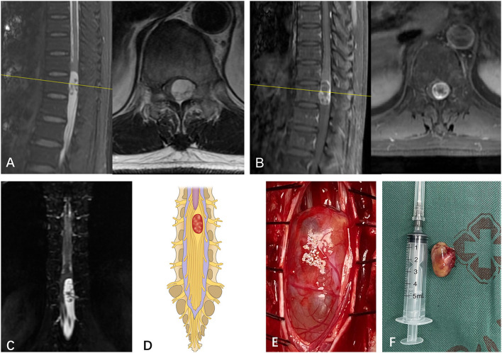
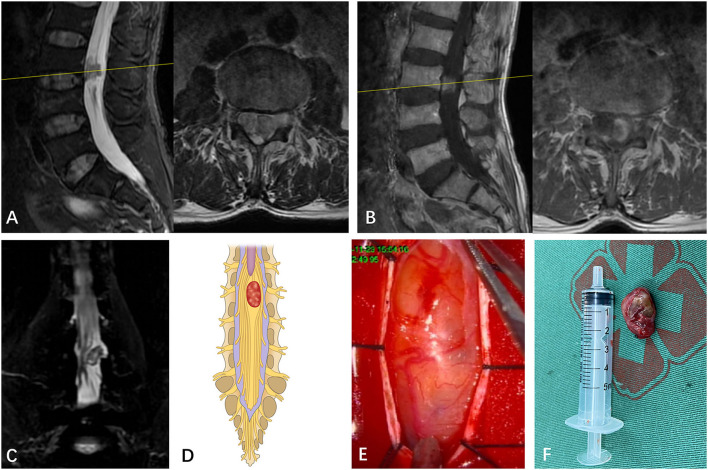
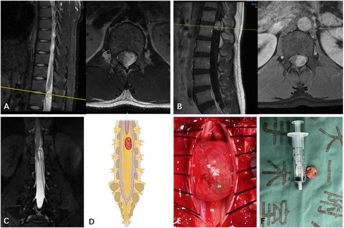
Purpose: In the present study, the authors intend to identify the spatial relationship between subdural tumors and spinal nerve fibers of cauda equina prior to operation using the coronal MRI of three-dimensional fast-field echo with water-selective excitation (CMRI).
Methods: In total, 30 case series with surgically and pathologically verified subdural tumors were enrolled in the present study. The spatial relationship between subdural tumors and spinal nerve fibers of the cauda equina was assessed via conventional MRI and CMRI by three experts prior to operation. The spatial relationship between subdural tumors and spinal nerve fibers of the cauda equina was classified using CMRI. The accuracy of imaging observation was determined via intraoperative observation.
Results: Though conventional MRI and gadolinium (Gd)-enhanced MRI (Gd MRI) cannot identify the spatial relationship between subdural tumors and spinal nerve fibers of cauda equina in all cases, CMRI can identify it prior to operation and divide the spatial relationship of spinal nerve fibers of cauda equina with subdural tumors into three types. CMRI shows higher sensitivity (97.44%) and specificity (90.47%) in identifying the spatial relationship of spinal nerve fibers of cauda equina with subdural tumors. Additionally, CMRI also showed a substantial agreement with a kappa value of 0.78.
Conclusion: Herein, the authors first describe a potential novel application that CMRI can successfully identify the spatial relationship between subdural tumors and spinal nerve fibers of cauda equina prior to operation, which play an essential role in making a prudent surgical plan and preventing postoperative nerve damage.
Summary: Intraoperative observation confirms spinal nerve fibers of cauda equina are often wrapped into subdural tumors of the thoracolumbar and lumbar region, which can result in a high rate of sensory and motor dysfunction after the operation due to the unconscious about the wrapping of nerves into subdural tumors prior to operation. To date, there is not an effective strategy to identify the wrapping before operation.
Keywords: cMRI; cauda equina; sensitivity; specificity; subdural tumors.
Copyright © 2022 Tang, Yuan, Yin, Zhu, Jia and Cheng.
Conflict of interest statement
The authors declare that the research was conducted in the absence of any commercial or financial relationships that could be construed as a potential conflict of interest.
Figures
Similar articles
-
Magnetic resonance imaging findings of redundant nerve roots of the cauda equina.World J Radiol. 2021 Jan 28;13(1):29-39. doi: 10.4329/wjr.v13.i1.29. World J Radiol. 2021. PMID: 33574992 Free PMC article.
-
A Case of Dural Herniation of the Cauda Equina Caused by Enlarged Spinal Subdural Extra-arachnoid Hygroma Following Lumbar Microsurgical Decompression: Case Report.NMC Case Rep J. 2021 Jun 17;8(1):261-265. doi: 10.2176/nmccrj.cr.2020-0301. eCollection 2021. NMC Case Rep J. 2021. PMID: 35079473 Free PMC article.
-
Circulatory dynamics of the cauda equina in lumbar canal stenosis using dynamic contrast-enhanced magnetic resonance imaging.Spine J. 2015 Oct 1;15(10):2132-41. doi: 10.1016/j.spinee.2015.05.014. Epub 2015 May 18. Spine J. 2015. PMID: 25998328
-
Acute Lumbar Spinal Subdural Hematoma Inducing Paraplegia After Lumbar Spinal Manipulation: Case Report and Literature Review.World Neurosurg. 2019 Aug;128:182-185. doi: 10.1016/j.wneu.2019.05.002. Epub 2019 May 9. World Neurosurg. 2019. PMID: 31078801 Review.
-
Magnetic resonance image findings of primary intradural Ewing sarcoma of the cauda equina: case report and review of the literature.Spine J. 2014 Apr;14(4):e7-e11. doi: 10.1016/j.spinee.2013.09.024. Epub 2013 Oct 11. Spine J. 2014. PMID: 24314762 Review.
Cited by
-
Evaluation of Safety and Efficacy of Preoperative Coronal MRI-Guided Minimally Invasive Surgery for Cervical Spondylotic Radiculopathy.Med Sci Monit. 2023 Dec 21;29:e942137. doi: 10.12659/MSM.942137. Med Sci Monit. 2023. PMID: 38124352 Free PMC article.
References
Publication types
LinkOut - more resources
Full Text Sources