

MRI Markers of Small Vessel Disease and the APOE Allele in Cognitive Impairment
- PMID: 35912087
- PMCID: PMC9326313
- DOI: 10.3389/fnagi.2022.897674
MRI Markers of Small Vessel Disease and the APOE Allele in Cognitive Impairment
Abstract
Objective: The apolipoprotein E (APOE) ε4 allele is the main genetic risk factor for dementia and Alzheimer's disease (AD), but the underlying mechanism for the increased risk is not well understood. Cerebral small vessel disease (SVD) is prevalent among patients with cognitive impairment and is thought to play an important role in the pathophysiology of dementia. We aimed to investigate the association between the APOE ε genotype and magnetic resonance imaging (MRI) markers of SVD in a memory clinic population.
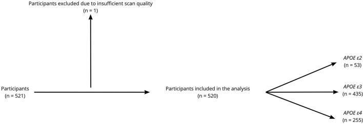
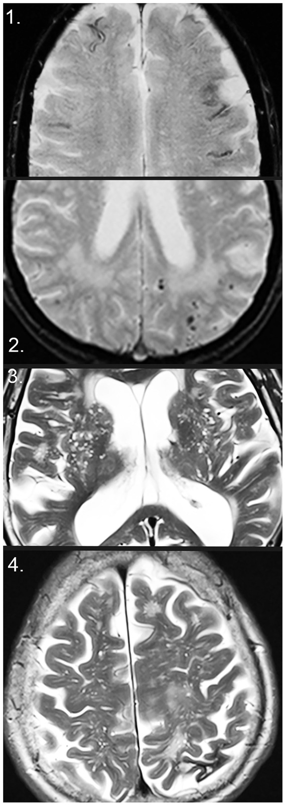
Material and methods: This is a cross-sectional study with a total of 520 patients undergoing dementia investigation, including an MRI brain scan and APOE genotyping in all patients enrolled, and cerebrospinal fluid (CSF) analysis for routine AD biomarkers in 399 patients. MR images were assessed for markers of SVD: cerebral microbleeds (CMBs), cortical superficial siderosis, intracerebral hemorrhage, white matter hyperintensities, lacunar infarcts, and enlarged perivascular spaces.
Results: Apolipoprotein E carriers with AD had a higher number of CMBs when looking at all brain regions and lobar brain regions (p < 0.001). A lower number of CMBs were seen in APOE ε2 (p < 0.05), ε3 and ε3/3 carriers (p < 0.001) when looking at all brain regions. A higher number of CMBs in deep and infratentorial regions were seen in APOE ε2 and ε3 (p < 0.05). In APOE ε4/4 carriers, CMBs, cortical superficial siderosis, white matter hyperintensities, and enlarged perivascular spaces were associated with lower levels of CSF amyloid β (Aβ) 42 in the whole cohort, and in individuals with AD and mild cognitive impairment (p < 0.05).
Conclusion: Apolipoprotein E ε4 is associated with MRI markers of SVD related to amyloid pathology, specifically CMBs and Aβ42 plaque formation in the brain, as reflected by decreased CSF Aβ42 levels, whereas APOE ε3 and ε2 are associated with the markers of hypertensive arteriopathy, as reflected by the association with CMBs in deep and infratentorial brain regions.
Keywords: Alzheimer's disease; apolipoprotein E; cerebral amyloid angiopathy; cerebral small vessel disease; dementia; hypertensive vasculopathy; magnetic resonance imaging.
Copyright © 2022 Shams, Shams, Martola, Cavallin, Granberg, Kaijser, Wintermark, Westman, Aspelin, Kristoffersen Wiberg and Wahlund.
Conflict of interest statement
The authors declare that the research was conducted in the absence of any commercial or financial relationships that could be construed as a potential conflict of interest.
Figures
Similar articles
-
Association of candidate genetic variants and circulating levels of ApoE/ApoJ with common neuroimaging features of cerebral amyloid angiopathy.Front Aging Neurosci. 2023 Apr 11;15:1134399. doi: 10.3389/fnagi.2023.1134399. eCollection 2023. Front Aging Neurosci. 2023. PMID: 37113571 Free PMC article.
-
Apolipoprotein E genotype and the diagnostic accuracy of cerebrospinal fluid biomarkers for Alzheimer disease.JAMA Psychiatry. 2014 Oct;71(10):1183-91. doi: 10.1001/jamapsychiatry.2014.1060. JAMA Psychiatry. 2014. PMID: 25162367
-
Topography and Determinants of Magnetic Resonance Imaging (MRI)-Visible Perivascular Spaces in a Large Memory Clinic Cohort.J Am Heart Assoc. 2017 Sep 22;6(9):e006279. doi: 10.1161/JAHA.117.006279. J Am Heart Assoc. 2017. PMID: 28939709 Free PMC article.
-
Cerebral small vessel disease and the risk of Alzheimer's disease: A systematic review.Ageing Res Rev. 2018 Nov;47:41-48. doi: 10.1016/j.arr.2018.06.002. Epub 2018 Jun 26. Ageing Res Rev. 2018. PMID: 29898422
-
Cerebral microbleeds: a review.Panminerva Med. 2012 Sep;54(3):149-60. Panminerva Med. 2012. PMID: 22801432 Review.
Cited by
-
Association of candidate genetic variants and circulating levels of ApoE/ApoJ with common neuroimaging features of cerebral amyloid angiopathy.Front Aging Neurosci. 2023 Apr 11;15:1134399. doi: 10.3389/fnagi.2023.1134399. eCollection 2023. Front Aging Neurosci. 2023. PMID: 37113571 Free PMC article.
-
Factors Associated With Post-Stroke Cognitive Impairment: A Narrative Review.Brain Neurorehabil. 2024 Nov 21;17(3):e20. doi: 10.12786/bn.2024.17.e20. eCollection 2024 Nov. Brain Neurorehabil. 2024. PMID: 39649710 Free PMC article. Review.
-
Cerebral white matter rarefaction has both neurodegenerative and vascular causes and may primarily be a distal axonopathy.J Neuropathol Exp Neurol. 2023 May 25;82(6):457-466. doi: 10.1093/jnen/nlad026. J Neuropathol Exp Neurol. 2023. PMID: 37071794 Free PMC article.
-
Imaging markers of cerebral small vessel disease are associated with Alzheimer's disease: a systematic review and meta-analysis.Front Aging Neurosci. 2025 Jan 28;17:1498636. doi: 10.3389/fnagi.2025.1498636. eCollection 2025. Front Aging Neurosci. 2025. PMID: 40071121 Free PMC article.
-
Role of white matter hyperintensity in effects of apolipoprotein E on cognitive injury.Front Hum Neurosci. 2023 May 19;17:1176690. doi: 10.3389/fnhum.2023.1176690. eCollection 2023. Front Hum Neurosci. 2023. PMID: 37275347 Free PMC article. Review.
References
-
- Brickman A. M., Honig L. S., Scarmeas N., Tatarina O., Sanders L., Albert M. S., et al. . (2008). Measuring cerebral atrophy and white matter hyperintensity burden to predict the rate of cognitive decline in Alzheimer disease. Arch. Neurol. 65, 1202–1208. 10.1001/archneur.65.9.1202 - DOI - PMC - PubMed
-
- Brickman A. M., Schupf N., Manly J. J., Stern Y., Luchsinger J. A., Provenzano F. A., et al. . (2014). APOE ε4 and risk for Alzheimer's disease: Do regionally distributed white matter hyperintensities play a role? Alzheimers Dement J Alzheimers Assoc 10, 619–629. 10.1016/j.jalz.2014.07.155 - DOI - PMC - PubMed
LinkOut - more resources
Full Text Sources
Miscellaneous