Review
. 2022 Jul 14:10:930355.
doi: 10.3389/fcell.2022.930355. eCollection 2022.

Moonlighting at the Poles: Non-Canonical Functions of Centrosomes

Affiliations
Review

Moonlighting at the Poles: Non-Canonical Functions of Centrosomes

Laurence Langlois-Lemay et al. Front Cell Dev Biol. .

Abstract

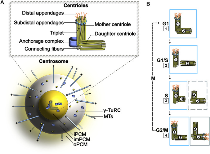
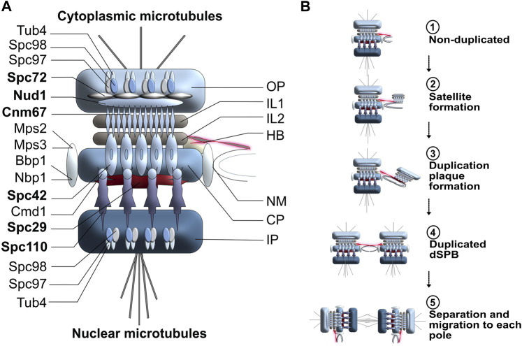
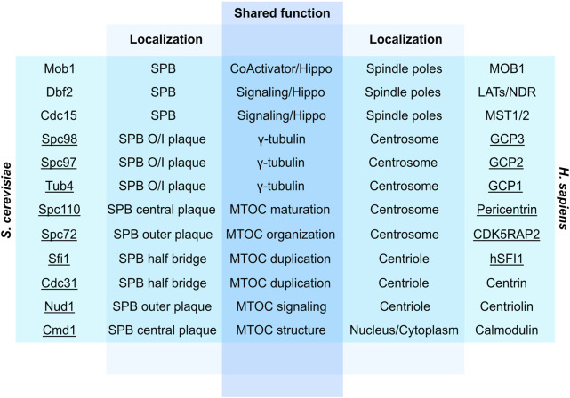
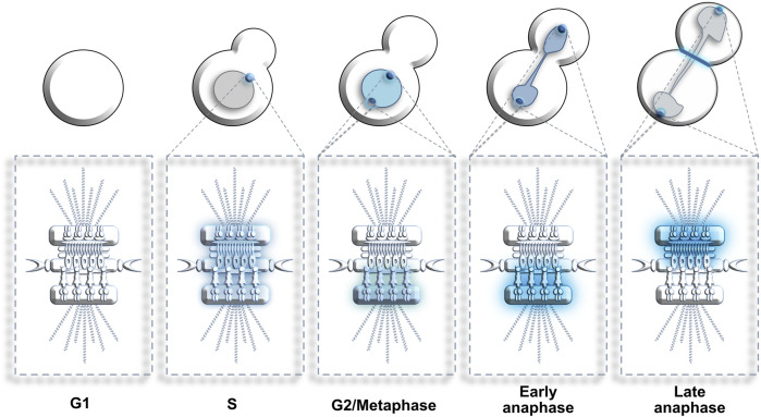
Centrosomes are best known as the microtubule organizing centers (MTOCs) of eukaryotic cells. In addition to their classic role in chromosome segregation, centrosomes play diverse roles unrelated to their MTOC activity during cell proliferation and quiescence. Metazoan centrosomes and their functional doppelgängers from lower eukaryotes, the spindle pole bodies (SPBs), act as important structural platforms that orchestrate signaling events essential for cell cycle progression, cellular responses to DNA damage, sensory reception and cell homeostasis. Here, we provide a critical overview of the unconventional and often overlooked roles of centrosomes/SPBs in the life cycle of eukaryotic cells.

Keywords: Cdc5; MTOCs; PLK1; cell cycle; centrosomes; spindle pole bodies.


Conflict of interest statement

The authors declare that the research was conducted in the absence of any commercial or financial relationships that could be construed as a potential conflict of interest.

Figures

FIGURE 1
FIGURE 1
(A) Schematic representation of the centrosome. PCM, Pericentriolar material; iPCM, Inner PCM; imPCM, Intermediate PCM; oPCM, Outer PCM (B) Centrosome duplication cycle. The duplication of centrosomes is termed semi-conservative, as each older centriole will generate a new centriole. 1–2. At the G1/S transition, the two centrioles separate. 3. In S phase, PCM forms around each parting centriole. 4. Daughter centrioles expand orthogonally and reach opposite poles. See text for more details.
FIGURE 2
FIGURE 2
(A) Schematic representation of budding yeast SPB organization and duplication cycle. OP, Outer plaque; IL1, Intermediate layer 1; IL2, Intermediate layer 2; HB, Half-bridge; NM, Nuclear membrane; CP, central plaque; IP; Inner plaque. Core SPB components are highlighted in bold. (B) SPB duplication cycle in budding yeast. The duplication process of the SPB is conservative and highly dynamic. Step 1: In early G1, the half-bridge is connected to the SPB central plaque and will act as a scaffold for SPB duplication. Step 2: The half-bridge elongates and the core of the daughter SPB (satellite) is generated on the cytoplasmic face of the half-bridge. Step 3: The duplication plaque, resulting from the elongation and growth of the satellite SPB, matures and mimics the cytoplasmic organization of a mature SPB. Step 4: The half-bridge retracts and fuses to the nuclear membrane. The daughter SPB is assembled and is embedded in the nuclear membrane next to the mother SPB. Step 5: The link between mother and daughter SPBs breaks, leading to the separation of the two organelles.
FIGURE 3
FIGURE 3
Overview of conserved yeast and human proteins involved in MTOC structure, signaling, duplication and function. Underlined are physical constituents of centrosomes/SPBs. SPB, Spindle pole body; O/I, Outer/Inner; Hippo, Hippo pathway; Pericentrin, Kendrin/CG-NAP (Fraschini, 2019).
FIGURE 4
FIGURE 4
Dynamic localization of Cdc5/Polo kinase at SPBs. G1: Cdc5 is absent from cells. S: Cdc5 enriches at the non-duplicated SPB. G2 to metaphase: Cdc5 decorates the nucleus and the nuclear surface of both SPBs. Early anaphase: Cdc5 concentration at the nuclear surface of both SPBs increases. Late anaphase: Cdc5 relocates from the inner to the outer surface of both SPBs (and bud neck) where it stimulates mitotic exit. Blue color represents enrichment of Cdc5. Color intensity represents Cdc5 concentration levels.
FIGURE 5
FIGURE 5
Centrosome-specific regulation of protein kinase A (PKA) signaling. (A) PKA is a tetrameric holoenzyme composed of two regulatory subunits and two catalytic subunits. Its activity relies on cyclic AMP (cAMP) cellular levels and is involved in many regulatory processes. (B) Regulation of PKA following G protein-coupled receptor (GPCR) activation. A ligand binds to the GPCR (step 1), initiating the signal transduction cascade. This signal induces a GDP to GTP exchange on a heterotrimeric G complex (step 2). The Gα subunit is released and binds to adenylyl cyclase (AC), an event that induces the formation of cyclic adenosine monophosphate (cAMP) from ATP. A subpopulation of PKA anchors at the centrosomes (step 3). The resulting AKAP450 complex increases PKA affinity for cAMP. Centrosomal PKA is selectively activated by cAMP, whilst cytosolic PKA (shown in grey) remains mostly inactive (step 4). A specialized cellular response is induced by the catalytic activation of PKA at centrosomes (step 5).
FIGURE 6
FIGURE 6
Visual representation of sporulation and ascus formation in budding yeast. (A) In response to environmental stressors, diploid yeast cells initiate the sporulation program. (B) Completion of meiosis I nuclear division. (C) After the second round of chromosome segregation, the prospore membrane (shown in orange) forms and expands around each duplicated SPB (shown in blue). (D) The membrane grows and encapsulates each haploid nucleus in the tetrad. (E) Spore wall assembly begins and the remnants of the mother cell breaks down.
FIGURE 7
FIGURE 7
Cellular changes associated with the quiescent state in yeast. These changes include the disappearance of cytoplasmic microtubules (MTs) and formation of a nuclear bundle of MTs (nMTs) that spans the entire nucleus. Centromeres (shown in yellow) normally cluster together at the end of nuclear MTs in interphase cells (left) but get redistributed along the length of the newly formed nMT bundle in quiescent cells (right). Chromosome arms are omitted from this figure to simplify the representation. See text for more details.

Similar articles

Cited by

References

    1. Adams I. R., Kilmartin J. V. (1999). Localization of Core Spindle Pole Body (SPB) Components during SPB Duplication in Saccharomyces cerevisiae . J. Cell Biol. 145 (4), 809–823. 10.1083/jcb.145.4.809 - DOI - PMC - PubMed
    1. Agircan F. G., Schiebel E., Mardin B. R. (2014). Separate to Operate: Control of Centrosome Positioning and Separation. Phil. Trans. R. Soc. B 369 (1650), 20130461. 10.1098/rstb.2013.0461 - DOI - PMC - PubMed
    1. Alcaide-Gavilán M., Lahoz A., Daga R. R., Jimenez J. (2014). Feedback Regulation of SIN by Etd1 and Rho1 in Fission Yeast. Genet. Feb 196 (2), 455–470. 10.1534/genetics.113.155218 - DOI - PMC - PubMed
    1. Alfa C. E., Booher R., Beach D., Hyams J. S. (1989). Fission Yeast Cyclin: Subcellular Localisation and Cell Cycle Regulation. J. Cell Sci. Suppl. 1989, 9–19. 10.1242/jcs.1989 - DOI - PubMed
    1. Andersen J. S., Wilkinson C. J., Mayor T., Mortensen P., Nigg E. A., Mann M. (2003). Proteomic Characterization of the Human Centrosome by Protein Correlation Profiling. Nature 426 (6966), 570–574. 10.1038/nature02166 - DOI - PubMed