Review
. 2022 Jul 13:10:910440.
doi: 10.3389/fcell.2022.910440. eCollection 2022.

Chromatin and the Cellular Response to Particle Radiation-Induced Oxidative and Clustered DNA Damage

Affiliations
Review

Chromatin and the Cellular Response to Particle Radiation-Induced Oxidative and Clustered DNA Damage

John M Danforth et al. Front Cell Dev Biol. .

Abstract

Exposure to environmental ionizing radiation is prevalent, with greatest lifetime doses typically from high Linear Energy Transfer (high-LET) alpha particles via the radioactive decay of radon gas in indoor air. Particle radiation is highly genotoxic, inducing DNA damage including oxidative base lesions and DNA double strand breaks. Due to the ionization density of high-LET radiation, the consequent damage is highly clustered wherein ≥2 distinct DNA lesions occur within 1-2 helical turns of one another. These multiply-damaged sites are difficult for eukaryotic cells to resolve either quickly or accurately, resulting in the persistence of DNA damage and/or the accumulation of mutations at a greater rate per absorbed dose, relative to lower LET radiation types. The proximity of the same and different types of DNA lesions to one another is challenging for DNA repair processes, with diverse pathways often confounding or interplaying with one another in complex ways. In this context, understanding the state of the higher order chromatin compaction and arrangements is essential, as it influences the density of damage produced by high-LET radiation and regulates the recruitment and activity of DNA repair factors. This review will summarize the latest research exploring the processes by which clustered DNA damage sites are induced, detected, and repaired in the context of chromatin.

Keywords: DNA repair; chromatin; clustered DNA damage; multiply-damaged sites; particle radiation.


Conflict of interest statement

The authors declare that the research was conducted in the absence of any commercial or financial relationships that could be construed as a potential conflict of interest.

Figures

FIGURE 1
FIGURE 1
A schematic of dose-depth curves of ion beams and low-LET photon and electron radiation illustrating the difference in peak energy deposition as the radiation travels through tissue.
FIGURE 2
FIGURE 2
Simplified illustration of energy deposition of equivalent doses of a high-LET particle and a low-LET photon generated secondary electron, with ionizing (large) and excitation (small) events along their trajectories, represented by solid yellow arrows. DNA damage can be generated either directly through ionization (large) or indirectly through generation of radicals after the ionization or excitation of water (small), where dashed yellow lines represent the trajectories of secondary electrons and delta rays.
FIGURE 3
FIGURE 3
Schematic of IR induced DNA damage. Simple damage includes base lesions such as oxidized bases, apurinic or apyrimidinic sites (AP sites), and isolated strand breaks. Clustered damage occurring within 1–2 helical turns can be comprised of ≥2 simple lesions which may be non-DSB oxidative clustered DNA lesions (OCDLs) or clustered DSBs. Strand breaks can also be complex, with additional base lesions comprising “dirty ends”.
FIGURE 4
FIGURE 4
Cumulative DNA damage observable with (daily) repetitive alpha particle exposure even at low doses (<100 mGy). 48BR cells were given a one-time (acute) dose of 500 or 1,000 mGy particle IR (light grey) or repetitively exposed to 35, 70 or 140 mGy particle IR once per day for 15 days (dark grey), with sham irradiated control (white). Repetitively irradiated cells were harvested 24 h after the final dose, whilst acutely irradiated cells were harvested 1 h post IR. All cells were then fixed, stained and imaged together, generating refined γH2AX signal; black bars = mean ± SEM of n = 3 (1,700 cells total per condition). ** = statistically significant (<0.01); **** = statistically significant (<0.0001). Refined γ-H2AX represents the average intensity of γ-H2AX objects per individual cell as a function of the size of the nucleus measured as DAPI volume, and corrected for signal expansion of γ-H2AX during DSB repair. For a more detailed explanation of refined γ-H2AX, see (Stanley et al., 2020).
FIGURE 5
FIGURE 5
Schematic of pathways for base lesion and single strand break (SSB) repair. Base damage is recognized by a DNA glycosylase and subsequent enzymatic processing generates an abasic site. Abasic sites (also called AP, for apurinic/apyrimidinic sites) are processed by bifunctional DNA glycosylases or AP endonucleases, generating a gap in the DNA strand that can have a variety of ends requiring processing to enable ligation (Kim and Wilson III 2012). The gap is then filled by Polβ or Polλ via short patch BER, and the nick is resolved by XRCC1-DNA Lig III complex (Beard, Prasad, and Wilson 2006; Lebedeva et al., 2005; Cappelli et al., 1997). In long patch BER, Polδ and Polε fill in 2–12 nucleotides, forming an intermediary flap structure that is resolved by flap endonucleases (Matsumoto, Kim, and Bogenhagen 1994; Liu, Kao, and Bambara 2004; Wang, Wu, and Friedberg 1993). Flap removal generates SSB which is sealed by DNA ligase I. In direct SSB repair, damage is signaled by the activity of PARP1 (Masson et al., 1998). Gap filling and ligation in SSB repair are then accomplished in similar fashion to BER.
FIGURE 6
FIGURE 6
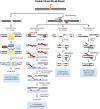
Schematic of end protected and resection dependent DNA double strand break (DSB) repair pathways. NHEJ is initiated by the binding of the Ku70/80 heterodimer which recruits DNA-PKcs, a kinase which functions to tether the break ends and recruit the factors involved in end processing and ligation shown here. In S- and G2, the increase in active cyclin dependent kinases (CDKs) in complex with cyclins phosphorylate key proteins like the resection mediating factor, CtIP which activates the nuclease activity of MRE11 in the MRN complex (Huertas et al., 2008; Makharashvili et al., 2014). The subsequent resection is the key event that commits a DSB to HR repair, producing single stranded 3′-DNA ends, which cannot be ligated by NHEJ (Williams et al., 2009; Himmels and Sartori 2016). Resected DNA is converted into a pre-synaptic filament with the replacement of RPA by the Rad51 recombinase protein, which then searches for homology (Thorslund et al., 2010; Ma et al., 2017; Sullivan and Bernstein 2018). Once homology is found, the filament invades the duplex DNA and the strand is elongated through synthesis (McVey et al., 2016; Wright, Shah, and Heyer 2018; Tavares et al., 2019). After the second end is captured and synthesized the resulting four strand structure, the double Holliday Junction, can then be dissolved or resolved (Svendsen and Harper 2010; Wyatt et al., 2013; Bizard and Hickson 2014; Wyatt and West 2014). In lieu of rad51 filament formation, in SSA Rad52 functions to anneal homologous DNA sequences and is always associated with the deletion of DNA between the two regions of homology. The resulting flap of DNA is subsequently removed by the XPF-ERCC1 nuclease complex after DNA ligation, which is enhanced by the presence of Rad52 (Motycka et al., 2004; Bhargava, Onyango, and Stark 2016). The occurrence of MMEJ has been correlated to the length of the 3′ overhang end, with longer ends of 45–100 bps being ideal for Polymerase θ (Polθ) helicase to act on the single stranded ends, facilitating the annealing of these ends as well as low fidelity polymerase activity for extension (Wyatt et al., 2016; Black et al., 2019). After extension, the 3′-flap overhangs are trimmed by the XPF-ERCC1 nuclease complex, and finally annealing is performed by Ligase I/III.
FIGURE 7
FIGURE 7
Schematic of DSB repair pathway choice for high-LET induced damage in the context of chromatin. In the repair of simple euchromatic DSBs NHEJ functions as the predominant repair pathway, with a portion of repair carried out by Artemis mediated NHEJ (G0/G1) or HR (S/G2). Initiation of NHEJ at sites of clustered damage is attempted, but due to the presence of short fragments which inhibit activation of DNA-PKcs, pathway choice shifts to resection mediated pathways. Hence, alternative, more error prone pathways are considered the major repair mechanism for high-LET induced clustered DSBs, but a more detailed mechanism for pathway choice in a chromatin context is still uncertain. Bolded arrows denote the ‘preferred’ pathway for repair. Detailed pathway schematics can be found in Figure 6.
FIGURE 8
FIGURE 8
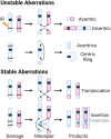
DSBs caused by exposure to IR (black bars) shown on chromosomes with centromeres (blue and red, and dark blue, respectively). Dotted arrows indicate where the breaks rejoin. Products of the various misrepair scenarios include unstable aberrations: acentric and dicentric chromosomes, centric rings; and stable aberrations: translocations, as well as insertions and inversions. For insertions and inversions, black arrows show where the fragments join during an insertion, and grey arrows show where the inverted chromosomal fragment joins. The inversion is further illustrated by inverting the color gradient of the inserted chromosome fragment and the final inversion product is enclosed in a grey box.

References

    1. Ahrabi S., Sarkar S., Pfister S. X., Pirovano G., Higgins G. S., Porter A. C. G., et al. (2016). A Role for Human Homologous Recombination Factors in Suppressing Microhomology-Mediated End Joining. Nucleic Acids Res. 44 (12), 5743–5757. 10.1093/nar/gkw326 - DOI - PMC - PubMed
    1. Alessio N., Esposito G., Galano G., De Rosa R., Anello P., Peluso G., et al. (2017). Irradiation of Mesenchymal Stromal Cells with Low and High Doses of Alpha Particles Induces Senescence And/or Apoptosis. J. Cell. Biochem. 118 (9), 2993–3002. 10.1002/jcb.25961 - DOI - PubMed
    1. Alexandrov L. B., Nik-Zainal Serena, Nik-Zainal S., Wedge D. C., Aparicio S. A. J. R., Behjati S., et al. (2013). Signatures of Mutational Processes in Human Cancer. Nature 500 (7463), 415–421. 10.1038/nature12477 - DOI - PMC - PubMed
    1. Alizadeh E., Orlando T. M., Sanche L. (2015). Biomolecular Damage Induced by Ionizing Radiation: The Direct and Indirect Effects of Low-Energy Electrons on DNA. Annu. Rev. Phys. Chem. 66, 379–398. 10.1146/annurev-physchem-040513-103605 - DOI - PubMed
    1. Allshire R. C., Madhani H. D. (2018). Ten Principles of Heterochromatin Formation and Function. Nat. Rev. Mol. Cell Biol. 19 (4), 229–244. 10.1038/nrm.2017.119 - DOI - PMC - PubMed

LinkOut - more resources