Case Reports
. 2022 Jun 28:12:Doc18.
doi: 10.3205/oc000205. eCollection 2022.

Combined central retinal artery and vein occlusion following trabeculectomy

Affiliations
Case Reports

Combined central retinal artery and vein occlusion following trabeculectomy

Albert John Bromeo et al. GMS Ophthalmol Cases. .

Abstract

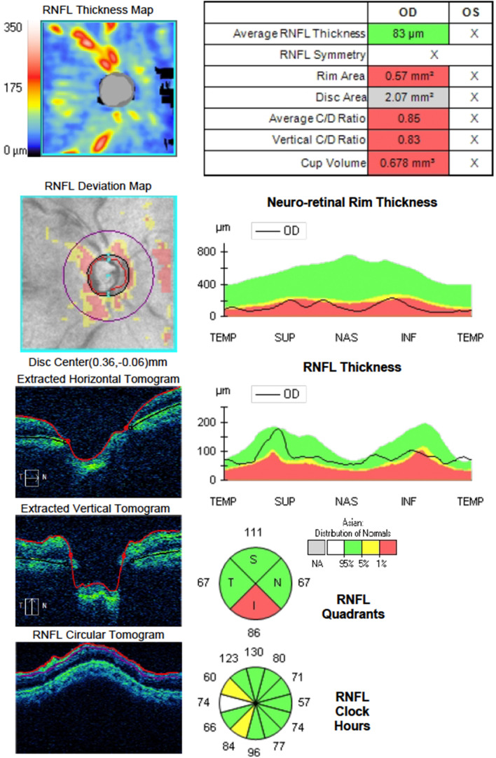
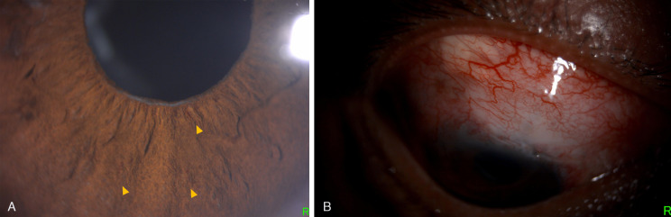
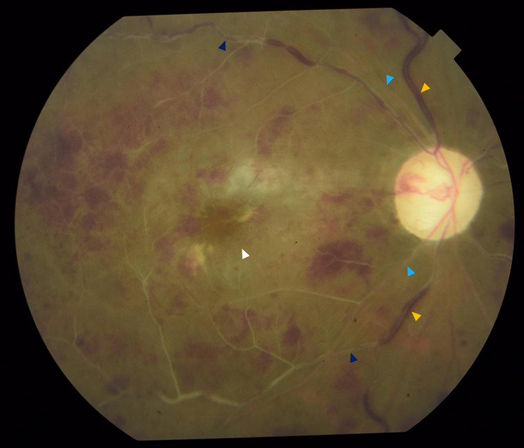
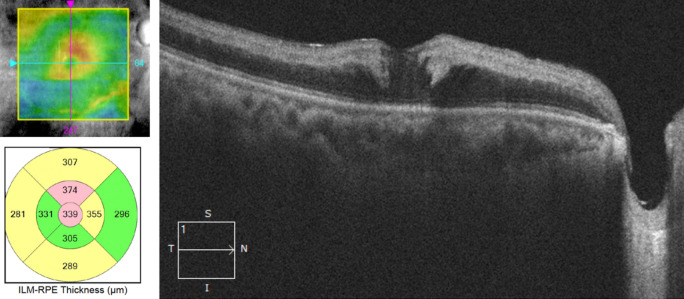
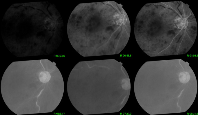
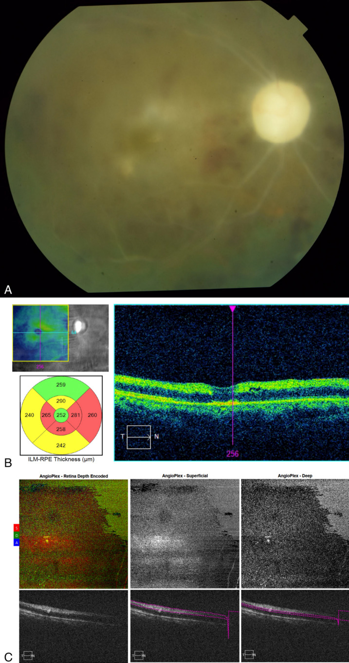
Retinal vascular events may occur as rare complications of glaucoma procedures due to various factors, including exacerbation of ischemia in patients with pre-existing vascular comorbidities, toxic effect of mitomycin-C, and decompression retinopathy. We present the case of a 47-year-old hypertensive male who underwent trabeculectomy for advanced glaucoma in his right eye. At 3 weeks postoperatively, he presented with a drop in visual acuity to light perception with a spike in intraocular pressure. On examination, there was increased bleb vascularity as well as rubeosis. Fundoscopy revealed findings consistent with both central retinal artery occlusion and central retinal vein occlusion. Combined central retinal artery and vein occlusion is a rare retinal vascular condition. Neovascular glaucoma can occur as a sequelae of the ischemic process in the retina. Despite treatment, there is a poor visual prognosis, with the affected eye usually becoming blind from optic atrophy and neovascularization.

Keywords: glaucoma; retinal artery occlusion; retinal vein occlusion; trabeculectomy.


Conflict of interest statement

The authors declare that they have no competing interests.

Figures

Figure 1
Figure 1. Preoperative optical coherence tomography of the optic nerve head showing cupping and thinning of the neuroretinal rim at its inferior aspect
Figure 2
Figure 2. Slit lamp photographs showing (A) fine rubeosis affecting the pupil edge and iris (yellow arrowheads) and (B) low rise and increased vascularity on the filtering bleb
Figure 3
Figure 3. Fundus photograph showing features consistent with both CRAO and CRVO, including dilated and tortuous veins (yellow arrowhead), ischemic retinal whitening with a cherry red spot (white arrowhead), and sclerosed arteries (light blue arrowhead), as well as sclerosed veins with boxcarring of red blood cells (dark blue arrowhead)
Figure 4
Figure 4. Optical coherence tomography scan showing hyperreflective and thickened inner retinal layers that correspond to retinal edema as well as mild cystoid changes in the fovea
Figure 5
Figure 5. Fluorescein angiogram showing delayed filling of arteries, increased arteriovenous transit time with subsequent delay in venous filling, widespread capillary nonperfusion, and minimal macular edema
Figure 6
Figure 6. Multimodal imaging after 3 months of treatment; (A) fundus photograph showing optic atrophy and sclerosis of all retinal vessels with residual intraretinal hemorrhages; (B) optical coherence tomography scan showing generalized retinal atrophy more prominent in the inner retinal layers; (C) optical coherence tomography angiography showing absence of blood flow within the retinal vessels in both the superficial and deep capillary plexuses

References

    1. Dev S, Herndon L, Shields MB. Retinal vein occlusion after trabeculectomy with mitomycin C. Am J Ophthalmol. 1996 Oct;122(4):574–575. doi: 10.1016/s0002-9394(14)72120-8. - DOI - PubMed
    1. Wang H, Chang Y, Zhang F, Yang R, Yan S, Dong J, Zhang M, Peng S. Clinical Features of Combined Central Retinal Artery and Vein Occlusion. J Ophthalmol. 2019 Oct;2019 doi: 10.1155/2019/720273.7202731 - DOI - PMC - PubMed
    1. Fischer C, Bruggemann A, Hager A, Callizo Planas J, Roider J, Hoerauf H. Vascular Occlusions following Ocular Surgical Procedures: A Clinical Observation of Vascular Complications after Ocular Surgery. J Ophthalmol. 2017;2017 doi: 10.1155/2017/9120892.9120892 - DOI - PMC - PubMed
    1. Nuyts RM, Van Diemen HA, Greve EL. Occlusion of the retinal vasculature after trabeculectomy with mitomycin C. Int Ophthalmol. 1994;18(3):167–170. doi: 10.1007/BF00915967. - DOI - PubMed
    1. Brown GC, Duker JS, Lehman R, Eagle RC., Jr Combined central retinal artery-central vein obstruction. Int Ophthalmol. 1993 Feb;17(1):9–17. doi: 10.1007/BF00918861. - DOI - PubMed

Publication types