. 2022 Jul 13:12:897839.
doi: 10.3389/fonc.2022.897839. eCollection 2022.

Highly Invasive Fluorescent/Bioluminescent Patient-Derived Orthotopic Model of Glioblastoma in Mice

Affiliations

Highly Invasive Fluorescent/Bioluminescent Patient-Derived Orthotopic Model of Glioblastoma in Mice

Diana Yuzhakova et al. Front Oncol. .

Erratum in

Abstract

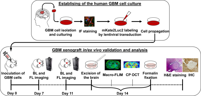
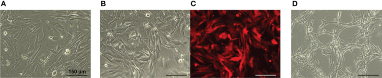
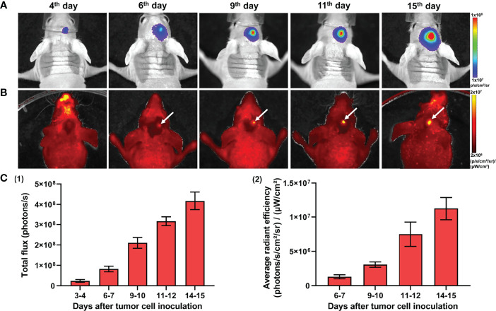
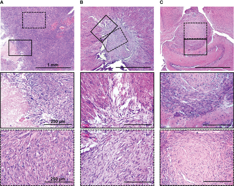
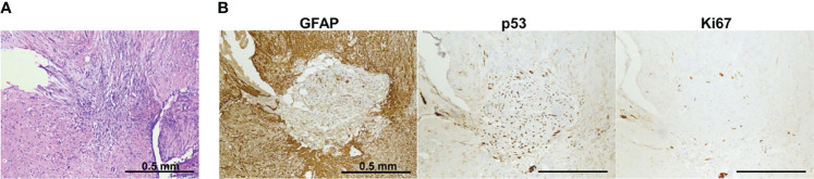
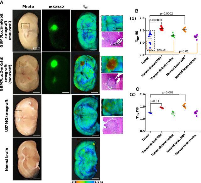
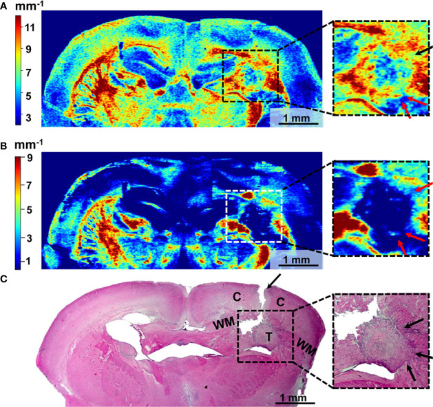
Development of the novel diagnostic and therapeutic approaches in neuro-oncology requires tumor models that closely reproduce the biological features of patients' tumors. Patient-derived xenografts (PDXs) are recognized as a valuable and the most "close-to-patient" tool for preclinical studies. However, their establishment is complicated by the factors related to both the surgical material and technique of the orthotopic implantation. The aim of this work was to develop a patient-derived glioblastoma multiform (GBM) model that stably co-expresses luciferase and a far-red fluorescent protein for monitoring of tumor progression in the brain and, using this model, to validate new diagnostic methods-macroscopic fluorescence lifetime imaging (macro-FLIM) and cross-polarization optical coherence tomography (CP OCT). The established model was similar to the original patient's GBM in terms of histological and immunohistochemical features and possessed reproducible growth in nude mice, which could be observed by both fluorescence and bioluminescence imaging. Our results demonstrated the high potential of macro-FLIM and CP OCT for intraoperative differentiation of GBM from the white matter. Thus, the dual-labeled PDX model of GBM proved to be an excellent approach for observation of tumor development by optical methods.

Keywords: FLIM (fluorescence lifetime imaging microscopy); fluorescence imaging; glioblastoma (GBM); patient-derived xenograft (PDX); primary cell line.


Conflict of interest statement

Author VS was employed by Becker&Hickl GmbH. The remaining authors declare that the research was conducted in the absence of any commercial or financial relationships that could be construed as a potential conflict of interest.

Figures

Figure 1
Figure 1
Design of the experiment. Establishing of the cell culture from the human tissue sample of GMB included isolation from the freshly resected brain tumor specimen, immunofluorescence (IF) staining, lentiviral transduction for dual labeling, cell culturing, and propagation. Validation and analysis of GBM7-Luc2-mKate2 xenograft included in vivo bioluminescence (BL) and fluorescence (FL) imaging, ex vivo macro-FLIM, CP OCT, histological evaluation, and immunohistochemistry (IHC).
Figure 2
Figure 2
Representative IF staining images of the primary human GBM cells. Cell nuclei were counterstained with DAPI. Bars are applicable to all images in the row.
Figure 3
Figure 3
Phase contrast (A, B, D) and fluorescence microscopy (C) of the GBM cell monolayer. (A) GBM7 cells. (B, C) GBM7-Luc2-mKate2 cells. (D) U87 MG cells. Bars are applicable to all images in the row.
Figure 4
Figure 4
In vivo bioluminescence and fluorescence imaging of GBM7-Luc2-mKate2 xenografts. Representative bioluminescence (A) and fluorescence (B) images of the tumor-bearing mice from the 4th to 15th days after tumor cell inoculation. (C) Quantification of the bioluminescence (1) and fluorescence (2) signal during the tumor development. Mean ± SEM, n = 5–7 tumors.
Figure 5
Figure 5
H&E staining of original patient’s tumor (A), dual-labeled human GBM (B), and U87 MG (C) xenografts. Enlarged regions are indicated by the black squares on the lower-magnification panel. Bars are applicable to all images in the row.
Figure 6
Figure 6
IHC characterization of GBM7-Luc2-mKate2 xenograft. Representative H&E (A) and IHC (B) stained sections of a whole tumor. Bars are applicable to all images in the row.
Figure 7
Figure 7
Macro-FLIM of human GBM xenografts and normal brain. (A) Representative auto fluorescence time-resolved images of GBM7-Luc2-mKate2 xenografts, U87 MG xenograft, and normal mouse brain without tumor. Enlarged regions with a tumor are indicated by the black squares on the lower-magnification panel. The corresponding H&E-stained section is presented under each enlarged region. (B) Quantification of the mean fluorescence lifetime tm in the NAD(P)H spectral channel in (1) dual-labeled human GBM xenografts and (2) U87 MG xenografts and normal brain. Scatter dot plot displays the measurements for individual animals (dots) and the mean and SEM (horizontal lines). WM is a white matter.
Figure 8
Figure 8
Wide-field OCT color-coded maps of the mouse brain with GBM7-Luc2-mKate2 tumor (A, B) and corresponding histology (C). Color-coded maps based on calculation of two optical coefficients: attenuation in co-channel (Attco-) (A) and in cross-channel (Attcross-) (B). Perifocal areas of high cancer density are marked with arrows (see enlarged fragments). T, tumor; C, cortex; WM, white matter.

Similar articles

Cited by

References

    1. Tan AC, Ashley DM, López GY, Malinzak M, Friedman HS, Khasraw M. Management of Glioblastoma: State of the Art and Future Directions. CA A Cancer J Clin (2020) 70:299–312. doi: 10.3322/caac.21613 - DOI - PubMed
    1. Oronsky B, Reid TR, Oronsky A, Sandhu N, Knox SJ. A Review of Newly Diagnosed Glioblastoma. Front Oncol (2021) 10:574012. doi: 10.3389/fonc.2020.574012 - DOI - PMC - PubMed
    1. Hicks WH, Bird CE, Traylor JI, Shi DD, El Ahmadieh TY, Richardson TE, et al. . Contemporary Mouse Models in Glioma Research. Cells (2021) 10:712. doi: 10.3390/cells10030712 - DOI - PMC - PubMed
    1. Haddad AF, Young JS, Amara D, Berger MS, Raleigh DR, Aghi MK, et al. . Mouse Models of Glioblastoma for the Evaluation of Novel Therapeutic Strategies. Neuro-Oncol Adv (2021) 3:vdab100. doi: 10.1093/noajnl/vdab100 - DOI - PMC - PubMed
    1. Golebiewska A, Hau A-C, Oudin A, Stieber D, Yabo YA, Baus V, et al. . Patient-Derived Organoids and Orthotopic Xenografts of Primary and Recurrent Gliomas Represent Relevant Patient Avatars for Precision Oncology. Acta Neuropathol (2020) 140:919–49. doi: 10.1007/s00401-020-02226-7 - DOI - PMC - PubMed