Case Reports
. 2022 Jul 14:12:914379.
doi: 10.3389/fonc.2022.914379. eCollection 2022.

Case Report: Monoclonal Gammopathies of Clinical Significance-Associated Myopathy: A Case-Based Review

Affiliations
Case Reports

Case Report: Monoclonal Gammopathies of Clinical Significance-Associated Myopathy: A Case-Based Review

Hongbin Yu et al. Front Oncol. .

Abstract

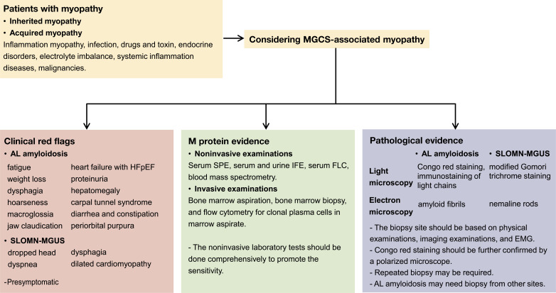
Monoclonal gammopathies of clinical significance (MGCS)-associated myopathy is a group of muscular MGCS-based rare manifestations. It mainly includes amyloid light chain (AL) amyloidosis and sporadic late-onset nemaline myopathy with monoclonal gammopathy of undetermined significance. When myopathy manifests as the initial or sole clinical symptom, it can often be delayed or misdiagnosed as other myopathies. We report the case of a 60-year-old man who initially presented with fatigue and muscle weakness of the symmetric proximal lower limbs. Muscle biopsy did not reveal mononuclear cell infiltration, atrophy, necrosis, or positive Congo red staining results. The results of serum protein electrophoresis and immunofixation electrophoresis were negative. No specific diagnosis was established. After 1 year, the patient was diagnosed with AL amyloidosis after myocardial and fat pad biopsies were performed and myopathy was diagnosed as AL amyloidosis-associated myopathy after reassessment. The patient received CyBorD regime chemotherapy and achieved hematological and organ remission. Therefore, we reviewed the clinical and pathological manifestations of MGCS-associated myopathies. Based on published articles and the present case, we conclude that comprehensive screening for MGCS in unexplained myopathy is essential to avoid misdiagnosis or delayed diagnosis.

Keywords: MGCS; MGUS; amyloidosis; myopathy; nemaline myopathy.


Conflict of interest statement

The authors declare that the research was conducted in the absence of any commercial or financial relationships that could be construed as a potential conflict of interest.

Figures

Figure 1
Figure 1
Magnetic resonance imaging results showing abnormality (A–D). Increased signal intensity could be detected in both T1-weighted (A, B) and T2-weighted with fat suppression (C, D) images, indicating muscle atrophy and edema. The pelvic muscles and inner and posterior muscles of both thighs were affected heavily. Pathology studies of the skeletal muscle showing amyloid deposits (E–J). Muscle biopsy obtained from the patient showing slight myofibers atrophy on hematoxylin and eosin (H&E)-stained section (E). Weak congophilic deposits within the blood vessel walls in Congo red-stained section visualized under light microscopy (F, G, black arrow). λ light chain immunofluorescent staining showed a positive staining in blood vessel walls and perimysium (H, white arrow), and the perimysium involvement was not detected in Congo red staining. κ light chain immunofluorescent staining showed a negative result (I), indicating a λ light chain restriction. Congo red-stained specimen viewed via polarized microscopy revealed positive apple-green birefringence of amyloid deposits (J, white arrow). Magnifications (×): 100 in (E, F, H, I); 400 in (G, J).
Figure 2
Figure 2
Time and events of the case report. The level of creatine kinase considerably dropped after the initiation of CyBorD regime chemotherapy and is within the normal range at present. Hematological remission was also achieved soon after chemotherapy. Clinical symptoms were alleviated after six cycles of chemotherapy.
Figure 3
Figure 3
Workup for patients suspected of MGCS-associated myopathy. Clinical red flags if presented may suggest AL amyloidosis or SLOMN-MGUS. Systemic damages are common in AL amyloidosis, and some presymptomatic signs should also be noted in AL amyloidosis. M protein screening (comprehensive noninvasive and invasive bone marrow examinations) is required for diagnosis. Pathological evidence is crucial for MGCS-associated myopathy diagnosis. Special staining methods such as Congo red staining for AL amyloidosis and modified Gomori trichrome staining for SLOMN-MGUS are useful. Light chain immunostaining is only useful for AL amyloidosis. Electron microscopy is indispensable for differential diagnosis.

References

    1. Dispenzieri A. Monoclonal Gammopathies of Clinical Significance. Hematology (2020) 2020(1):380–8. doi: 10.1182/hematology.2020000122 - DOI - PMC - PubMed
    1. Merlini G, Dispenzieri A, Sanchorawala V, Schönland SO, Palladini G, Hawkins PN, et al. . Systemic Immunoglobulin Light Chain Amyloidosis. Nat Rev Dis Primers (2018) 4(1):38. doi: 10.1038/s41572-018-0034-3 - DOI - PubMed
    1. Muchtar E, Derudas D, Mauermann M, Liewluck T, Dispenzieri A, Kumar SK, et al. . Systemic Immunoglobulin Light Chain Amyloidosis–Associated Myopathy: Presentation, Diagnostic Pitfalls, and Outcome. Mayo Clinic Proc (2016) 91(10):1354–61. doi: 10.1016/j.mayocp.2016.06.027 - DOI - PubMed
    1. Pinto MV, Dyck PJB, Liewluck T. Neuromuscular Amyloidosis: Unmasking the Master of Disguise. Muscle Nerve (2021) 64(1):23–36. doi: 10.1002/mus.27150 - DOI - PubMed
    1. Lundberg IE, Tjärnlund A, Bottai M, Werth VP, Pilkington C, de Visser M, et al. . European League Against Rheumatism/American College of Rheumatology Classification Criteria for Adult and Juvenile Idiopathic Inflammatory Myopathies and Their Major Subgroups. Arthritis Rheumatol (2017) 69(12):2271–82. doi: 10.1002/art.40320 - DOI - PMC - PubMed

Publication types

LinkOut - more resources