. 2022;89(2):535-551.
doi: 10.3233/JAD-220333.

The Clinical Use of Alzheimer's Disease Biomarkers in Patients with Mild Cognitive Impairment: A European Alzheimer's Disease Consortium Survey

Affiliations

The Clinical Use of Alzheimer's Disease Biomarkers in Patients with Mild Cognitive Impairment: A European Alzheimer's Disease Consortium Survey

Camilla Caprioglio et al. J Alzheimers Dis. 2022.

Abstract

Background: Recent advances occurred in the field of Alzheimer's disease (AD) biomarkers and the introduction of a research framework grounded on a biomarker-based definition of AD might have fostered an increased clinical use of AD biomarkers. For this reason, an up-to-date depiction of the clinical use of AD biomarkers is needed.

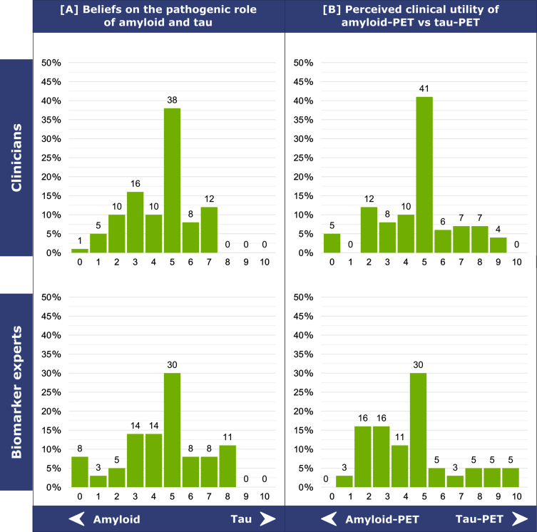
Objective: To investigate the clinical use of the main AD biomarkers in patients with mild cognitive impairment (MCI) by examining the beliefs and preferences of professionals (clinicians and biomarker experts) of the European Alzheimer's Disease Consortium (EADC).

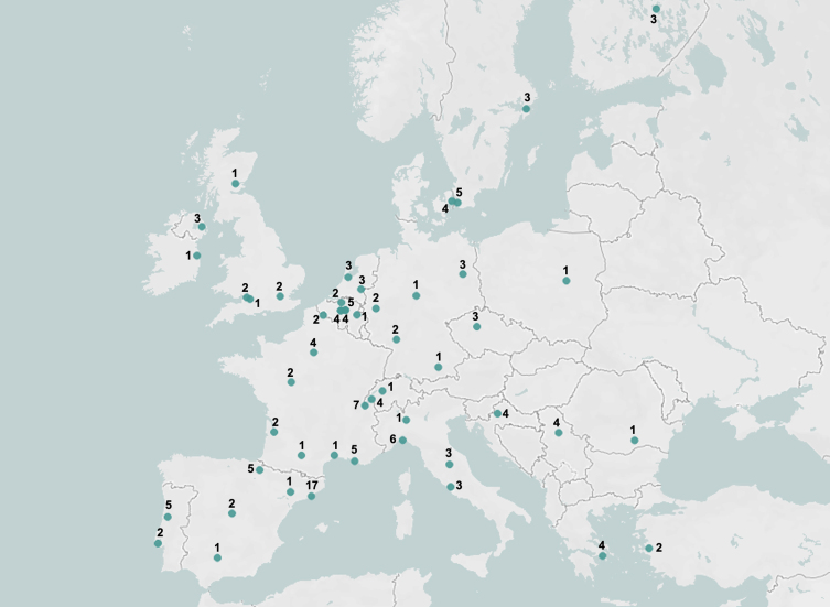
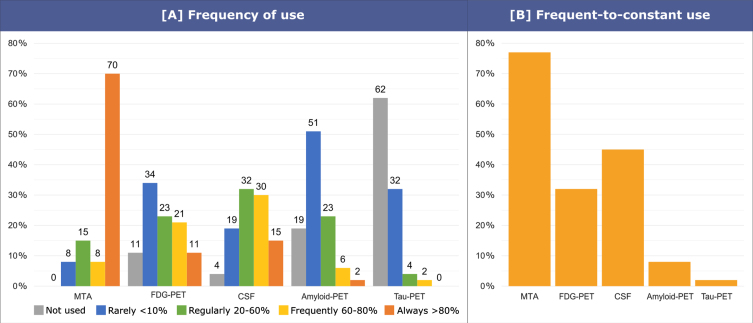
Methods: 150 professionals filled in an online survey from May to September 2020. The investigated biomarkers were medial temporal lobe atrophy score (MTA) on structural MRI, typical AD (i.e., temporoparietal and posterior cingulate) hypometabolism on FDG-PET, CSF (Aβ42, p-tau, t-tau), amyloid-PET and tau-PET.

Results: The frequency of responders reporting a frequent-to-constant use of MTA (77%) is higher than that of those reporting a frequent-to-constant use of the other AD biomarkers (i.e.

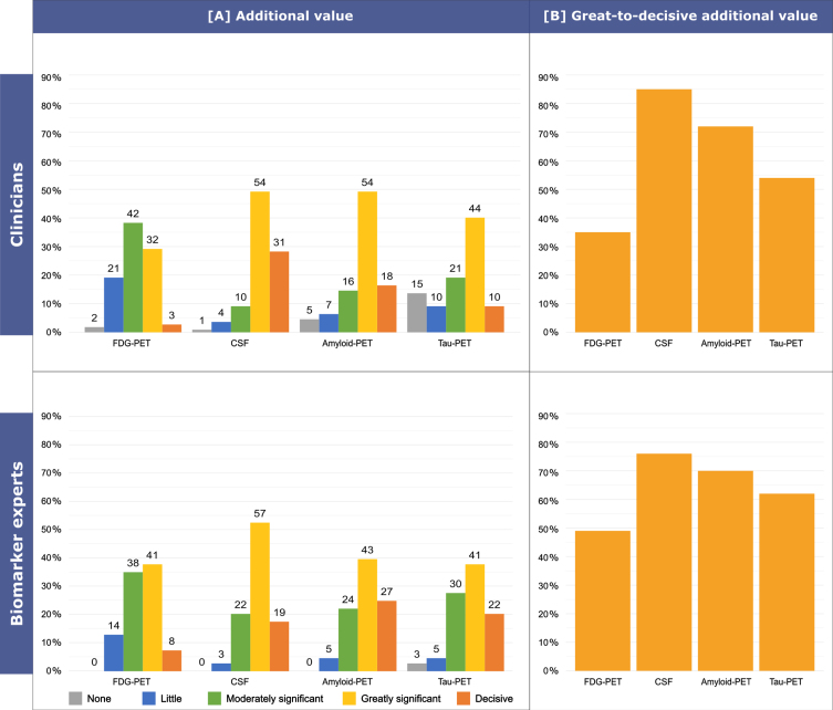
, csf: 45%, p = 0.014; FDG-PET: 32%, p < 0.001; amyloid-PET: 8%, p < 0.001; and tau-PET: 2%, p < 0.001). CSF is considered the most valuable biomarker in terms of additional diagnostic value, followed by amyloid-PET, tau-PET, and typical AD hypometabolism on FDG-PET.

Conclusion: AD biomarkers are widely used across European memory clinics with a clinical research background for the diagnosis of MCI. Overall, we observed that CSF is currently considered as the most useful biomarker, followed by amyloid-PET.

Keywords: APOE; Alzheimer’s disease; FDG-PET; amyloid-PET; biomarkers; cerebrospinal fluid; clinical use; magnetic resonance imaging; mild cognitive impairment; tau-PET.


Conflict of interest statement

Authors’ disclosures available online (https://www.j-alz.com/manuscript-disclosures/22-0333r2).

Figures

Fig. 1
Fig. 1
Geographical distribution of the responders and number of responders per center.
Fig. 2
Fig. 2
Beliefs on the pathogenic role of amyloid and tau in AD [A] and perceived clinical utility of amyloid-PET versus tau-PET in MCI and mild dementia [B]. (A) The question asked to responders was: “What is your belief/opinion about the pathogenic role of amyloid and tau in Alzheimers disease pathology and symptoms?”. Answers were grouped into three categories: 0–4, favoring the abnormal accumulation of amyloid as the initial cause of AD; 5, amyloid and tau have the same relevance in causing AD, or neither amyloid nor tau are the initial cause of AD; 6–10, favoring the abnormal accumulation of tau as the initial cause of AD. (B) The question posed to responders was: “Independent of any specific patients feature and based on your clinical experience with patients usually seen in your memory clinic, what is, in your opinion, the most clinically useful exam for etiological diagnosis of MCI and mild dementia?”. Answers were grouped into three categories: 0–4, favoring amyloid-PET; 5, neutral; 6–10, favoring tau-PET.
Fig. 3
Fig. 3
Frequency of use of AD biomarkers in MCI patients. The question asked to clinicians was: “In MCI, in your clinical practice, please state frequency of use for medial temporal lobe atrophy (MRI), FDG-PET, CSF (e.g., A β42, p-tau, t-tau), amyloid-PET, tau-PET”. Possible answers were: not used (0%), rarely (<10%), regularly (20–60%), frequently (60-80%), always (>80%). Answers were grouped into three categories: no use, rare-to-regular use (rarely or regularly), frequent-to-constant use (frequently or always).
Fig. 4
Fig. 4
Use of AD biomarkers to support etiological diagnosis in MCI. The question asked to clinicians was: “Do you use imaging biomarkers / CSF collection (e.g., A β42, p-tau, t-tau) / APOE genotyping to support your etiological diagnosis in MCI?”. Possible answers were yes or no. Clinicians reported to use imaging to support their etiological diagnosis in MCI in 90% of cases, CSF in 87% of cases and APOE in 27% of cases.
Fig. 5
Fig. 5
Additional value over neuropsychological testing and structural MRI in MCI. The question asked to responders was: “Assuming that clinical examination with neuropsychological testing and brain structural MRI are the most feasible procedures in most memory clinics, please rate the additional diagnostic value (i.e., the ability to provide diagnostic information in excess of that already provided by neuropsychological testing and brain structural MRI) in an MCI patient of FDG-PET, CSF markers (e.g., Aβ42, p-tau, t-tau), amyloid-PET, tau-PET”. Possible answers were: none, little, moderately significant, greatly significant, decisive. Answers were grouped into three categories: none-to-little (none or little), moderate (moderately significant), great-to-decisive (greatly significant or decisive).
Fig. 6
Fig. 6
Confidence in an etiological diagnosis of AD in MCI. The following case vignette was proposed to responders: “A 75 years old person comes into your office complaining of memory deterioration in the past 612 months, he/she is in good physical health, has no problems in his/her daily chores, but is clearly worried. Routine labs are normal, but he/she performs 1.5 SD below the age-and education-adjusted mean on a test of verbal or non-verbal recall. How confident would you be with a diagnosis of MCI due to AD (or prodromal AD) on the basis of i) evidence of clear-cut medial temporal lobe atrophy alone, ii) clear-cut temporoparietal and posterior cingulate hypometabolism on FDG-PET alone, iii) clearly abnormal CSF levels of Aβ and tau alone, iv) clearly positive amyloid-PET, v) clearly positive tau-PET, vi) at least one clearly positive amyloid marker and at least one clearly positive neuronal injury marker.”. Possible answers were: not at all comfortable, moderately comfortable, comfortable, very comfortable, extremely comfortable. Answers were grouped into three categories: not comfortable, sufficiently comfortable (moderately comfortable or comfortable), very-to-extremely comfortable (very comfortable or extremely comfortable).

References

    1. Dubois B, Hampel H, Feldman HH, Scheltens P, Aisen P, Andrieu S, Bakardjian H, Benali H, Bertram L, Blennow K, Broich K, Cavedo E, Crutch S, Dartigues JF, Duyckaerts C, Epelbaum S, Frisoni GB, Gauthier S, Genthon R, Gouw AA, Habert MO, Holtzman DM, Kivipelto M, Lista S, Molinuevo JL, O’Bryant SE, Rabinovici GD, Rowe C, Salloway S, Schneider LS, Sperling R, Teichmann M, Carrillo MC, Cummings J, Jack CR (2016) Preclinical Alzheimer’s disease: Definition, natural history, and diagnostic criteria. Alzheimers Dement 12, 292–323. - PMC - PubMed
    1. Jack CR, Bennett DA, Blennow K, Carrillo MC, Dunn B, Haeberlein SB, Holtzman DM, Jagust W, Jessen F, Karlawish J, Liu E, Molinuevo JL, Montine T, Phelps C, Rankin KP, Rowe CC, Scheltens P, Siemers E, Snyder HM, Sperling R, Elliott C, Masliah E, Ryan L, Silverberg N (2018) NIA-AA Research Framework: Toward a biological definition of Alzheimer’s disease. Alzheimers Dement 14, 535–562. - PMC - PubMed
    1. Ramusino MC, Garibotto V, Bacchin R, Altomare D, Dodich A, Assal F, Mendes A, Costa A, Tinazzi M, Morbelli SD, Bauckneht M, Picco A, Dottorini ME, Tranfaglia C, Farotti L, Salvadori N, Moretti D, Savelli G, Tarallo A, Nobili F, Parapini M, Cavaliere C, Salvatore E, Salvatore M, Boccardi M, Frisoni GB (2020) Incremental value of amyloid-PET versus CSF in the diagnosis of Alzheimer’s disease. Eur J Nucl Med Mol Imaging 47, 270–280. - PubMed
    1. Altomare D, Caprioglio C, Assal F, Allali G, Mendes A, Ribaldi F, Ceyzeriat K, Martins M, Tomczyk S, Stampacchia S, Dodich A (2021) Diagnostic value of amyloid-PET and tau-PET: A head-to-head comparison. Eur J Nucl Med Mol Imaging 48, 2200–2211. - PMC - PubMed
    1. Barthel H, Sabri O (2017) Clinical use and utility of amyloid imaging. J Nucl Med 58, 1711–1717. - PubMed

Publication types